JANUARY 2020
VOLUME 1 // ISSUE 1
THE DORSAL COLUMN SHARING NEUROSCIENCE RESEARCH WITH OUR BROADER COMMUNITY parietal cortex
WELCOME TO OUR FIRST ISSUE
occipital cortex
BY RAMINA ADAM
It is a pleasure to welcome you to the very first issue of The Dorsal Column – a quarterly publication that will share stories about brain research with the broader London community. Each issue will feature articles regarding neuroscience research carried out in affiliation with Western University and will be written and reviewed by a team of graduate students. Our mission is to strengthen public engagement and enthusiasm for science by bringing research out of the lab and into the community.
IN THIS ISSUE Welcome Letter By Ramina Adam
1
‘Micro’ Cells with Major Functions: Scientists discover new pathway for brain immunity By Vasiliki Tellios
4
Alzheimer’s disease: Losing behavioural flexibility By Ariel Frame
5
PET project to highlight the trace of death in life By Simon Benoit
8
Small RNA Molecules May Explain a Hallmark of ALS By Dika Ojiakor
9
Hair, Cortisol, and Mummies By Sam Mestern
11
Major Assumption of fMRI Likely Correct By Nicholas Handfield-Jones
13
“Hand-in-Hand”: How the brain handles a missing body part By Kartik Pradeepan
15
Epilepsy Provides Insight into Déjà Vu Phenomenon By Julia Sunstrum
17
frontal cortex temporal cortex
brainstem
cerebellum
spinal cord Illustration of a human brain.
The origin behind our name, The Dorsal Column, is quite fitting: “the dorsal column” is a pathway in your spinal cord that is made up of a bundle of nerves that bring information from your body to your brain. Try this: ask someone to trace a number on the palm of your hand using their index finger while you look away or close your eyes. Can you guess what number they traced on you? You can thank your dorsal column for that! Those sensations you felt on your skin were sent to your brain through nerves that travel through the dorsal column in your spine. Much like the dorsal column, we are here to bring information from one area (the research lab) to another (our community)!
THE IMPORTANCE OF SCIENCE COMMUNICATION
Since taxpayers fund the majority of our research, scientists and trainees have a responsibility to publicly share research findings to showcase the exciting and valuable progress achieved to date. We ...continued on pg. 3
SOCIETY OF NEUROSCIENCE GRADUATE STUDENTS
1
JANUARY 2020
VOLUME 1 // ISSUE 1
VOLUNTEER TEAM Founders Ramina Adam Faraj Haddad Editor in Chief Ramina Adam Senior Editor Faraj Haddad Reviewing Editors Aaron Cecala Niveen Fulcher Zachary Hawley Rachel Lackie Megan Roussy Web Editor Simon Benoit Social Media Vasiliki Tellios
2
Layout Editor Josephine Pham Community Liaisons Indra Bishnoi Krystyna Wieczerzak Visual Communications Kelly Bullock Editor Writers in this issue Simon Benoit Nicholas Handfield-Jones Sam Mestern Dika Ojiakor Kartik Pradeepan Julia Sunstrum Vasiliki Tellios
THE DORSAL COLUMN believe it is important to educate others about the small advances in our research area that usually fly under the radar for any non-specialist. Scientific discovery is rarely defined by a single “eureka” moment – instead, research findings build on each other over time and that is what has typically led to groundbreaking discoveries. We hope that if there is an understanding of the slow but incremental progress garnered by basic research, public trust in scientists and the value of research to society long-term will be better appreciated. But beyond the responsibility to share science with taxpayers, science communication is especially important at a time when pseudoscientific beliefs are rampant. It is absolutely crucial for scientists to engage with a wide audience and to share research findings outside of the echo chamber of academic conferences. We hope that by engaging everyone in science, we are building a community that appreciates the scientific method and can recognize and question pseudoscience in the media.
OUR ORIGIN STORY
Faraj Haddad and I are PhD students in the Neuroscience program at Western University and only recently learned that we share a common interest in science communication and outreach. We both attended the 2019 London Health Research Day and were completely inspired by the keynote talk on the importance of science communication by Timothy Caulfield, a professor at the University of Alberta who specializes in health law and science policy and who is a fierce advocate for debunking pseudoscience. We felt that we had no choice but to take action and so we put
VOLUME 1 // ISSUE 1
together a ‘Science Communication and Outreach Committee’ within the Society of Neuroscience Graduate Students and thus was born The Dorsal Column. Since graduate students receive minimal, if any, training in effective science communication, we envisioned The Dorsal Column as an opportunity for us all to sharpen our writing skills and become engaging scientific storytellers. On that note, we owe many thanks to Emily Leighton, Communications Specialist with Schulich School of Medicine & Dentistry, and Crystal Mackay, the Media Relations Officer at Western University, for hosting an engaging and insightful workshop on the art of science writing to get us started. We will be publishing four issues per year that feature peer-reviewed research summary articles using a double-blinded review process. The articles summarized in The Dorsal Column involve research conducted by scientists affiliated with Western University and published in peer-reviewed scientific journals within the past five years. We chose to focus on local research in order to highlight the strong scientific community residing in London and to foster a dialog between scientists and the wider community.
SOCIETY OF NEUROSCIENCE GRADUATE STUDENTS
3
JANUARY 2020
VOLUME 1 // ISSUE 1
‘MICRO’ CELLS WITH MAJOR FUNCTIONS SCIENTISTS DISCOVER NEW PATHWAY FOR BRAIN IMMUNITY BY VA S I L I K I T E L L I O S
T
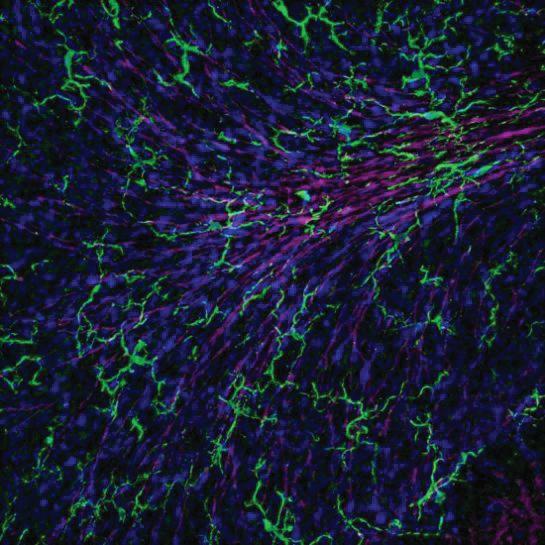
hink of your brain like a bustling city. Neurons – the principle functional cells in the brain – act as roads, carrying important information from one area of town to the other. Like any complex city, the key to efficient transportation is road maintenance and infrastructure. This is where microglia, a supporting brain cell, comes into play. Like their name suggests, microglia are tiny cells – no larger than 10 microns in length, or the equivalent of one tenth the diameter of a grain of salt. These cells work behind the scenes to Fluorescent tagging of microglia (green) shows intricate ensure healthy neuronal function. processes interacting with neuronal structures (pink) and cell bodies (blue). Image supplied by the Lu Lab, Just like construction workers, unpublished. microglia function to modify neurons and clean up any harmful debris neighbouring cells, including neurons. like bacteria, dead cells or faulty neurons, “Neurons are regulated by glia, and by that might affect overall neuronal studying these supporting cells of the activity. brain, we can provide new insight into brain disorders we don’t fully Traditionally, brain research has focused understand,” said lead author Matthew on understanding how neurons work. Maksoud, a PhD Candidate in the This research is warranted, considering Graduate Program of Neuroscience at neurons are the basic functional unit of Western University. the nervous system, responsible for relaying critical signals throughout the Specifically, the research team led by Dr. brain that may allow one to think, move, Wei-Yang Lu proposed a new link and interact with their surrounding between nitric oxide – a well-known environment. However, neuroscientists mediator of brain inflammation – and are only just starting to understand how microglia’s ability to ‘clean up’ harmful microglia support and regulate neuronal bacteria in the brain. Microglia can function. A recent paper published this remove bacteria through a process called year by researchers at Western University phagocytosis, which results in these cells in the journal, Glia, presents a new destroying and engulfing unwanted signaling pathway in microglia that has debris within the brain. The authors the potential to affect the function of
4
THE DORSAL COLUMN
VOLUME 1 // ISSUE 1
showed the ability of a microglial cell to perform phagocytosis relies heavily on the presence of nitric oxide, an inflammatory molecule produced by microglia. “Nitric oxide is an interesting molecule, because in the past it has been looked at as solely detrimental. It’s not that this molecule is negative, but there needs to be a balance.” Think back to our city. Microglia work to keep our neuronal ‘roads’ clean and debris-free. An abundance of nitric oxide results in microglia working overtime – over-modifying neuronal structures until they are rendered ineffective. A lack of nitric oxide leads to excessive clutter, again rendering neurons ineffective at transporting signals.
microglia, respectively. “What we essentially discovered was that two individual processes already known to affect microglia phagocytosis – endogenous nitric oxide production, and a specific calcium ion channel – are actually part of the same signaling pathway,” said Maksoud. “Phagocytosis is such an important part of brain function and dysfunction, so discovering a new mechanism of action can hopefully uncover new ways to modulate microglia function in health and disease.” This new mechanism behind phagocytosis is just the start of emerging microglia research. With a solid foundation underlying the relationship between nitric oxide and calcium ion channels, Maksoud hopes to see this research field progress using a disease model. “From the literature we also see similar trends with nitric oxide and calcium ion channels in diseases such as brain tumors. Examining this pathway with respect to this type of pathology could be beneficial in uncovering new, potent therapeutic treatments.”
Using various microscopic cellular imaging methods, Maksoud examined phagocytosis in microglia that were unable to produce nitric oxide. Interestingly, microglia lacking nitric oxide production were not as successful in removing bacterial debris compared to normal microglia. Less phagocytosis was not only attributed to decreases in nitric oxide, but also decreased levels of calcium ion channels, which are proteins on the surface of microglia. These calcium ion channels transport calcium in and out of the cell and are LOSING BEHAVIOURAL FLEXIBILITY necessary for cleaning up those BY ARIEL FRAME potentially harmful bacteria.
ALZHEIMER’S DISEASE
If microglia act as city workers, calcium ion channels work to provide essential resources to microglia, such as the ability to change shape and structure in order to better clean up debris within their environment. Just like nitric oxide, too much or too little calcium ion channel function can overwork or underwork
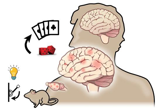
On board game night, you embark on a
night of fun, moving tiny figurines around a table, picking up various playing cards, and trying to outcompete your friends. This is a familiar setting. Every week the same friends join you to play the same three games you happen to
SOCIETY OF NEUROSCIENCE GRADUATE STUDENTS
5
JANUARY 2020 own, each with rules you have learned by heart. That is until, a new player arrives. This novel companion claims to know these games you have been playing for years, but that they play by different rules. How well will you be able to compete under these circumstances? Will you be able to adapt your well learned strategies to accommodate the new rules? If this does not sound like a challenge to you, you probably have high behavioural flexibility. Behavioural flexibility is a function of the brain that allows an animal to learn rules of a game then switch their strategy for success when the rules change. If you find yourself struggling to accommodate new rules during board game night, you may be having a hard time switching strategies and this could be an indication of a problem in the brain. Alzheimer’s disease (AD) is a neurodegenerative disease most commonly occurring in the elderly, with a brutal decline in cognitive ability over the course of decades. A new study published in the journal Brain Behaviour and Immunity by Alexander Levit, an MD/PhD Candidate working in Dr. Shawn Whitehead’s research laboratory at Western University, shows how impaired behavioural flexibility could be an indication of AD onset caused by particular changes in the brain’s immune system. To make this discovery, Dr. Whitehead’s group used a special breed of genetically modified rats, TgAPP21, that have the human version of an AD associated gene expressed in their brain. In fact, these rats’ brains have the same abnormal gene found in people who have AD onset extremely early; typically younger than 65. Whereas many studies
6
VOLUME 1 // ISSUE 1
Adapted from image by CNX OpenStax under Creative Commons Attribution 3.0 Unported license
have used rodent models with severe brain pathology and age-related memory decline to study AD, Dr. Whitehead’s group chose to use this particular breed of rats at an age where these hallmarks of late stage AD are not present. Instead, these rats exhibit brain pathology and behaviour more reminiscent of early AD. Modeling early AD allows for understanding of a stage of AD where therapeutic intervention is more feasible compared to late stages in the disease where too much of the brain is already lost. A test of behavioural flexibility was done with the genetically modified rats. This test involved training rats to press a lever only upon appearance of a light above it to receive a sugar reward. Training continues until this rule is just as easy to remember for the rat as it is to remember doubles in the game Monopoly means roll again. Then, Dr. Whitehead’s group changed the rules. Now, the rats can only receive a reward by pressing the lever to one side, regardless of the light’s position. If rats are unable to adhere to this new rule, then this is an indication of a behavioural flexibility impairment.
THE DORSAL COLUMN The results of Dr. Whitehead’s study demonstrated that this specific breed of genetically modified rats exhibit impaired behavioural flexibility - these rats could not switch their strategy well. This has a clear similarity to the cognitive impairments of AD patients. Just as cognitive impairments in AD patients generalize to many daily tasks, the deficits shown by these rats were generalized to other measures of behavioural flexibility. Finally, Dr. Whitehead’s group sought to investigate the underpinnings of behavioural flexibility impairments and decided that brain inflammation was a good candidate because it has been shown to occur in the brain of AD patients. Just as a grazed knee becomes hot, red, and swollen, so does the brain when it is injured. This physiological response is called inflammation and it is mediated by immune cells. In the brain, microglia are the immune cells which are activated when certain regions are inflamed. To draw a connection between the two phenomena, behavioural inflexibility and brain inflammation, Dr. Whitehead’s group sought after evidence of inflammation in the brain of their genetically modified rats to see if there was a correlation with behavioural flexibility. Dr. Whitehead’s group uncovered high levels of brain inflammation in these rats, quantified as level of activated microglia, that could significantly predict how impaired each rat’s behavioural flexibility was in the lever pressing task. There are many reasons why one might want to stick to the classics and continue to play old games like Monopoly and Clue. But, if there is any way to help
VOLUME 1 // ISSUE 1 prevent AD patients from being unable to learn new rules to games and generally help prevent decline in cognitive abilities such as behavioural flexibility, then discovering the molecular underpinnings of AD is crucial. These findings from Dr. Whitehead’s lab demonstrate that brain inflammation occurs in a model of early stage AD and correlates well with impaired behavioural flexibility, which means that inflammation could be part of what triggers AD initially and warrants further investigation if preventative measures that reduce brain inflammation are to be discovered.
PET PROJECT TO HIGHLIGHT THE TRACE OF DEATH IN LIFE BY SIMON BENOIT
T
here is a little known fact that the average adult loses about 10 billion cells per day through a process called apoptosis or “cell death”, which is where cells undergo a form of programmed death. Normally, this highly predictable and controlled process is necessary to maintain normal functioning of the human body. For example, during human development in the womb, the fingers of our hand are formed after programmed death of the cells in between them. However, abnormal
SOCIETY OF NEUROSCIENCE GRADUATE STUDENTS
7
JANUARY 2020
VOLUME 1 // ISSUE 1
increases in cell death have been linked to neurodegenerative diseases like Alzheimer’s and Parkinson’s disease. Alternatively, decreased cell death can lead to abnormal cell growth and potentially to the development of various cancers. As such, developing adequate tools to study apoptosis is essential to gain a better understanding of a wide spectrum of human diseases. Until now, no adequate method existed to study this process in living beings, where early detection of abnormal cell death could mean major advances in the treatment of neurodegenerative diseases. A multi-disciplinary team of researchers led by Marco Prado and Robert Bartha from the Robarts Research Institute at Western University have been able to accomplish exactly this. In their recently published work in the journal Contrast Media and Molecular Imaging, the team developed a radioactive molecule called a ‘tracer’ which is almost exclusively retained in cells undergoing apoptosis.
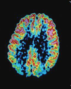
Example of PET imaging using a glucose tracer in the brain of an Alzheimer’s disease patient. Areas in red have the highest glucose uptake while blue depicts the lowest.
promising collection of data that will be useful in the development of novel clinical approaches. Thus far, the researchers showed that cells undergoing programmed cell death are efficiently labelled by the tracer. They performed experiments using brain cell cultures, brain cells that have been grown under controlled conditions that mimic those in the body (optimal temperature, pressure, supply of important nutrients, etc.). The researchers exposed those cultures first to a compound that directly induced cell death and to two forms of physiological stress that mimic the conditions in stroke and Alzheimer’s disease (both of which induce apoptosis).
This tracer, which functions like a neon sign pointing directly at these cells, makes them visible using the widely available positron-emission tomography (PET) scanner. Dying cells can be observed as a bright highlighted area using this technology (see image for an example), which is already widely used in the hospital setting for testing heart function, brain function and to help locate and identify tumours. PET can also be combined with other imaging techniques to simultaneously obtain Keep in mind that physiological stress is information about human anatomy. This different from the types of stress that you combination of techniques can provide a and I may be more familiar with in our
8
THE DORSAL COLUMN day-to-day (hopefully not too familiar!), like when work deadlines are approaching. For the stress imitating stroke, brain cell cultures were deprived of glucose and oxygen, two essential elements to sustain life, which led to cell death. In the second form of stress, the cultured cells were exposed to a protein that is typically associated with Alzheimer’s disease, which also led to cell death.
VOLUME 1 // ISSUE 1
SMALL RNA MOLECULES MAY EXPLAIN A HALLMARK OF ALS BY DIKA OJIAKOR
Amazingly, the tracer labelled dying cells in all three conditions. This led the researchers to try it in models of stroke and Alzheimer’s disease. Again, the tracer allowed clear identification of the loss of brain cells in areas that are typically associated with these disorders. Taken together, the results of this teams’ work hold promise for applications to many human conditions, including earlier diagnosis of neurodegenerative diseases based on the location of cell death or to measure the effect of treatments on apoptosis. For example, the authors suggested that this tracer could be used to see the impact of cancer treatments on tumours, where an increase in signal from the tracer would indicate that the cancer cells are dying as hoped. However, despite the tracer not showing any negative side effects in the brain cells or in this study, it will be very important to ensure safety in humans before we can hope to use it. This important work brings us much closer to seeing that reality.
In
the summer of 2015, a year after millions of people dumped buckets of ice water over their heads to promote awareness of amyotrophic lateral sclerosis or ALS, Zachary Hawley made the decision to join a lab at Western University focused on understanding the cellular biology of ALS. Nearly five years later, Hawley is the lead author on a recent study published in Brain Research that might explain the molecular origins of the disease. ALS is a progressive neurodegenerative disease caused by the death of motor neurons – long and stringy brain cells that control some of our most basic operations, such as the ability to breathe, walk, speak or swallow. Perhaps no statistic to do with ALS is more telling than that the life expectancy of a person with the disease ranges from 2-5 years after diagnosis. One of its major hallmarks is the formation of sticky, microscopic aggregates caused by changes in the abundance of specific proteins, called intermediate filaments. These proteins are needed to maintain the structure and integrity of motor neurons. “The question to us became why do we see these changes in intermediate
SOCIETY OF NEUROSCIENCE GRADUATE STUDENTS
9
JANUARY 2020
VOLUME 1 // ISSUE 1
filaments such that they’re pooling,” said Hawley, referring to the aggregation of intermediate filaments that occurs in ALS. “And that’s a critical point because intermediate filaments are as important to a neuron as the skeleton is to a human being – they provide that nice structure so that neurons can function normally and send signals.”
The study found that two microRNAs – small genetic molecules that control the amount of intermediate filament produced by motor neurons – are reduced within the spinal cord of ALS patients. This triggers a chain of events that leads these proteins to pool within motor neurons, eventually causing them to lose their strand-like structures and die.
“Intermediate filaments are as
Based on these findings, the research team suggests that restoring normal levels of these microRNAs (called miR-105 and miR-9) could slow down or reverse the progression of ALS by preventing intermediate filaments from pooling within motor neurons. Such an intervention could have life-altering consequences for ALS patients, as the only drugs currently approved for ALS treatment show minimal health benefits. However, Hawley believes there is work still to be done before the development of microRNA-based clinical interventions can occur.
important to a neuron as the skeleton is to a human being”
“MicroRNAs are a relatively new field of study in the context of ALS so we still know very little about them,” said Hawley. “The problem is that we still don’t understand why these microRNAs are dysregulated. We know they’re causing a lot of issues, but now we need to figure out why they’re causing these issues, or why they’re so dysregulated in ALS patients.”
Doing the ALS Ice Bucket Challenge: a young boy dumps a pail of ice water over his head to promote awareness of amyotrophic lateral sclerosis or ALS
10
Now in the final year of his PhD, Hawley is soon joining the lab of Kevin Eggan at Harvard University as a visiting scholar, where he hopes to gain a better understanding of the causes of microRNA dysregulation in ALS. “Our models are very limited,” he said. “So
THE DORSAL COLUMN I’m going there to test some of my hypotheses in actual motor neurons from ALS patients to see if what I’m thinking is actually correct.” Hawley says he is still interested in studying neurodegenerative diseases like ALS after completing his doctoral studies. The research team hopes to follow up this study by identifying the specific features that drive microRNA dysregulation and protein aggregation in the disorder.
HAIR, CORTISOL & MUMMIES HOW YOUR BRAIN’S STRESS RESPONSE CAN LEAVE MARKS ON YOUR BODY THAT LAST A MILLENNIUM BY SAM MESTERN
A
VOLUME 1 // ISSUE 1 a sudden plummet in stress levels. Researchers theorize that this may be the result of failing organs – the disease had progressed too far, and the individual could no longer mount a proper stress response. But, with no written record of this individual, how do we know so much about him more than 1,000 years following his death? As it turns out, researchers at Western University can utilize hormones found in human hair to reconstruct a ‘timeline’ of experienced stress in the months leading up to death. The above example comes from the study “Integrating cortisol and isotopic analyses of archeological hair: Reconstructing individual experiences of health and stress”, the result of a collaboration between the Longstaffe and Van Uum labs at Western University. In this study, researchers took samples from mummified remains in the Nazca region of Peru. In addition to assessing stress hormones found in hair, the researchers measured levels of carbon and nitrogen in the hair, which differ in response to a changing diet. Combining dietary and stress measures from hair composition allowed researchers to correlate dietary changes with stress levels, which may inform them as to whether the stress is resulting from famine or otherwise. In total, hair samples were gathered from 14 individuals (ranging from 1,000 to 2,000 years old), and researchers then took on the task of measuring stress in all of these samples. Surprisingly, this record of stress can be maintained in hair many centuries after death.
round 1,000 – 2,000 years ago, an individual in the Nazca region of Peru succumbed to an illness that had been afflicting them for months. Due to poor record-keeping, we are not sure exactly when this individual – identified as CAH493 – lived, nor do we know exactly where he was found. But what researchers can reconstruct may surprise you. We know, for example, in the months preceding his death, the individual experienced mounting physiological stress – likely the rumblings of the illness that would take his life. Two months before his death, timeline reconstruction was stress levels were peaking, they were ill. This accomplished by measuring levels of the Very ill. One month prior, things took a stress hormone ‘cortisol’ incorporated turn for the worse – CAH493 experienced SOCIETY OF NEUROSCIENCE GRADUATE STUDENTS
11
JANUARY 2020
VOLUME 1 // ISSUE 1
Cortisol ‘tagging’ hair strands. Image by Sam Mestern, Composite illustration. Map from [1] released by British Library under public domain. Released under Public Domain..
into human hair. Cortisol represents the body’s hormonal response to stress. In the event of a stressor. like a final exam or a tight project deadline at work, cortisol blood levels will rise. As hair grows, its soaks up whatever bodily levels of cortisol are present. Moreover, cortisol does not spread within the hair strand. This means that a hair strand can act as metaphorical ‘tree-rings’ for stress levels. As levels peak – say as a result of illness – hair grown at that time will have a strong presence of cortisol compared to further down the hair strand. Cortisol release is the endpoint of the brain’s stress signaling cascade, known as the hypothalamic-pituitary-adrenal axis (HPA). A small region located at the base of the brain, called the hypothalamus, serves as the central integrator of the brain stress response. Signals from across the brain inform the hypothalamus about changes to the body’s physiological (illness, violence, food availability), or psychological balance. In response to a stressor, the hypothalamus initiates the hormonal stress response, eventually resulting in the release of cortisol. Cortisol wears many hats in order to prepare the body for challenges; it regulates immune function, blood pressure, and metabolism. Importantly, because excessive stress is harmful,
12
cortisol also provides negative feedback on the HPA axis. In essence, cortisol keeps itself (and in-turn the stress response) in check, and it will turn off ‘the tap’ (the HPA axis) once levels of cortisol have risen high enough. Loss of this crucial feedback may result in a system that runs haywire, cascading into un-remitting un-controlled stress response. Loss of negative feedback has been implicated in illnesses like depression. All-in-all, body levels of cortisol may be correlated with physiological and/or psychological stress. In this way, hair cortisol represents a potential biological marker of the brain’s stress response — a semi-permanent chronicle of the nervous system state. With this in mind, primary author Emily Webb and colleagues segmented the hair into 1cm chunks. Each segment represents approximately one month of growth, and therefore a snapshot of cortisol over that same period. The researchers also measured levels of carbon and nitrogen in the hair to characterize the individual’s shifting diet and nutrition. In some individuals, the researchers found differences in cortisol associated with changes in diet. The researchers proposed that these
THE DORSAL COLUMN individuals’ shifting diet & cortisol were the result of uprooting and moving elsewhere. In CAH493’s case, the shift in nutrition was not consistent with starvation or diet overhaul, but with an immune response, which is typical when the body attempts to fight things like the flu. Cortisol is also a potent suppressor of the immune system, and CAH493’s mounting cortisol response was likely a result of heightened immune signals in the hypothalamus. In other words, the brain was releasing cortisol as an attempt to slow down the immune response. Other individuals experienced an elevation in cortisol levels at specific points, independent of changes in diet. Webb et al. propose this is likely the result of violence or another psychological stressor present in this person’s life shortly before death. Recently, the Van Uum lab collaborated on a hair-cortisol study regarding current-day youths exposed to high levels of trauma. In their study “Hair cortisol concentrations in war-affected adolescents: A prospective intervention trial” Rana Dajani and colleagues sampled hair cortisol from Jordanian and Syrian youths. They found that individuals that presented high levels of insecurity expressed high levels of cortisol. Those exposed to multiple traumas displayed abnormal cortisol levels, indicative of a breakdown of proper stress responses. Importantly, following an intervention, the researchers found a normalization of cortisol levels for all participants. These findings underline the importance of mental health interventions in erasing or reducing the potential harm that long-term high cortisol levels may leave on the body.
VOLUME 1 // ISSUE 1 Overall, the discussed study highlights a novel method of reconstructing a timeline of stress. Since this study was published, other researchers have investigated timeline reconstruction. For example, a 2019 study replicated the Peruvian technique in Egyptian mummies dated to 50-450CE. Moreover, a 2017 study reconstructed stress timelines in whales by analyzing their baleen, which is a part of their filter-feeding system that is similar in composition to human hair . These studies give us insight into how stress and diet are reflected throughout the body and give us a fascinating look into how science can reconstruct portraits of people’s lives from microscopic remnants of the past.
MAJOR ASSUMPTION OF fMRI LIKELY CORRECT BY NICHOLAS HANDFIELD- JONES
Functional Magnetic Resonance Imaging, more commonly known as fMRI, has revolutionized the way researchers study the brain. By using a magnet that is tens of thousands times stronger than the Earth’s magnetic field, scientists can peer into the deep recesses of the brain and see how it works.
Despite its widespread use, the interpretation of fMRI data relies on assumptions, some of which remain untested. Recently, researchers at Western University have shown that one important assumption in modern fMRI analysis is
SOCIETY OF NEUROSCIENCE GRADUATE STUDENTS
13
JANUARY 2020
VOLUME 1 // ISSUE 1
valid. Assumptions in science are things we accept as true, even if there is no concrete evidence for them—if an assumption is wrong, it might mean that our scientific conclusions are wrong. Researchers must make assumptions when using fMRI because it does not directly measure brain activity. Rather, it measures the oxygen content of the blood in the brain, which is related to brain activity. The idea is that more oxygen will be required by active brain areas during an fMRI experiment. This advanced technology presents some potential problems.
activity in fMRI misses the fine details of activity within each region.
Today, researchers do not average, but instead decode the pattern of activity within a region. They look at all the colours, so to speak. “We think that this “There is some disassociation between pattern represents something, some what the neurons are actually doing and information. The key word is represents,” what we’re actually measuring,” says says Arbuckle. Spencer Arbuckle, a PhD student in neuroscience and lead author of this To understand what these patterns research study that was published last represent, scientists examine how activity year in the journal NeuroImage. patterns change between conditions, for example, when patients are doing In the early days of fMRI experiments in different activities. But new assumptions the 90s, and even now, researchers would come with these new techniques. One examine the average activity within a important assumption that is commonly brain region and observe if there were made is that these activity patterns are any overall increases or decreases of the same, regardless of the average activity in response to some task. Results activity in a region. In other words, of this method proved limited, leading to scientists assume that the same pattern of modern fMRI analysis, which focuses on activity is measured whether the brain the differences between activity patterns region is only moderately active versus within the brain regions. Think of this very active. If we consider the rainbow way: if you averaged all the colours of the again, imagine we wanted to measure the rainbow, you would get white light, and colours at daytime versus at night. you would miss the beauty of each colour. Scientists have assumed that the patterns In much the same way, averaging brain we measure are the same. But what if they
14
THE DORSAL COLUMN
VOLUME 1 // ISSUE 1
are not? What if it’s like comparing apples to oranges? This assumption is critical to test if, for example, one wants to make inferences between patient HOW THE BRAIN HANDLES A populations and healthy controls, who MISSING BODY PART may have differences in overall B Y K A RT I K P R A D E E PA N activity. Arbuckle and his colleagues aimed to make sure that this assumption is a reasonable one to make. To do this, they looked at the brain n the 1990s, V.S. Ramachandran activity patterns of people pressing released a series of experiments that buttons. described a phenomenon whereby, despite Arbuckle explains it as such: when you a limb being amputated, a number of move one finger, you have a certain patients continued to vividly experience pattern of activity in the motor cortex in the presence of that limb – a phantom the brain. These patterns are largely sensation. This phantom sensation would unique to each finger. If you move the often occur as a phantom pain, whereby same finger twice within the same excruciating sensation would feel like it’s time-frame, the overall activity coming from the body part that is no increases, but ideally the same longer there. fine-grained activity pattern is still present. Therefore, if the assumption This experiment lent itself to the idea of about the stability of fMRI activity widespread reorganization of the sensory patterns is valid, you would expect to see circuits of the brain, even after adulthood. similar patterns for the same finger early and even current whether it was pressed at slow speeds or Many neuroscientists believe that our perception fast speeds. of touch originates from a topographical Through a series of analyses, they found map of our body parts in the brain. Much that the patterns of the finger presses like a map that tells you where restaurants, were measurably quite stable as the schools, hospitals are in the city, the pressing speed increased. This provides topographical maps inform us where the strong evidence in support of this feelings of hunger or control of hand assumption. This is good news for a movements are located in the brain. When large body of studies and analysis visualized, it creates a distorted approaches that rely on measuring brain human-like representation of our lips, feet, hands, etc. on the surface of the brain, patterns for their fMRI analysis. called a homunculus. “We are a bit more confident,” Arbuckle says. “We think that these Accordingly, when a limb undergoes an representations actually, truly do amputation, adjacent areas are believed to represent something meaningful and take over the now missing-limb representation, thus changing the informative.” organization of the homunculus. In other words, the brain real-estate previously
“HAND-IN-HAND”
I
SOCIETY OF NEUROSCIENCE GRADUATE STUDENTS
15
JANUARY 2020
VOLUME 1 // ISSUE 1
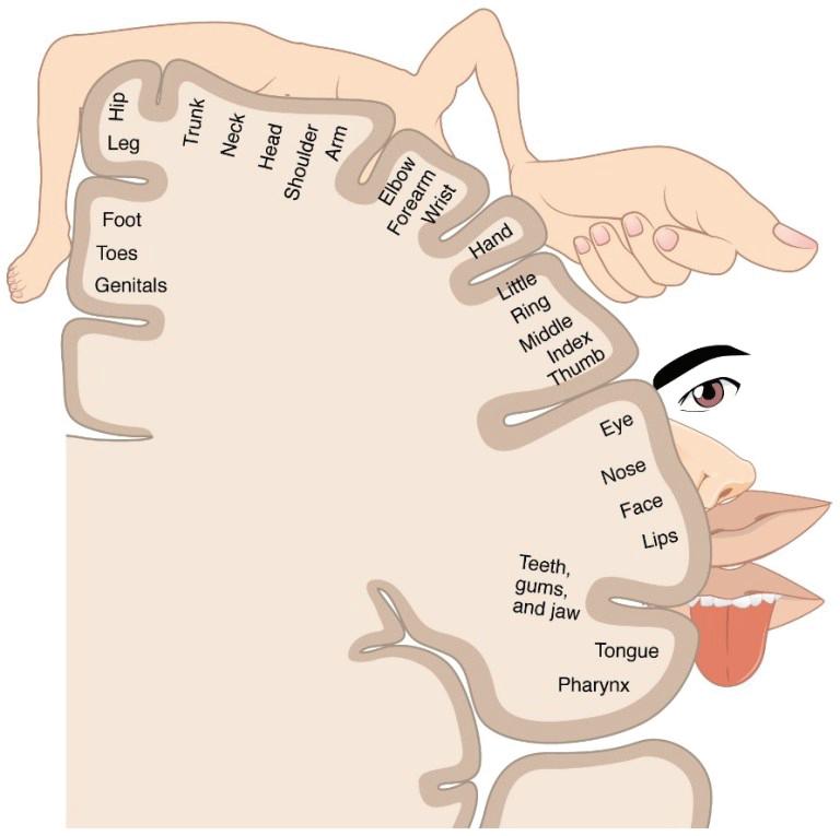
occupied by the missing limb becomes occupied by other brain functions. Traditionally, these body map representations have been visualized using functional magnetic resonance imaging (fMRI). This technique detects brain activity by measuring blood flow throughout the brain. Since active brain regions require more oxygen and nutrients, increased blood flow can be used as an indicator of increased brain activity. By measuring the activity of brain regions when individuals are performing a particular activity, scientists could identify which regions may be involved in mediating the activity. While amputees and congenital one-handers share the common feature of missing a limb, the benefit of studying individuals born with one hand is that it allows researchers to investigate the extent of brain remapping. If one hand was missing from birth, would we still see brain activity in the brain region that would typically control the hand? Just like if the restaurants can no longer be built in a city, would the brain map still leave their spots open or will they be filled up by banks and houses. A recent paper from Weizmann Institute of Science in collaboration with Western University’s Scott N. Macdonald in Dr. Jody Culham’s lab aimed to answer these questions.
Image from “Sensory Homunculus Wikipedia
territory based on the way an individual uses their other limbs to compensate. Indeed, they observed an increase in connections of the missing-hand territory with the entire brain. The researchers suggested that during early brain development, since the missing-hand territory lacks input from the missing-hand, it begins to weakly connect with other non-specific inputs as well as the body parts involved in compensatory movements, such as using feet to replace typical hand movements like reaching and grasping. One of the limitations of previous studies was the use of passive tasks, where the researchers would stimulate various parts of the body, by touching or probing, and see differences in blood flow to the missing-hand territory. This suggested that body representations located far from the missing-hand region did not rewire into the brain region.
Prior to MacDonald’s study, Tamar Makin and colleagues in 2013 suggested that the brain representation of the missing-hand territory lends itself to the intact-hand, depending on how the intact hand is used day-to-day. Macdonald’s study expanded on the idea that other body parts, and not just the intact However, this study used an active task. hand, can be linked to the missing-hand When one-handers were asked to perform
16
THE DORSAL COLUMN
VOLUME 1 // ISSUE 1
everyday tasks, they relied on their residual arm less than expected but instead, exhibited increased use of their face, lower limbs, and other objects to compensate for their missing hand’s function. The functional MRI data showed this was BY JULIA SUNSTRUM related to increased blood flow to the missing-hand territory during the movements of other, originally unexpected and sometimes quite ave you ever been overcome by the distant, body parts. overwhelming sensation that you have experienced this exact situation before? Remapping in “the missing hand This is the phenomenon of déjà vu – the territory was selective to those body two-fold feeling that something is familiar parts used for compensatory purposes”, but the knowledge that it shouldn’t be. said the authors. “The typical hand territory may not necessarily represent Most people, approximately 2/3rds, the hand per se, but rather any other report experiencing the fleeting sense of body part that can mimic the missing déjà vu, which is French for “already hand’s functionality.” seen”. Interestingly, it is most common
EPILEPSY PROVIDES INSIGHT INTO DÉJÀ VU PHENOMENON
H
This idea may be groundbreaking because it changes the subtle misunderstanding of the homunculus as “body part” specific to “function” specific. Instead of brain regions representing individual body parts like the lip, hands, and leg, they represent specific types of functional movements. With this new understanding of body representations in the brain, treatments that are used for phantom limb pain like the mirror box, which uses a mirrored illusion to trick the brain into believing the residual limb is actually the amputated limb, can be optimized by focusing on specific movements, and appropriate therapies can be developed to maximize the ability of the residual limb territory to re-wire itself.
between the ages of 15-25 and is more likely to occur during times of stress or exhaustion. Despite the high prevalence, scientists do not understand the underlying cause. The feeling itself is hard to study because spontaneous déjà vu cannot be induced or created by researchers.
So, what causes this feeling? Research suggests it may be the result of a momentary hiccup in a basic memory process. The brain is constantly scanning the environment to identify familiar or new things. This process, termed recognition memory, depends on the brain’s ability to recognize something stored in our memory. Recognition memory is an essential function that helps us navigate and interact with the world. Recognition happens in two ways: familiarity, where we have a feeling that we have seen or experienced something before but might not remember when or
SOCIETY OF NEUROSCIENCE GRADUATE STUDENTS
17
JANUARY 2020
VOLUME 1 // ISSUE 1
where, and recollection, where we remember the precise details of a specific situation or event. For example, imagine visiting a coffee shop and realizing you’ve been there before. If you recognize the décor and layout but cannot remember when or why you were there, your recognition is based on familiarity (i.e., “you know this place”). In contrast, if you know right away that you were there on a certain date with a certain friend, your recognition would be based on recollection of that event (i.e., “you remember this place”). Although controversial, research suggests that familiarity and recollection rely on two different structures within the temporal lobe of the brain, namely the rhinal cortex and hippocampus, respectively. One theory suggests that déjà vu is caused by abnormal activity of these memory related brain structures, resulting in an incorrect feeling of familiarity (e.g., “I feel like I’ve been here before”). This suggests that abnormal activity in the rhinal cortex, which is responsible for assessing familiarity, is likely involved in déjà vu. However, déjà vu is not only the feeling of familiarity. It is also accompanied by the feeling that this familiarity is wrong (e.g., “I feel like I’ve been here before, but I know I have not”), which may be based on recollection of our past experiences. What is not yet known is whether this recollection process is needed in order to experience déjà vu. In other words, does déjà vu rely on recollection, and the hippocampal structures that support it, to create the feeling of déjà vu?
This is a transparent 3D brain model highlighting the two main areas affected by epilepsy: the rhinal cortex (brown) and the hippocampus (yellow). Modified image from Jordan DeKraker.
déjà vu comes from a disorder of abnormal brain activity: epilepsy. Specifically, patients with one type of epilepsy, called medial temporal lobe epilepsy (TLE), experience déjà vu immediately before seizures. Importantly, déjà vu is not experienced in all individuals with epilepsy. As different forms of epilepsy result from unusual activity in specific brain structures, investigating how individuals with different kinds of epilepsy experience déjà vu can provide unique insight into this complicated phenomenon.
Research at Western University, led by Dr. Stefan Köhler, has previously shown that patients with unilateral TLE (seizures that occur on one side of the brain) who experience déjà vu are more likely to have deficits in their ability to assess whether something is familiar or new (Martin et al., As déjà vu cannot be readily induced, 2012). Further, they are more likely to figuring out how it works has eluded have abnormalities in the rhinal cortex – scientists, until recently. A possible the medial temporal lobe structure window into the brain mechanisms of
18
THE DORSAL COLUMN involved in familiarity recognition. These findings support the idea that déjà vu may involve dysfunctions in familiarity recognition and hint at the rhinal cortex as a key structure underlying this phenomenon. However, patients with damage to one side of the temporal lobe (unilateral TLE) may still have functioning recollection processes. While this study implicates the rhinal cortex and familiarity recognition in déjà vu, it is still unknown whether recollection (and the hippocampus) plays a role. In contrast to unilateral TLE, patients with bilateral TLE (seizures that occur on both sides of the brain) have more widespread damage to the temporal lobes and tend to have more difficulty with recollection. In a new study published in the journal Memory, Dr. Köhler and his team investigated whether patients that experience déjà vu and have bilateral TLE show similar deficits to patients with unilateral TLE. Studying bilateral TLE patients allowed researchers to examine whether déjà vu could occur with impaired recollection. The authors reasoned that if the process of recollection was essential to the identification of false familiarity during déjà vu, patients that experience déjà vu should not have problems in recollection. On the contrary, if recollection is not essential in creating the feeling of déjà vu, patients that have deficits in recollection should still be able to experience déjà vu. The authors analyzed recollection and familiarity using recognition memory tasks. For example, the Remember-Know task involved showing participants sets of pictures in an initial session and a second session and asking them whether the pictures were new or old and whether they specifically remembered it or if they just knew. The latter question aimed to
VOLUME 1 // ISSUE 1 assess whether they were working off recollection or familiarity, respectively. The authors also used magnetic resonance imaging (MRI), a technique that produces detailed images of the brain, to assess the volume of medial temporal lobe brain structures. Large reductions in volume can indicate dysfunction in specific regions. They found that, in addition to deficits in familiarity, bilateral TLE patients also struggled with recollection memory and accordingly had abnormalities in the hippocampus – a brain structure in the temporal lobe region that is implicated in recollection. Importantly, despite the deficits in recollection memory performance, these patients still experienced déjà vu. Therefore, the researchers concluded that déjà vu likely does not depend on recollection, and by extension may also not involve the hippocampus. While the results do not completely rule out a role for recollection in déjà vu, they strongly support the involvement of abnormal familiarity assessment, a process likely dependent on the rhinal cortex – another brain structure in the temporal lobe. The authors add that more research investigating the connections between different brain areas is needed to tease out the specific involvement of the hippocampus versus the rhinal cortex in déjà vu. So, the next time you experience the eerie sensation of déjà vu, remember that it is just a byproduct of your brain trying to help you process memories and figure out what is familiar in the world. The study, “Relationship between déjà vu experiences and recognition-memory impairments in temporal-lobe epilepsy”, was published in July 2019 in Memory.
SOCIETY OF NEUROSCIENCE GRADUATE STUDENTS
19
The human brain has about 86 billion neurons, which are cells that carry electrical signals to communicate with each other. Shown above is a ‘pyramidal neuron’ – the largest and most common type of neuron in our brain. Pyramidal neurons play an important role in cognition (for example, paying attention, memory, and learning) and sensory processing (for example, seeing and hearing). For reference, the average size of a pyramidal neuron's cell body is about 0.2 millimeters, which is about the thickness of a strand of hair, and its length can reach up to several centimeters. The pyramidal neurons seen here were modelled and textured in 3D, based on the Layer 5 Pyramidal Cell from Open Source Brain. Image © Kelly Bullock Art, 2020.
THE DORSAL COLUMN Send us an email: thedorsalcolumn@gmail.com
Follow us on twitter:
Visit our website:
@thedorsalcolumn
songsuwo.com/thedorsalcolumn
SOCIETY OF NEUROSCIENCE GRADUATE STUDENTS