4 minute read
Hand-in-Hand: How the brain handles a missing body part
By Kartik Pradeepan
In the 1990s, V.S. Ramachandran released a series of experiments that described a phenomenon whereby, despite a limb being amputated, a number of patients continued to vividly experience the presence of that limb – a phantom sensation. This phantom sensation would often occur as a phantom pain, whereby excruciating sensation would feel like it’s coming from the body part that is no longer there.
This experiment lent itself to the idea of widespread reorganization of the sensory circuits of the brain, even after adulthood.
Many early and even current neuroscientists believe that our perception of touch originates from a topographical map of our body parts in the brain. Much like a map that tells you where restaurants, schools, hospitals are in the city, the topographical maps inform us where the feelings of hunger or control of hand movements are located in the brain. When visualized, it creates a distorted human-like representation of our lips, feet, hands, etc. on the surface of the brain, called a homunculus.

Sensory Homunculus.
Image from “Sensory Homunculus" page on Wikipedia.
Accordingly, when a limb undergoes an amputation, adjacent areas are believed to take over the now missing-limb representation, thus changing the organization of the homunculus. In other words, the brain real-estate previously occupied by the missing limb becomes occupied by other brain functions.
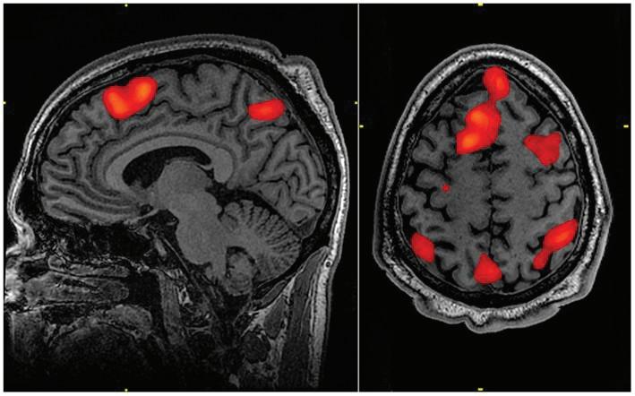
Traditionally, these body map representations have been visualized using functional magnetic resonance imaging (fMRI). This technique detects brain activity by measuring blood flow throughout the brain. Since active brain regions require more oxygen and nutrients, increased blood flow can be used as an indicator of increased brain activity. By measuring the activity of brain regions when individuals are performing a particular activity, scientists could identify which regions may be involved in mediating the activity.
While amputees and congenital one-handers share the common feature of missing a limb, the benefit of studying individuals born with one hand is that it allows researchers to investigate the extent of brain remapping. If one hand was missing from birth, would we still see brain activity in the brain region that would typically control the hand? Just like if the restaurants can no longer be built in a city, would the brain map still leave their spots open or will they be filled up by banks and houses. A recent paper from Weizmann Institute of Science in collaboration with Western University’s Scott N. Macdonald in Dr. Jody Culham’s lab aimed to answer these questions.
Prior to MacDonald’s study, Tamar Makin and colleagues in 2013 suggested that the brain representation of the missing-hand territory lends itself to the intact-hand, depending on how the intact hand is used day-to-day. Macdonald’s study expanded on the idea that other body parts, and not just the intact hand, can be linked to the missing-hand territory based on the way an individual uses their other limbs to compensate. Indeed, they observed an increase in connections of the missing-hand territory with the entire brain. The researchers suggested that during early brain development, since the missing-hand territory lacks input from the missing-hand, it begins to weakly connect with other non-specific inputs as well as the body parts involved in compensatory movements, such as using feet to replace typical hand movements like reaching and grasping.
One of the limitations of previous studies was the use of passive tasks, where the researchers would stimulate various parts of the body, by touching or probing, and see differences in blood flow to the missing-hand territory. This suggested that body representations located far from the missing-hand region did not rewire into the brain region.
However, this study used an active task. When one-handers were asked to perform everyday tasks, they relied on their residual arm less than expected but instead, exhibited increased use of their face, lower limbs, and other objects to compensate for their missing hand’s function. The functional MRI data showed this was related to increased blood flow to the missing-hand territory during the movements of other, originally unexpected and sometimes quite distant, body parts.
Remapping in “the missing hand territory was selective to those body parts used for compensatory purposes”, said the authors. “The typical hand territory may not necessarily represent the hand per se, but rather any other body part that can mimic the missing hand’s functionality.”
This idea may be groundbreaking because it changes the subtle misunderstanding of the homunculus as “body part” specific to “function” specific. Instead of brain regions representing individual body parts like the lip, hands, and leg, they represent specific types of functional movements.
With this new understanding of body representations in the brain, treatments that are used for phantom limb pain like the mirror box, which uses a mirrored illusion to trick the brain into believing the residual limb is actually the amputated limb, can be optimized by focusing on specific movements, and appropriate therapies can be developed to maximize the ability of the residual limb territory to re-wire itself.
