
ADVOCATE MYELOPATHIES: It Isn’t Always a Disc Christine Senneca, DVM, DACVIM (Neurology) | Page 10 MEET THE PRESIDENT Dr. Jaqueline S. Shellow | Page 8 FVMA ANNUAL CONFERENCE 2023 Recap | Page 22 Exotic Small Mammal SEDATION AND ANESTHESIA Lorelei D’Avolio, LVT, VTS (Clinical Practice-Exotics), CVPM | Page 16 ISSUE 2 | 2023
7207 Monetary Drive
Orlando, Florida 32809
Phone: 407-851-3862
Toll-free: 800-992-3862
Fax: 407-240-3710
info@fvma.org
www.fvma.org
BOARD OF GOVERNORS
DR. JACQUELINE S. SHELLOW President

DR. SCOTT RICHARDSON President-elect
DR. ALEX 'STEVE' STEVERSON Treasurer
DR. MARTA P. LISTA
Past President
BOARD OFFICERS
DR. ERNEST C. GODFREY
Trustee Emeritus
DR. RICHARD B. WILLIAMS
AVMA Delegate
DR. RICHARD C. SUTLIFF
AVMA Alternate Delegate
DR. JULIE MOODOYAN
District 1 – Big Bend
DR. THOMAS E. HESTER
District 2 – Northeast
DR. TODD FULTON
District 3 – Central
DR. DONALD S. HOWELL
District 4 – Tampa Bay
DR. BETH KESER
District 5 – Treasure Coast
DR. ROBERT L. SWINGER
District 6 – South Florida
DR. BARBARA LEWIS
District 7 – Southwest
DR. JOHN R. WIGHT
District 8 – Northwest
DR. CHRISTINE M. STORTS
District 9 – Space Coast
DR. SALLY DENOTTA

FAEP Representative to the FVMA Executive Board
President's MESSAGE
Greetings,
It is an honor for me to continue to serve our great profession and our professional organization, and now as the 95th FVMA President. I want to thank our outgoing President Dr. Marta Lista, our outgoing Past President Dr. Mary Smart, the FVMA Executive Board, the FVMA staff, and our Executive Director Jim Naugle. I look forward to continuing to work with all of them.
I also want to give a big thank you to Dr. Richard Williams for all his work with the FVMA’s Legislative Advocacy Committee who work hard to advocate and protect our profession and the animals we take care of. The FVMA will continue to work to protect the Veterinary Patient Client Relationship (VPCR).
Thank you to Dr. Ernest Godfrey for representing us so well as our AVMA Delegate. Dr. Richard Williams will take over as AVMA Delegate, and Dr. Richard Sutliff is our new AVMA Alternate Delegate. Thank you to Dr. Terry Clekis, who heads the FVMA’s Disaster Preparedness Committee, for all of his work coordinating the relief and recovery efforts for Hurricane Ian. I want to congratulate this year's Power of Ten graduates and welcome the new Power of Ten class. Our young leaders are the future of our profession, and I want to continue to empower them and encourage them to be involved.
This year we will continue to focus on offering high quality continuing education in attractive venues. Those of you that attended FVMA Annual Conference in Orlando – thank you. It was a great program, as usual, put together by Dr. Ernest Godfrey. Our next meeting, The Gulf Atlantic Veterinary Conference (TGAVC), will be in Tampa on Sept. 14-17, 2023, and I hope to see you there. We also have our equine-exclusive Promoting Excellence Symposium in West Palm Beach on Oct. 19-22, 2023.
Lastly, we would like our members to become more involved in the FVMA. Feel free to contact me or your District Representative who works hard for you or our Executive Director, Jim Naugle, or any of the rest of the amazing FVMA staff. "We are here for you. How can we help?"
Warm regards,
Jackie Shellow, DVM, MS
To advance the veterinary medical profession, promote animal health and well-being, and protect public health.
Opinions and statements expressed in FVMA Advocate reflect the views of the contributors and do not represent the official policy of the FVMA, unless so stated. Placement of an advertisement does not represent the FVMA's endorsement of the product or service.

2 | FVMA Advocate
FVMA MISSION:
YOU ARE THE CENTER OF OUR FOCUS.

A lot of tasks compete for your time as you manage your practice. From administrative work and equipment maintenance to client communications, appointment scheduling and reminder phone calls, it’s easy to lose sight of what’s most important. Patterson’s experienced support teams help you take care of these details so you can get back to what you love – helping your patients.
SKIN BARRIER:

| 3 www.fvma.org @thefvma @the__fvma @thefvma spleH I n h ibitInfa m matory M e srotaid To learn more, visit DERMAQUIN.COM Available in SOFT CHEWS for cats and dogs
A HARDY
the best defense against allergens
We focus on you so you can focus on them.
23PV662053d (8/22)
TRUSTED EXPERTISE. UNRIVALED SUPPORT.™
JIM NAUGLE Executive Director
& Editor-in-Chief
ERIKA MEYER
Administrative Manager
NICOLE ALVAREZ
Marketing & Communications Manager

JESSICA KEATTING
Graphic Designer
KATHERINE PEARCE
Senior Creative Lead
JILLIAN SINCLAIR
Marketing Specialist
SOFIA GONZALEZ
Jr. Graphic Designer
CYNDI WHITAKER
Director of Conferences & Strategic Partnerships
RALPH HUBER
Business Development Manager
JASON SMITH
External Relations Manager
DAMON PATAI
Meetings & Events Planner
MELISSA MEDAL
Meetings & Events Planner
PAIGE VOGT
Member Services Representative
LIAM CROSS
Database Clerk
MARIBEL BLUM
Finance & Accounting Manager
2 | President's Message 7 | Farewell to Dr. Lista 8 | Meet the President Dr. Shellow 10 | Myelopathies: It Isn’t Always a Disc 16 | Exotic Small Mammal Sedation And Anesthesia 22 | FVMA Annual Conference 2023 Recap 28 | Navigating the (Sometimes Treacherous) Waters of Social Media 30 | Five Steps to Start Your Exit Strategy: Don’t Be Caught Off Guard 32 | Practice Pulse 34 | Classified Advertisements IN THIS ISSUE
Staff
4 | FVMA Advocate
100% manufactured following FDA enforced regulations
Available for dispensing and unlimited hospital administration
FDA Federal SEC. 503B. [21 U.S.C. 353b]

All drugs ready to ship same day with 2 days in transit
Scan the QR code for a list of available drugs

20% OFF YOUR ORDER
of Epicur Pharma’s 503B Medications
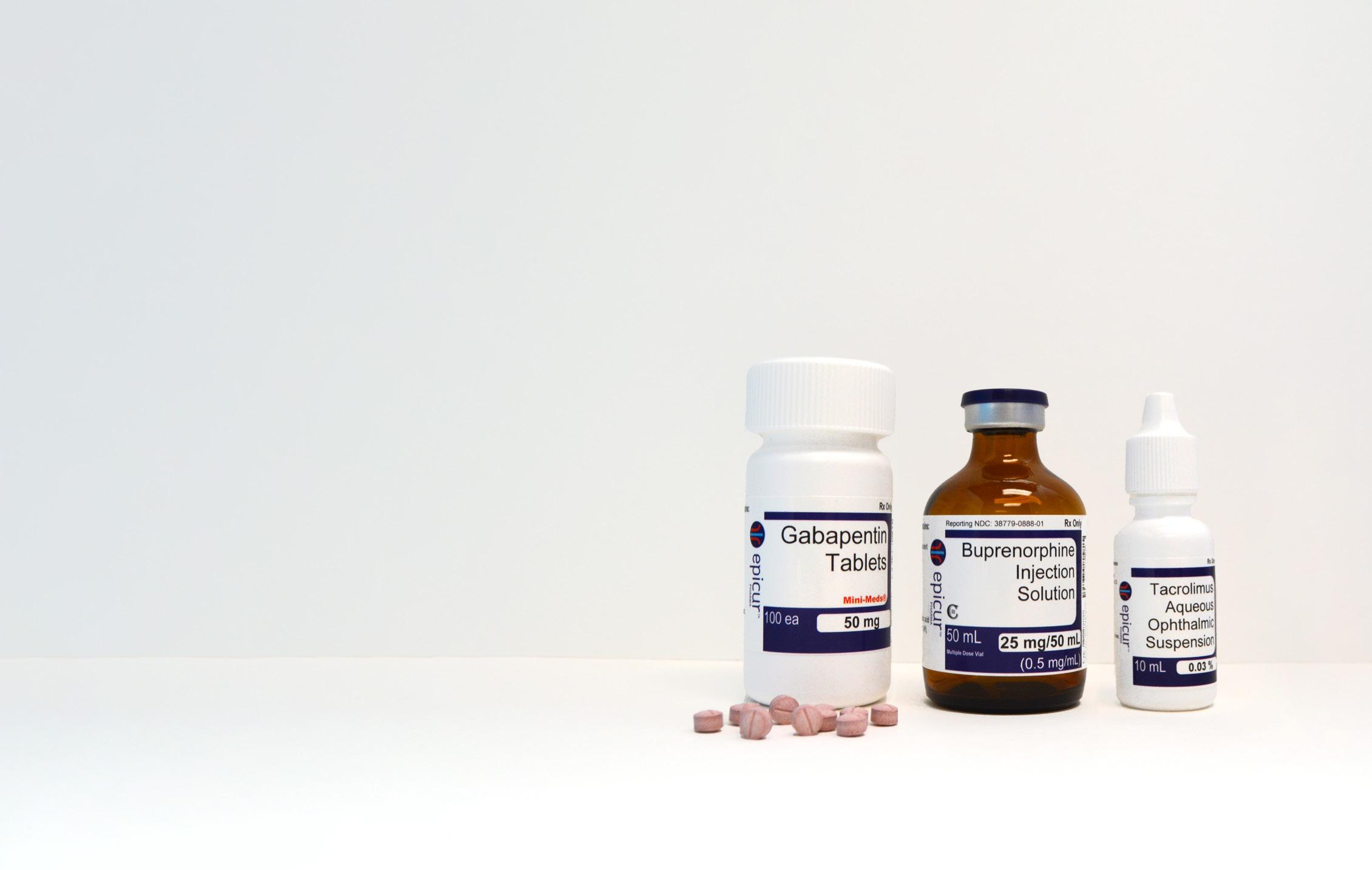
Order today! 888-508-5032
Mention promo code: FLVMA23
Florida Practices for Sale
FL, Sarasota: Here It Is! Solo Practice Grossing $1.3M+ & growing. Buyer’s after debt income ≈$320K. Stunning place to live & work near Siesta Key. Leased facility in a busy plaza for 18 years. PRX Only (FL25S)
PRICE REDUCED! FL, Titusville, East Coast: Open 4 days a week. Solo Doctor. 2940SF free-standing facility located on .60 acre. 3 exam rooms. Abaxis in-house blood machines. Revenues of $1.1M. Buyer’s after debt income ≈$230K+. PRX & RE (FL40T)
FL, Palm Beach County: Solo, DVM, $1.7M+ Gross, 5 -day work week, No weekends, 2142SF Free-standing facility. Loaded with equipment. Seller willing to stay employed if needed. PRX & RE (FL53L)
FL, New Listing! West Coast, Clearwater: 1 DVM, SA practice with real estate, grossing $900K+, well equipped, and growing. PRX & RE (FL20C)
FL, New Listing! Florida Panhandle - 4 season paradise. 1 DVM, SA practice. 30 years and growing. PRX & RE (FL32P)
For
Simmons Says: Practice AppraisalNow Is The Time! You’ve already done the hard work of gathering financial information for tax prep. Now get an accurate diagnosis on your practice health. Call Simmons today 800-333-1984
nationwide, add yourself to Practice Watch from our website. 800.333.1984 | southeast@simmonsinc.com | simmonsinc.com
listings
*One time use. Discount valid via phone only. Offer expires 7/31/23.
| 5 www.fvma.org @thefvma @the__fvma @thefvma


Worldwide Nationwide airanimal.com https://socialordeals.com/fvma/ sales@socialordeals.com BOOK YOUR COMPLIMENTARY DIGITAL AUDIT TODAY! Does Your Practice Face Challenges In These Areas? Local Search Rankings Online Reviews Social Media SEO (Search Engine Optimization) Directories/GPS/Voice Listings are Inconsistent Website is Outdated Digital Advertising (Adwords, Social Ads)
FAREWELL TO Dr. Lista
Jillian Sinclair | Marketing Specialist Florida Veterinary Medical Association
Dr. Marta Lista officially handed over the title of FVMA president to Dr. Jaqueline S. Shellow at the 2023 FVMA Annual Conference and transitioned to the past president position. She originally became involved with the FVMA when the FVMA helped to stop tax-funded veterinary facilities from being built in Miami where Dr. Lista lives and practices. Together, they implemented a plan that still exists 10 years later, where existing, privately-owned veterinary hospitals are funded to provide veterinary care. She continued to lobby in Tallahassee on behalf of the FVMA and served as District 6 Representative for six years.
HER GOALS
One of Dr. Lista’s goals for her presidency was to advocate for mothers working in the veterinary industry. As a mother herself, she is passionate about highlighting the challenges of having a veterinary career while having young children.
“It’s a challenge. Working moms basically have two full-time jobs – raising their children and being a veterinarian. I wanted to bring to light the challenges we go through.”
Thanks to Dr. Lista, FVMA Annual Conference 2023 offered child care for all attendees this year. She wanted working mothers to be able to attend the conference and fully participate without worrying about their children.
Another cause Dr. Lista wanted to focus on during her presidency was establishing committees within the FVMA. She feels it is important for FVMA members to be involved with the association and be able to voice their opinions and ideas through committees. When asked what she was most proud of accomplishing during her time serving as president Dr. Lista said,
“We were able to enhance our involvement with the University of Florida College of Veterinary Medicine, including establishing scholarships. This was very near and dear to my heart because UF is our state school and where I attended. Having a good relationship with the school and its students is extremely important to me.”
Dr. Lista also worked to expand the FVMA Legislative Committee, which recently attended FVMA’s Legislative Action Days in Tallahassee. They advocated for several bills that affect veterinary medicine that are being voted on this year.
THE NEXT STEP
Now that Dr. Lista has transitioned to the past president position, she will begin working with the FVMA Foundation.
“They’re not getting rid of me yet! I’ll be leading the Foundation now. My goal is for the Foundation to become more involved with community projects that members are a part of. I’d love to see it help our members throughout the state.”

| 7 www.fvma.org @thefvma @the__fvma @thefvma
MEET THE PRESIDENT
Dr. Jaqueline S. Shellow
Jillian Sinclair | Marketing Specialist Florida Veterinary Medical Association
At FVMA Annual Conference 2023, Dr. Jaqueline S. Shellow officially transitioned to her new role as president of the FVMA after taking the reins from now-past president Dr. Marta Lista.
Dr. Shellow has been heavily involved with organized veterinary medicine throughout her career. Her experience as president of the Florida Association of Equine Practitioners (FAEP) (2010), FAEP council member (2006-present), FAEP representative to the Executive Board of the FVMA (2017-2022), FVMA Legislative Committee member (2008-2022), and other leadership roles have prepared Dr. Shellow for her new role as president of the FVMA.

HER PLATFORM
During her presidency, Dr. Shellow plans to focus on the FVMA’s continuing education efforts. She would like to continue to improve FVMA conferences and deliver the highest quality events for our members and attendees. She believes that continuing education is an essential part of improving veterinary medicine and wants to help the FVMA be able to offer exceptional CE for veterinary professionals. She also wants to continue to share the work of the FVMA with the public and other veterinary professionals.
“I want to work to elevate the veterinary profession by educating the public on the important and varied types of work veterinarians do to protect both animal and human health and safety. My goal is to continue to get the word out about what the FVMA does to help veterinarians and animals,” Dr. Shellow said.
ABOUT DR. SHELLOW
While growing up on an avocado grove in Miami, FL, Dr. Shellow began interacting with a variety of animals from a young age. Her dad told her to always do something she loves, so she chose a career where she can work with animals every day. Dr. Shellow attended Stephens College in Columbia, MO, and then earned her master's in animal nutrition at the University of Kentucky. She attended veterinary school at the University of Florida College of Veterinary Medicine and graduated in 1987.
Dr. Shellow spent most of her career as an equine racetrack veterinarian as part of TFB Equine. Now, she works with small animals at Embassy Lakes Animal Hospital part-time. She also worked with racing Greyhounds as a track veterinarian and fell in love with the breed. In addition to adopting one of her own, she fosters and works closely with a Greyhound adoption group
8 | FVMA Advocate
that finds retired racers forever homes. Dr. Shellow is grateful to be able to work with animals every day and help the people that love them.

Her dedication to veterinary medicine and organized medicine has not gone unnoticed over the years. Dr. Shellow was a 2010 Gold Star Award winner for her work as the liaison between the FVMA and the FAEP. She was also the FVMA’s Veterinarian of the Year in 2014.

INVOLVEMENT WITH ORGANIZED VETERINARY MEDICINE
Dr. Shellow has been a member of the FVMA since she was in veterinary school. Her late husband, Barry Faske, and her dad were both very involved in their professional organizations, which inspired her to do the same. Dr. Shellow says they taught her the importance of these kinds of associations and are a large part of why she accepted the position of president of the FVMA. She is excited about the opportunity to advocate for the veterinary profession and work with FVMA members; especially younger veterinarians. She is passionate about encouraging and mentoring young professionals because they are the future of veterinary medicine. Dr. Shellow hopes to work with young veterinarians in her new role and encourages everyone to find a good mentor when first starting out.
“I have had some great mentors throughout my career and am very grateful. Many were also involved in their professional associations or in organized veterinary medicine and encouraged me to be involved to give back to my profession — and, of course, the animals.”
"My goal is to continue to get the word out about what the FVMA does to help veterinarians and animals."
MYELOPATHIES: It Isn’t Always a Disc
Christine
Southeast Veterinary Neurology of Jupiter
 Senneca, DVM, DACVIM (Neurology)
Image courtesy of Shutterstock.
Senneca, DVM, DACVIM (Neurology)
Image courtesy of Shutterstock.
Intervertebral disc disease (IVDD) is the most common problem affecting the spine and spinal cord in dogs, resulting in a range of clinical signs from pain to complete paralysis with loss of deep pain perception. Disc disease also occurs in our feline patients, though the incidence is far less often.
It is very important to be familiar with the many other causes of myelopathy which can mimic clinical signs of IVDD. In this lecture, several common causes of myelopathy will be reviewed, reminding the practitioner to consider other differentials when managing a patient presenting with neurologic dysfunction localized to the spinal cord.
VASCULAR DISEASE: FIBROCARTILAGINOUS EMBOLIC MYELOPATHY
Fibrocartilaginous embolic myelopathy (FCEM) is due to acute spinal cord infarction after a small volume of fibrocartilage, suspected to be from an intervertebral disc, embolizes within cord vasculature, resulting in ischemic necrosis to dependent regions of spinal cord parenchyma. It occurs commonly in dogs but rarely in cats, and it has been reported in various other species including humans. Though many possible mechanisms for this process have been discussed, the exact pathophysiology is not definitively known. FCEM can occur anywhere along the spinal cord. In cats, a predilection for the cervical spine is reported. In general, large and giant breed dogs are more commonly affected compared to small breed dogs, though the Miniature Schnauzer is the most frequently identified individual breed.
Clinical signs of FCEM are peracute in nature and have a distribution and severity referable to the site and extent of the spinal cord infarction. Usually, signs can be localized to a single neuroanatomical region such as C1-C5 myelopathy or T3-L3 myelopathy, etc. Interestingly, however, there are some reports of multifocal localizations, which may be due to sequelae such as spinal shock or a shower of emboli to multiple areas of spinal cord vasculature. Neurologic signs are typically non-progressive, and patients are non-painful, though some patients may exhibit transient initial discomfort. Activity or trauma do not always precede an FCEM. A hallmark finding in patients with FCEM is lateralization of clinical signs due to the asymmetric distribution of spinal cord vasculature. Some patients may only experience weakness, whereas others may show complete loss of motor function or even deep pain perception.
A definitive diagnosis of FCEM can only be made post-mortem. Grossly, the spinal cord may appear normal, but visible swelling, malacia, and hemorrhage are possible. Confirmation of FCEM is via histopathologic identification of fibrocartilaginous material within spinal arteries or veins, within or near a focal
area of myelomalacia with or without concurrent hemorrhage. Grey matter is more often affected than white matter, and lesions tend to be well-demarcated.
Presumptive diagnosis is typically based on history, clinical signs, and advanced imaging findings. MRI is by far the imaging modality of choice in cases of suspected FCEM. Images typically reveal a focal, sharply demarcated, intramedullary, often lateralized and often linear, intramedullary lesion predominantly affecting the gray matter. Lesions are T2W, STIR, FLAIR, hyperintense, and T1 iso- or hypointense with possible mild heterogeneous post-contrast enhancement. Lesions are most often longer than the length of a vertebral body and not centered over an intervertebral disc. The spinal cord may appear focally swollen with attenuation of the epidural fat and cerebrospinal fluid surrounding the spinal cord at the level of the lesion. Cerebrospinal fluid analysis may be normal or may show mild elevations in microprotein and white blood cell counts; however, a recent case report described a dog with a marked CSF pleocytosis.
FCEM is a non-surgical disease, and, therefore, treatment is solely supportive care driven with prognosis being good to excellent in many cases. Prognosis is guarded in those patients with loss of deep pain perception, particularly if the patient is bilaterally and symmetrically affected. Corticosteroid or nonsteroidal therapy typically does not impact clinical recovery; yet physiotherapy should be provided.
INFLAMMATORY DISEASE: MENINGITIS, MYELITIS, AND MENINGOMYELITIS OF UNKNOWN ETIOLOGY
Meningitis is the term used to describe inflammation of the meninges, the three connective tissue layers which line the spinal cord; whereas myelitis is inflammation of the spinal cord itself. Therefore, meningomyelitis refers to concurrent inflammation of both the meninges and the spinal cord. Although inflammation can be caused by infectious agents including viral, bacterial, fungal, protozoal, rickettsial, and parasitic organisms, many cases of meningitis and meningomyelitis, particularly in dogs, do not have a discernible etiology and are thought to be immune-mediated in nature. Often, meningitis, myelitis, or meningomyelitis occur in combination with inflammation of the brain (encephalitis), hence the terms, meningoencephalitis, encephalomyelitis, and meningoencephalomyelitis. However, inflammation restricted to the spinal cord is possible.
In dogs, the most common form of immune-mediated inflammatory disease affecting the spinal cord is Steroid Responsive Meningitis-Arteritis (SRMA), which primarily affects young adult dogs with a breed predisposition for the Beagle, Boxer, Bernese Mountain Dog, Petit Basset Griffon
INTRODUCTION
| 11 www.fvma.org @thefvma @the__fvma @thefvma
Vendeen, Nova Scotia Duck Tolling Retriever, and Weimaraner. Inflammation is particularly found within the leptomeninges and associated vessels and not within the spinal cord. Clinical signs include pain, stiffness, reluctance to move the neck, and fever. Signs may be episodic. Most dogs do not have any neurologic deficits as the spinal cord is spared.
and have variable degrees of post-contrast enhancement. Fairly recently, STIR hyperintensity within the paraspinal musculature was reported in a cohort of cases with cervical spinal cord involvement. CSF analysis most often reveals a mononuclear or mixed pleocytosis with elevated protein levels. Results can vary widely, and a small percentage of cases may have normal CSF. Treatment of non-infectious meningomyelitis also involves long-term immunosuppression with the use of corticosteroids, and many cases require multimodal therapy for successful outcomes. Treatment courses are often longer than in cases of SRMA with some cases requiring life-long therapy. Prognosis is good, but relapses are relatively common, and, unfortunately, some patients do not respond to even the most aggressive treatments.
In feline patients, immune-mediated inflammatory disease is far less common than it is in dogs. Feline Infectious Peritonitis myelitis is the most common infectious/inflammatory cause of myelopathy in cats, followed by Cryptococcosis.
INFECTIOUS DISEASE: DISKOSPONDYLITIS AND EMPYEMA
As mentioned above, infectious causes of meningitis, myelitis, and meningomyelitis are possible in veterinary patients, with cats being more often affected than dogs. As such, infectious disease screening on serum, CSF, or both is often performed when clinical suspicion for infectious disease is high.
Figure 2.
Diagnosis is typically made via CSF analysis, which shows a marked, non-degenerate neutrophilic pleocytosis; however, MRI may show meningeal enhancement and enlarged vessels in some cases. Acute phase proteins, specifically C-reactive protein, may be elevated in both serum and CSF. Treatment involves the use of immunosuppressive corticosteroid therapy over a period of four to six months. In severe cases, additional immunosuppressive agents may be required. In very mild cases, non-steroidal therapy may be effective. Prognosis is typically excellent, and relapse is rare.
Unlike SRMA, patients suffering from other non-infectious causes of meningomyelitis will experience neurologic deficits as the pathology occurs within the spinal cord. Clinical signs are variable depending on the extent of the disease and the region of the spinal cord affected. MRI findings are also variable, as images may be normal, or may reveal changes within the spinal cord and/or meninges. Inflammatory lesions typically appear hyperintense on T2W, FLAIR, and STIR images and iso- or hypointense on T1W images. Lesions are often poorly defined
Far more likely to present to the general practitioner are cases of diskospondylitis, in which there is an infection of the cartilaginous vertebral end plates with secondary involvement of the intervertebral disc. Sternebrae may also be affected. Diskospondylitis is far more common in dogs than cats, but several reports of this disease in cats are in the literature. Mature adult male dogs are most often affected, though females and juveniles are also reported. Bacterial infections are the primary culprit; though other agents, particularly fungal, are possible. The most commonly identified bacteria include Staphylococcus spp., Streptococcus spp., Escherichia coli, and Brucella canis; however, various other infectious agents have been reported. Hematogenous spread of the infectious agent is considered the most likely cause of diskospondylitis, but isolation and identification of the agent often proves to be challenging.

Diagnosis of diskospondylitis can often be achieved via plain film radiography, though more advanced imaging modalities are sometimes required. Radiographic changes include irregular vertebral end plate lysis with extension into the vertebral body and collapse of the disk space. In later stages, sclerosis of the end plates and ventral spondylosis is seen. The disease may be so severe that vertebral subluxation occurs. Cultures of blood, urine, and synovial fluid as well as imaging-guided fine needle aspiration and culture of the affected disc can be performed, but

12 | FVMA Advocate
Figure 1. Sagittal and transverse T2W MRI images of the cervical spine. Spanning the length of the C5 vertebral body, there is a poorly defined, left lateralized, intramedullary hyperintensity, consistent with a fibrcartilaginous embolism (FCE).
Image courtesy of Dr. Christine Senneca.
Sagi&al and transverse T2W MRI images of the cervical spine. Spanning the length of the C5 vertebral body there is a poorly defined, leB lateralized, intramedullary hyperintensity, consistent with a fibrcarFlaginous embolism (FCE) .
L
Figure 2.
Sagi&al and transverse T2W MRI images of the cervical spine. Spanning the length of the C5 vertebral body there is a poorly defined, leB lateralized, intramedullary hyperintensity, consistent with a fibrcarFlaginous embolism (FCE) .
L
results may be negative. Fungal and Brucella serology as well as other specific testing for highly suspected organisms should be performed. Serum c-reactive protein measurement may be useful in early disease detection and may provide a means to monitor response to treatment. Antibiotic therapy is based on the results of bacterial or fungal organism isolation, and therapy should last no less than 12 weeks. In those cases where the causative agent is not identified, a first-generation cephalosporin should be initiated. Non-steroidal anti-inflammatory medications and analgesics such as gabapentin are beneficial in the early course of treatment.

Spinal epidural empyema refers to bacterial infection within the epidural space. Though it occurs rarely in small animal patients, spinal epidural empyema should be considered as a differential for animals presenting with acute myelopathy associated with fever and severe spinal hyperpathia. Empyema may be associated with diskospondylitis, but it can also occur without the involvement of the discs or vertebrae. Non-surgical treatment with antibiotic therapy may be effective; however, surgical decompression may be necessary in severely affected cases.
TRAUMA
Traumatic myelopathies occur in almost all veterinary species, and hence it is prudent to question the client for any potential history of trauma. Trauma should remain on the differential list even when there is no reported history of a traumatic event, particularly in feline patients. Trauma to the spine may cause only mild, contusive injury to the spinal cord for which rest and supportive care are sufficient. Alternatively, traumatic injury can cause catastrophic and irreversible spinal cord injury, with a grave prognosis for return of function even despite immediate surgical intervention.
When trauma is high on the differential list, radiography should be used initially for rapid assessment of spinal injury, but extreme care must be taken with patient handling and restraint. Computed tomography (CT) is an excellent diagnostic modality as it not only will provide for identification of traumatic spinal injury but can help to rule out other external or internal injuries. Furthermore, CT is extremely valuable for surgical planning if spinal stabilization is necessary.
Spinal stability is often based on the three-compartment model as disruption to any two of the three compartments will result in instability. The dorsal compartment is composed of the articular processes, dorsal laminae, pedicles, and dorsal spinous process. The middle compartment is composed of the dorsal longitudinal ligament, the dorsal portion of the vertebral body, and the dorsal portion of the annulus fibrosus. Finally, the ventral component is composed of the ventral longitudinal ligament, the nucleus pulposus, and the remaining portions of
the annulus fibrosus and vertebral body. This model should be used to help to guide clinical decision-making regarding surgery for traumatic spinal injury.
NEOPLASIA
Neoplasms affecting the spinal cord may be primary or metastatic from another site. Neoplasia may involve the spinal cord, the dura mater, the peripheral nerves, or the paraspinal tissues. A plethora of neoplastic conditions causing myelopathy in dogs and cats have been reported, some being exceedingly rare with only individual case reports documented. To discuss all reported tumor types is beyond the scope of this discussion. Instead, focus will be given to the more common and welldocumented tumor types.
Tumors causing myelopathy may either be extradural (completely outside of the spinal cord), intradural-extramedullary (inside the dura mater but outside of spinal cord parenchyma), or intramedullary (within spinal cord parenchyma). The location of the neoplasm in relation to the spinal cord can aid in determining the tumor type; yet histopathology is always necessary for definitive diagnosis. Clinical signs of spinal
| 13 www.fvma.org @thefvma @the__fvma @thefvma
Figure 2. Cerebrospinal fluid from a patient with Steroid Responsive Meningitis-Arteritis (SRMA). Note the cloudy appearance, indicative of a marked pleocytosis.
Image courtesy of Dr. Christine Senneca. Cerebrospinal fluid from a paFent with Steroid Responsive MeningiFs-Arteri appearance, indicaFve of a marked pleocytosis.
neoplasia vary from pain to complete paralysis depending on the lesion’s location and size.
Extradural tumors account for the majority of spinal tumors in small animal patients, with the most frequently reported neoplasms in the dog being osteosarcoma, fibrosarcoma, chondrosarcoma, hemangiosarcoma, multiple myeloma, liposarcoma, and lymphosarcoma. In regard to intraduralextramedullary neoplasia, the two most common primary tumor types in canine patients are meningioma and nerve sheath tumors, and the most common metastatic neoplasms include lymphoma, mammary carcinoma, and prostatic carcinoma. In juvenile canine patients, nephroblastoma should be considered as the primary differential for an intradural- extramedullary mass; though intramedullary nephroblastoma is also reported. Finally, intramedullary spinal cord neoplasia includes primary tumors, such as astrocytoma, oligodendroglioma, ependymoma, and metastatic diseases such as hemangiosarcoma. In general, spinal neoplasia in feline patients differs from canines as lymphoma and osteosarcoma are the most commonly reported tumors.
Diagnosis of spinal neoplasia can sometimes be made with plain film radiography given the osteolytic and osteoproliferative nature of many bone tumors. Still, some radiographic changes may be identified in cases of expansile extramedullary lesions such as peripheral nerve sheath tumors. Advanced imaging modalities (CT and/or MRI) are typically necessary for the identification of many spinal neoplasms. A hallmark finding for intradural-extramedullary tumors on MRI is a “golf-tee” sign. Myelography may be useful in situations where advanced imaging is not available, but this test requires proper training and has associated risks.
Treatment of spinal neoplasia may involve surgery, radiation therapy, chemotherapy, or a combination of these. Palliative therapy, through the use of anti-inflammatory corticosteroid and analgesic medications, is indicated in cases in which surgery cannot be performed because of the extent of disease or owner’s preference. Prognosis largely depends on neurologic status at the time of diagnosis, location of the tumor, degree of spinal cord involvement, and extent of surgical resection, if performed.
REFERENCES:
1. Bagley RS. Spinal neoplasms in small animals. Vet Clin N Amer Small Anim Pract. 2010;40(5):915-927.
2. Bartholomew KA, Stover KE, Olby NJ, Moore SA. Clinical characteristics of canine fibrocartilaginous embolic myelopathy (FCE): a systematic review of 393 cases (19732013) Vet Rec. 2016;179(25):650.
3. Besalti O, Caliskan M, Can P, et. al. Imaging and surgical outcomes of spinal tumors in 18 dogs and 1 cat. J. Vet. Sci. 2016;17(2):225-234.

4. Cornelis I, Van Ham L, Gielen I, DeDecker S, Bhatti SFM. Clinical presentation, diagnostic findings, prognostic factors, treatment and outcome in dogs with meningoencephalomyelitis of unknown origin: a review. Vet J. 2019;244:37-44.
5. DiRisio L, Platt R. Fibrocartilaginous embolic myelopathy in small animals. Vet Clin N Amer Small Anim Pract. 2010;40(5):859-869.
6. DeRisio L. A review of fibrocartilaginous embolic myelopathy and different types of peracute non-compressive intervertebral disk extrusions in dogs and cats. Front Vet Sci. 2015;doi.org/10.3389/fvets.2015.00024.

7. Eminaga S. Cherubini GB, Villiers E, Targett M, Caine A. STIR muscle hyperintensity in the cervical muscles associated with inflammatory spinal cord disease of unknown origin. J Small Anim Pract. 2013;54(3):137-142.
8. Granger N, Carwardine D. Acute spinal cord injury. Vet Clin N Amer Small Anim Pract. 2014;44(6):1131-1156.
14 | FVMA Advocate
al and transverse T1W post-contrast images of the thoracic spine of a 3 year old masFff with an
Sagi&al and transverse T1W post-contrast images of the thoracic spine of a 3 year old masFff with an extradural spinal mass at the level of T13. Histopathology confirmed osteosarcoma.
Figure 3. Sagittal and transverse T1W post-contrast images of the thoracic spine of a three-year-old mastiff with an extradural spinal mass at the level of T13. Histopathology confirmed osteosarcoma. Image courtesy of Dr. Christine Senneca.
9. Griffin JF, Levine JM, Levine G, Fosgate GT. Meningomyelitis in dogs: a retrospective review of 28 cases (1999-2007). J Small Anim Pract. 2008;49(10):509-517.
10. Kirberger RM. Early diagnostic imaging findings in juvenile dogs with presumed diskospondylits:10 cases. (2008-2014). J Amer Vet Med Assoc. 2016;249(5):539-546.
11. Kortum A, Freeman P. Fibrocartilaginous embolism and marked cerebrospinal fluid pleocytosis in a dog. Vet Rec Case Rep. 2018;6(2):e000608.
12. Lacassagne K, Hearon K, Berg J, et. al. Canine spinal meningiomas and nerve sheath tumors in 34 dogs (20082016): distribution and long-term outcome based on histopathology and treatment modality. Vet Comp Onc. 2018;16(3):344-351.
13. Monforte Monteiro SR, Gallucci A, Rousset N, et. al. Medical management of spinal epidural empyema in five dogs. J Am Vet Med Assoc. 2016;249(10):1180-1186.
14. Moore MP. Discospondylitis. Vet Clin North Am Small Anim Pract. 1992;22(4):1027– 1034.
15. Packer RA, Coates JR, Cook CR, et al. Sublumbar abscess and diskospondylitis in a cat.
16. Vet Radiol Ultrasound. 2005;46:396–399.

17. Pancotto TE, Rossmeisl JH, Zimmerman K, et. al. Intramedullary spinal cord neoplasia in 53 dogs (1990-
CHRISTINE SENNECA DVM, DACVIM (Neurology)

Dr. Christine Senneca is originally from Long Island, New York, but is excited to be calling South Florida home. After obtaining her bachelor's degree in biology from the State University of New York at Geneseo, Dr. Senneca went on to graduate with high honors from Ross University School of Veterinary Medicine in 2008 and then completed a one-year rotating internship with VCA Aurora and VCA Berwyn Hospitals in the Chicago suburbs. From 2010-2014, Dr. Senneca practiced as an emergency clinician at hospitals in New York and New Jersey and then went on to pursue a three-year neurology and neurosurgery residency at the University of Florida, which she completed in 2017. Dr. Senneca achieved board certification in veterinary neurology and also obtained her advanced neurosurgery certificate from the American College of Veterinary Medicine that same year. She enjoys all aspects of neurology, including a particular interest in neurosurgery and treating spinal cord injuries.
2010): distribution, clinicopathologic characteristics, and clinical behavior. J Vet Intern Med. 2013;27:1500-1508.
18. Park EH, White GA, Tieber LM. Mechanisms of injury and emergency care of acute spinal cord injury in dogs and cats. J Vet Ermer Crit Care. 2012;22(2)160-178.
19. Rissi DR, Barber R, Burnum A, Miller AD. Canine spinal cord glioma: a case series and review of the literature. J Vet Diag. 2016;29(1)126-132.
20. Tipold A, Stein V. Inflammatory diseases of the spine in small animals. Vet Clin N Amer Small Anim Pract. 2010;40(5):871-879.
21. Trub SA, Bush WW, Paek M, Cuff DE. Use of c-reactive protein concentration in evaluation of diskospondylitis in dogs. J Vet Intern Med. 2021;35(1):209-2016.
Veterinary Practice Sales & Valuations
Florida Practices for Sale
PINELLAS COUNTY - FL122
2 DVM practice with real estate. 5,000 SF facility with 4 exam rooms. 2021 gross revenue $1.36M. Huge growth potential!
DUVAL COUNTY - FL121
Solo practice with real estate. 2,800 SF facility with 3 exam rooms. 2021 gross revenue $650K. Room for growth!
BROWARD COUNTY - FL120
Solo practice in a 1,600 SF leasehold facility with 2 exam rooms. 2022 gross revenue $700K.
HILLSBOROUGH COUNTY - FL119
Solo practice with real estate. 2,400 SF facility with 2 exam rooms and the ability for a 3rd. 2021 gross revenue $1.05M.
BREVARD COUNTY - FL118
Solo practice with real estate. 2,500 SF facility with 3 exam rooms. 2021 gross revenue $898K. Excellent location!
| 15 www.fvma.org @thefvma @the__fvma @thefvma
800.636.4740
psbroker.com |
| info@psbroker.com
PS Broker has more listings available Nationwide!
Exotic Small Mammal SEDATION AND ANESTHESIA

Lorelei D’Avolio, LVT, VTS (Clinical Practice-Exotics), CVPM
Cape Cod Veterinary Specialists
Image courtesy of Canva.
INTRODUCTION
Technicians who are confident in anesthesia protocols for dogs and cats can use much of their knowledge for exotic small mammal anesthesia; however, there are some very important differences that they must learn first. Exotic small mammals have some physiological, anatomical, and behavioral differences that make anesthesia much more challenging than in other mammals. But once these unique traits are understood, anesthesia can be a safe event for even the smallest exotic pet mammal. This manuscript and lecture will describe anesthetic techniques for rabbits, guinea pigs, small rodents, and ferrets.
PRE-ANESTHETIC PREPARATION
Exotic small mammals that are prey species — such as rabbits, guinea pigs, chinchillas, and small rodents, — are much more susceptible to stress-related complications than dogs and cats. While ferrets may tolerate a lot of noise, commotion, and activity, prey species will do better in a quiet, dimly lit environment away from loud noises. Technicians should check vital signs before any medications are administered. This includes checking temperature, pulse, respiration rate (RR), and body weight. Because handling can artificially elevate some of these parameters, obtaining the RR through observing in the enclosure is recommended. Depending on the patient, diagnostic lab tests should be acquired prior to anesthesia to correct any abnormalities or adjust anesthesia. Rabbits and rodents are prone to underlying respiratory problems, and thoracic radiographs are helpful at understanding anesthetic risk. Ferrets are prone to hypoglycemia and may require a glucose CRI. Guinea pigs often have underlying reproductive or urinary tract issues and may be anemic.
Exotic small mammals, other than ferrets, should not be fasted. Rabbits and small rodents have an anatomical valve restricting food from passing from the stomach into the esophagus, so they cannot vomit. For herbivores, like rabbits, guinea pigs, and chinchillas, it is important to have food in their stomach, cecum, and intestines to prevent hypoglycemia, dehydration, and ileus. For omnivorous small rodents, like rats, mice, gerbils, and hamsters, fasting is exceptionally dangerous and can contribute to hypoglycemia under anesthesia. Removing food for an hour prior to anesthesia is acceptable and will help ensure that the oral cavity is clear, but fasting for longer periods should be avoided. Ferrets are also susceptible to hypoglycemia, and they have a very short gastrointestinal transit time of three to four hours. But because they can vomit, they should be fasted, but for no longer than three to four hours.
Intravenous or intraosseous catheters should be used whenever possible. Rabbits, ferrets, guinea pigs, and chinchillas can have 22, 24, or 26g IV catheters easily placed in the cephalic
veins. Guinea pigs and ferrets can have very tough skin and may require a small nick in the skin to facilitate threading the catheter. Guinea pig cephalic veins are very lateral in comparison to other species. If IV access is not possible, IO should be considered. These are relatively easy to place using a spinal needle, IV catheter, or regular hypodermic needle. Most guinea pigs or chinchillas require a 20 to 22-g needle while a gerbil or hamster may require 25 to 27g. The length of the needle should be long enough to extend 1/3 to 1/2 the length of the bone. A sterile piece of cerclage wire or stainless-steel suture may be needed to clear out boney material if a stylette is not used. IO catheter placement is considered painful and appropriate local analgesia or anesthesia/sedation is mandatory. Placement also requires proper aseptic technique and should be sutured in place for stability. When placed properly, risks of complications are rare but could include infection or emboli of fat or bone marrow. The sites for placement in rodents include the proximal humerus, proximal tibia, and proximal femur. IO catheters should not remain in place for more than 72 hours.
If IV or IO fluids are not possible, warm fluids should be administered prior to any surgical procedure and, prior to or immediately after induction subcutaneously. Fluids should be warmed to prevent hypothermia and can easily be given in the subscapular area. Veterinary technicians will need to draw predetermined doses into syringes rather than using a hanging bag because doses are sometimes only a few milliliters.
PRE MEDICATION AND INDUCTION
Pre-anesthetic sedatives and anxiolytic medication is recommended for rabbits, guinea pigs, and chinchillas as these species release systemic catecholamines and cortisol under stress, which can result in anesthetic complications. For these species, sedation or tranquilization is often required to allow IV catheterization and endotracheal intubation. There are some controversial opinions about whether to use premedication with small rodents or not because of their small size. In the event one of the potential adverse side effects of a premedication occurs, one must be able to detect and manage the problem. For example, if a gerbil is given acepromazine and ketamine and begins to seize, there is little a veterinary technician can do without having IV/IO access. Conversely, the benefits of pre medicating animals that are so prone to stress-related side effects can be profound. Decisions should be made with the doctors on an individual basis based on the ability of the staff to be able to monitor and treat potential premedication side effects. Each patient should be evaluated to determine what the appropriate premedication may include rather than a standard “cocktail” for all animals. One attribute unique to rabbits is that many of them produce atropinase, an enzyme that renders atropine useless, and other anticholinergics less effective. If a rabbit requires an anticholinergic, it is recommended to try glycopyrrolate.
| 17 www.fvma.org @thefvma @the__fvma @thefvma
The following drug options for premedication, sedation, and often secondary induction can be species and dose-dependent:
• Teaming a full mu opioid (such as hydromorphone or fentanyl) with a benzodiazepine (such as midazolam) can provide effective and safe analgesia and sedation in most healthy or ill patients. These combinations can be given IM or SQ; however, diazepam can have erratic absorption and is painful when given via the SQ or IM route. Therefore, choosing a more water-soluble benzodiazepine such as midazolam is preferred.
• Alpha-2 adrenergic agonists such as dexmedetomidine are used in exotic animal species; however, just as in small animals, they have profound cardiovascular side effects and are not appropriate for all patients. They are best used in young and/or healthy rabbits.
• Mixtures such as dexmedetomidine and ketamine are commonly used in combination with midazolam to pre medicate rabbits. Ketamine should be used with caution in animals with cardiac or renal issues. Ketamine should not be used alone as convulsions can occur.
• Acepromazine can be combined with an opioid; however, it is used less frequently than in dogs and cats due to concern over its side effects and reliability.
• The neuroactive steroid alflaxalone has a growing body of literature on its use in small mammals, and dosing for exotic small mammals is currently being extrapolated from cats and dogs. Reports on alflaxalone use in rabbits indicate minimal negative cardiovascular effects and rapid recoveries; however, there is little scientific research on these species.
• Propofol can be used as an induction agent for patients with an IV catheter placed. It is not recommended to use this if the patient is not going to be intubated due to the possibility of apnea with this drug.
These drug options can be used in combinations and at varying doses. Often, they will create a deep enough plane of sedation to allow for IV catheter placement and/or intubation. Once sedated, they can be put on Isoflurane or Sevoflurane for the transition from induction to maintenance anesthesia.
If patients are not sedated enough from premedications to be intubated or maintained on a mask without induction, they will require an induction agent. Using a mask with oxygen and Isoflurane or Sevoflurane is historically considered a very safe induction method as opposed to adding more injectable drugs. However, technicians must be diligent in preventing additional
stress to these patients. If it is determined that a patient is going to have a mask or box induction, it is important that technicians advocate for the patient and as part of the anesthetic team and help determine if other anxiolytics, sedatives, or analgesics can be used. Directly placing a mask over a patient’s head or placing the entire patient in an induction chamber without any premedication should be avoided.
INTUBATION
Some exotic small mammals can be easily intubated, such as the ferret. The procedure is like that of intubating a cat, but with a smaller diameter ET tube. Rabbits and rodents have very small chest cavities in comparison to their large abdominal cavities. When in dorsal recumbency, the viscera can put added pressure on the thoracic cavity. They also may have undiagnosed respiratory illnesses. Combining these factors with the use of drugs that may suppress the respiratory system creates a potential respiratory disaster if these patients are not intubated. Unfortunately, intubating them has more challenges than intubating ferrets.
Because rabbits primarily breathe through their nose, many practitioners do not routinely intubate them and instead use a mask covering the nose to deliver inhalant anesthetic agents. If

18 | FVMA Advocate
Image courtesy of Lorelei D’Avolio.
there are no anesthetic complications, this technique will suffice. However, in the event of respiratory arrest, there is very little one can do to ventilate the patient. It is possible that by pushing oxygen through a tight-fitting mask over the nose, the lungs may become inflated; however, it is also possible that the stomach will become dilated with oxygen rather than the lungs. The oral cavity of rabbits is very long and narrow with a large fleshy tongue and sharp angular teeth. These features make it almost impossible to visualize the glottis. Because of their anatomy, endotracheal intubation in rabbits is commonly done blindly and requires the animal to be very sedated or anesthetized. This procedure generally requires at least two people.
• While administering passive supplemental oxygen, hold the head up to straighten the trachea as much as possible.
• Apply dilute local anesthesia to the supraglottic region to reduce laryngeal spasm.
• Insert an appropriate-sized endotracheal tube.
• Listen for the loudest and clearest breath sounds through the endotracheal tube. While listening, attempt to feed the tube into the presumed trachea.
• Hair plucked from the patient can be used to detect whether the endotracheal tube is in the trachea.
• Visualization of condensation created by breath on the walls of a clear endotracheal tube is also a confirmation of proper placement.
• Using rigid or flexible endoscopes is also an option for practices with this equipment. These techniques often require a third person to drive the camera and can take longer than an experienced technician with excellent blind intubation skills.
A commercially available modified laryngeal mask that covers the supraglottic region (V-gel™ [Docsinnovent, Inc]) has been developed for rabbits and felines. Although endotracheal intubation is preferred, V-gel offers an easier approach to the airway when endotracheal intubation proves too difficult or time-consuming. V-gel is not ideal for animals requiring oral surgery as it takes up a significant amount of space in the mouth, and there is potential for fluid leakage around the endotracheal tube if the patient is undergoing a dental procedure. Providing positive pressure ventilation is also not considered a guarantee with the V-gel, as the potential for forced air slipping around the seal is present.

Another use of this device is to run a guide tube through the V-gel into the trachea, remove the V-gel, and then slide an endotracheal tube over the guide tube for intubation.
Guinea pigs are one of the most difficult to intubate. Their anatomy is unique with the presence of a palatal ostium. This is a tiny opening in the caudal portion of the oral pharynx which is made of tissue that is easily damaged by the prodding of an endotracheal tube and can bleed profusely, even causing asphyxiation. Additionally, the oral cavity is long and narrow with prominent cheek teeth. One way to potentially intubate a guinea pig is by using a tiny laryngoscope blade such as a size 0 Miller to provide visualization of the palatal ostium. If an endotracheal tube is passed through the opening, blind intubation of the trachea may be possible. Alternatively, using an appropriately sized ear cone can be used in a similar fashion. In hospitals where small-sized endoscopes are utilized, they can also be used to assist in visualization. If guinea pigs are not intubated, technicians need to continually swab out their oral cavity to prevent aspiration of mucosal secretions or food/feces they may be holding in their oral cavity.
The smallest patients most veterinary hospitals have the ability and equipment to intubate are chinchillas and rats. These rodents can be intubated in a similar fashion to the rabbit, in that the technique is mostly blind. Proper positioning with the ventral neck flexed is essential. One technique that may help visualize the glottis is the use of a syringe tube with the plunger taken out and cut at a 25-degree angle. Once visualization is
| 19 www.fvma.org @thefvma @the__fvma @thefvma
Image courtesy of Lorelei D’Avolio.
made, a tiny tube or even an IV catheter can be used to establish a patent airway.
MAINTENANCE AND MONITORING
For shorter procedures, injectable anesthetic premedication and induction agents may produce anesthesia of sufficient duration, especially when utilizing proper multimodal analgesia. For longer procedures, delivery of inhalant anesthetic agents such as isoflurane and sevoflurane via a non-rebreathing system is best. Exotic small mammals generally require higher levels of gas anesthesia than dogs and cats. Pre medications such as ketamine and dexmedetomidine will decrease inhalant anesthetic agent requirements while more painful procedures may increase the need for higher dosages. The key is for the veterinary technician to constantly be monitoring the patient and adjusting as needed. These small patients should have their chest and head slightly elevated above their abdominal cavity to allow the chest to expand easier.

Veterinary technicians should be comfortable assessing anesthetic depth by observing and recording reflexes. In rabbits, loss of pedal withdrawal, auricular (ear pinch), and palpebral reflexes indicate appropriate depth, while loss of corneal reflex and dilated pupils can be a sign of deep anesthesia and cerebral hypoxia. Mucous membrane color, capillary refill time, and temperature of extremities can be used to gauge cardiovascular function and tissue perfusion. Another valuable and easy monitoring technique is to use an ultrasonic Doppler to monitor the heart rate and rhythm. Placing the probe directly over the
heart works well. Some small rodents, such as hamsters and mice, are at risk of eye proptosis and technicians should monitor this using lubrication and by applying pressure against the eyes.
Conventional monitoring equipment can be used for exotic small mammals; however, pediatric settings that can read a heart rate up to at least 350 bpm should be selected. An SPO2 monitor that reads higher heart rates will work well on ears or thin extremities. They do not work well on rabbit feet as rabbits have very thick fur and no hairless pads. Alligator clamps on ECG leads can cause pressure necrosis or tear thin skin. Using needles, specially manufactured flattened clips, or modified alligator clips is important. Mainstream capnography can be useful in monitoring trends in intubated animals. The approximate normal range for end-tidal carbon dioxide (EtCO2) is between 35 to 45mmHg.
Maintaining normothermia is important and warming devices such as forced air warming blankets, water circulating pads, or other convective heat sources that provide heat while preventing burns are ideal. Other techniques include using aluminum foil or bubble wrap around the extremities, delivering heated fluids, and radiant heat sources. Pediatric rectal thermometers work well to monitor body temperature.
Blood pressure monitoring is a great tool to assess cardiac function under anesthesia. Due to their small size, placing direct arterial lines to invasively monitor blood pressure is very challenging. However, using a cuff and sphygmomanometer of a Doppler to non-invasively monitor peripheral blood pressure in some larger species is easy and should be part of any long anesthetic procedure. An appropriately sized cuff placed on an appendage or tail works well. Shaving small patches of hair will help obtain better results. As with many other devices, veterinary technicians should pay attention to trends rather than exact numbers, recognizing that mean arterial pressure should remain over 60 mmHg and systolic blood pressure (SaP) over 90 mmHg.
20 | FVMA Advocate
Image courtesy of Lorelei D’Avolio.
RECOVERY
Post-operative oxygen is recommended until the pet is conscious. Once jaw tone returns, the patient can be extubated. Technicians should examine the glottis to make sure it is clear of debris or secretions that could be aspirated or obstruct the airway. Patients should be placed in sternal recumbency and housed in a warm, quiet, dimly lit room away from predators to minimize stress. Rodents will chew at anything that is irritating to them, including uncomfortable IV/IO catheters and incisions. But unfortunately, exotic small mammals do not tolerate E-collars well and become highly stressed, sometimes refusing food. Others will adeptly wriggle out of even the most secure E-collar. Rather than taking these risks, careful monitoring of patients without an E-collar to observe inclination to chew at incision sites or catheters is critical. Additional analgesics should be administered or added, such as NSAIDs or repeated doses of short-acting opioids, that may have been eliminated from the body. Depending on the surgical procedure, these animals can and should be offered food immediately after awakening to prevent/correct potential hypoglycemia as well as distract the animal from chewing or barbering its incision. Sometimes bandages need to be applied to prevent chewing sutures or staples. Temperatures should be monitored, and patients should be kept in a heated enclosure until normothermia has returned.
REFERENCES AND SUGGESTED READING
• Vella D, Donnelly T: Rabbits: Basic Anatomy, Physiology, and Husbandry. Quesenberry K, Carpenter J (eds): Ferrets, Rabbits, and Rodents: Clinical Medicine and Surgery, ed 3, St. Louis, 2012, Elsevier/Saunders, p 162
• Lennox A, Bauck L: Small Rodents: Basic Anatomy, Physiology, Husbandry, and Clinical Techniques. Quesenberry K, Carpenter J (eds): Ferrets, Rabbits, and Rodents: Clinical Medicine and Surgery, ed 3, St. Louis, 2012, Elsevier/Saunders, p 361
• Lennox A: Rabbits: Respiratory Disease and Pasteurellosis. Quesenberry K, Carpenter J (eds): Ferrets, Rabbits, and Rodents: Clinical Medicine and Surgery, ed 3, St. Louis, 2012, Elsevier/Saunders, p 207
• Hawkins M, Pascoe P: General Topics: Anesthesia, Analgesia, and Sedation of Small Mammals. Quesenberry K, Carpenter J (eds): Ferrets, Rabbits, and Rodents: Clinical Medicine and Surgery, ed 3, St. Louis, 2012, Elsevier/Saunders, p 429


• Morrisey J, Carpenter J: Formulary. Quesenberry K, Carpenter J (eds): Ferrets, Rabbits, and Rodents: Clinical Medicine and Surgery, ed 3, St. Louis, 2012, Elsevier/ Saunders, p 571-573
• Longley L: Anaesthesia of Exotic Pets, ed1, St. Louis, 2008, Elesvier, p 27-28, 36-38, 60-62, 96-99
LORELEI D’AVOLIO LVT, VTS (Clinical PracticeExotics), CVPM
Lorelei D'Avolio is the practice administrator of New York City's only stand-alone exotic pet practice, the Center for Avian and Exotic Medicine. She received her BA in journalism from Boston University, and after earning a second bachelor's degree in Veterinary Technology from Mercy College in 2001, she has been working exclusively with exotic pets. Lorelei has lectured at many national and international veterinary symposiums and is an educator at a veterinary assistant program in Cape Cod. She also has authored and edited textbook chapters, journal articles, and professional publications. Lorelei was a founding member and is the past president of the Academy of Veterinary Technicians in Clinical Practice (AVTCP). She is also a Certified Veterinary Practice Manager (CVPM)and in 2016, she was awarded the Veterinary Technician of the Year award from the New York State Association of Veterinary Technicians.

| 21 www.fvma.org @thefvma @the__fvma @thefvma
Image courtesy of Lorelei D’Avolio.
Image courtesy of Lorelei D’Avolio.
FVMA ANNUAL CONFERENCE 2023 Recap


FVMA Annual Conference returned to Signia by Hilton Orlando Bonnet Creek for its 94th year on April 20-23, 2023, providing cutting-edge continuing education (CE) to veterinary professionals from around the world. Veterinarians, veterinary technicians, veterinarian assistants, and practice managers attended the conference not only to earn necessary RACE-approved CE hours but also to network with a variety of veterinary professionals and industry partners.
The flagship event offered 180+ lecture sessions as well as 13 hands-on wet labs where veterinarians and technicians practiced various techniques and clinical procedures such as abdominal ultrasound and dental extractions. Renowned speakers like Dr. Ralph Harvey, Dr. Gary Oswald, and Dr. Jacqueline Whittemore shared their insights on anesthetic techniques, diagnostic testing options, and a variety of specialized topics trending in veterinary medicine. Of the many different lecture sessions offered, Technician Emergency Medicine, Crisis Management and Anesthesia, and Feline Chronic Bronchitis and Asthma saw the highest attendance.


New and unique wet labs were offered this year including Backyard Chicken Medicine, which gave veterinarians and technicians the opportunity to learn about treating companion chickens. Some of the most popular wet labs included Soft Tissue Surgery Boot Camp, Basic Small Animal Abdominal Ultrasound, and Dental Nerve Blocks Cleaning, Probing & Charting.

CONFERENCE at a Glance 900 VETERINARY PROFESSIONALS REGISTERED 297 HOURS OF RACE-APPROVED CE 180+ LECTURE SESSIONS 13 HANDS-ON WET LABS
Jillian Sinclair | Marketing Specialist Florida Veterinary Medical Association
EVENING OF Excellence
The conference also hosted the FVMA’s Evening of Excellence, a service award ceremony which honors veterinarians, team members, citizens, and animals in the state of Florida who have made outstanding contributions to the association, veterinary medicine, animal health, and the human-animal bond.


The award honorees were selected through a peer-nomination process and attended the ceremony from across the state to be recognized in the presence of their families, friends, and peers. In a world where tireless dedication often goes unnoticed, the FVMA's Evening of Excellence offered a fitting stage to honor their outstanding contributions.
A change of leadership also took place at the 2023 Evening of Excellence. Past President, Richard Sutliff, DVM, ceremoniously transitioned into his new role as the AVMA Alternate Delegate. Now Past President Marta Lista, DVM, handed the gavel to the new President, Jacqueline Shellow, DVM.
Distinguished Service Award
Terry Clekis, DVM
Veterinarian of the Year
Sharon Powell, DVM

Gold Star AWARD RECIPIENTS
Lifetime Achievement Award
Richard Carpenter, DVM
Certified Veterinary Technician of the Year
Jessica Gagliano, CVT
Distinguished Service Award
Heidi Ward, DVM
Citizen of the Year
LeiAnna Tucker President’s Award

Julie Moodoyan, DVM
Pet Hall of Fame


Hero Lieutenant Hope
Morgan Watkins Tiefenthal, DVM, Julie Mosher, DVM, Glenn Kalick, DVM, Lawrence Garcia, DVM, Gerardo Diaz, DVM, Crystal Berarducci, DVM.
Not Pictured: David Ball, DVM, Kim BallSchemmer, DVM,
Power of TEN
The Power of Ten is an annual leadership program for which FVMA Representatives and Board Members select recent graduates in the veterinary medicine field, who are FVMA members, to undergo a specially designed curriculum. This curriculum is designed to foster and support leadership potential in the profession through learning experiences that will engage and develop foundational skills in leadership, communication, and business. The Power of Ten Class of 2022 graduated at the 2023 Evening of Excellence and the incoming class was inducted.
Rita Wehrman, DVM, MS, DACVO, Victoria Tomasino, DVM, Morgan Watkins Tiefenthal, DVM, Taylor McLendon, DVM, Julie Mosher, DVM, Angel Laws-Barnes, DVM, Brooke Eubanks, DVM, Paul DiBiase, DVM. Not Pictured: Victoria Harris, Senior Veterinary Student.
IN THE Exhibit Hall


Sounds of chatter and excitement could be heard through the doors of the large exhibit hall which hosted many events throughout the conference. Over 100 exhibitors attended including Patterson Veterinary, Simmons Veterinary Practice Sales & Appraisals, and Nutramax Laboratories Veterinary Sciences, INC.
Cover-all Bingo was offered to all conference attendees and numerous prizes were awarded throughout Saturday afternoon. Players had to take their bingo card to participating exhibitor booths to receive a stamp or sticker and then turned their completed card in to be eligible for the prize raffle. Awards given away included Apple Watches, Apple Home Pods, Amazon Alexas, and even a check for $1,000.
Among other festivities in the exhibit hall was the Wine Toss booth which acted as a fundraiser for the FVMA Foundation. The foundation maintains a scholarship fund to support the studies of Florida veterinary medical and veterinary technician students — awarding nearly $20,000 in student scholarships annually. At the Wine Toss booth, players were challenged with trying to capture bottles of wine and liquor by tossing rings at the bottles. Many bottles were won and over $1,500 was raised for the foundation.

A Welcome Reception took place in the exhibit hall to welcome conference attendees and give them a place to mingle and network. Also in the exhibit hall, attendees (and their pets!) took fun pictures at the FVMA photo booth.


Coming to an END
FVMA Annual Conference 2023 came to an end on Saturday evening with the festive Spring Fling party. Attendees enjoyed delicious food and desserts, beverages, and entertainment and danced the night away thanks to the live band. The Spring Fling was the perfect way to close out an exciting weekend of enjoyable learning and networking for veterinary professionals.
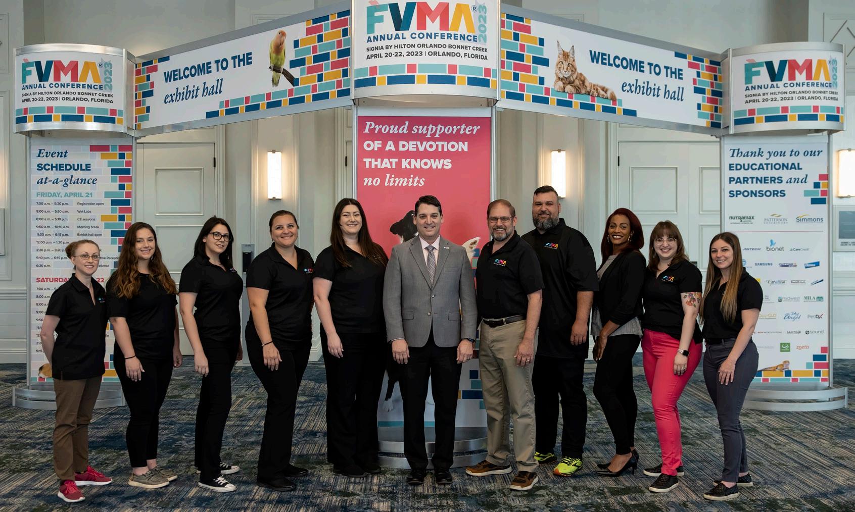
FVMA Staff
What’s NEXT
The FVMA staff had a great time setting up the conference, meeting conference attendees, and getting to know our members. The excitement of FVMA Annual Conference is something staff members look forward to each year. We can’t wait to see you at the next FVMA event!


Join the FVMA at our next event, The Gulf-Atlantic Veterinary Conference (TGAVC), which is returning to Tampa for the first time since 2019! This world-class veterinary conference will be held September 1417, 2023, at the Tampa Marriot Water Street.


We’ll be offering the three-hour Florida Laws and Rules & Dispensing of Legend Drugs course during this event, which satisfies Florida’s mandatory continuing education requirement for license renewal. A new wet lab will be offered, Stifle Exploratory and Extracapsular ACL repair, in addition to numerous other planned wet labs. Plus, the number of RACE hours attendees can earn is being increased for this conference.

Registration is capped to ensure a better learning environment, so don’t miss your chance to save your spot. Registration will be open this June, so stay tuned for more details coming soon!


SEPTEMBER 14-17, 2023
TAMPA MARRIOTT WATER STREET
Join us at the 10th Annual The Gulf-Atlantic Veterinary Conference (TGAVC), returning to Tampa for the first time since 2019! Presenting the best in small animal veterinary medical continuing education (CE) in a world-class destination, TGAVC is your chance to “experience the difference.”


TO LEARN MORE, VISIT FVMA.ORG/TGAVC2023 OR CALL 800-992-3862
EARLY BIRDS SAVE!
Register before August 16, 2023, for up to $100 off!
Featuring
• Florida Laws and Rules & Dispensing of Legend Drugs course

• 13 wet labs, including a new Stifle Exploratory and Extracapsular ACL Repair wet lab
• Over 230 hours of continuing education sessions
Registration is capped to ensure a better learning environment, so don’t miss your chance to save your spot.


TAMPA MARRIOTT WATER STREET 505 Water Street Tampa, FL 33602


Special Rate: $239**
CALL: 813-221-4900
Mention Group Code: FVMA
** Subject to applicable taxes. Resort fee included.
Discounted rates are valid until August 16, 2023, or until the room block is SOLD OUT!
REGISTRATION NOW OPEN | TGAVC 2023
26 | FVMA Advocate
Online Course:
SW Florida Coast- Solo Dr. small animal in a great location just minutes from the Gulf. Beautiful 5000 Sq ft. custom built hospital , 4 exam rooms, 2022 just over $1M. Owner willing to stay on for the right buyer.
N.E Florida- Solo Dr. now, but has been a multi Dr. practice in recent history. $1M gross in 2022. 3500 Sq. ft. custom built hos p.

S.E. Florida- 1.5 Dr. practice in a lease space. 202 2 gross of $1. 24 M, very profitable, 2 exam rooms with an option for a 3rd
S.E. Florida- ! Dr. small animal Practice that has served the area for over 30 year in Palm Beach County. Lease space with 2 exam rooms and room for a 3rd. $1.1M gros s, after debt income $242K.

New- Eastern Panhandle- Solo Dr. with $1.05M gross. 2300 sq.ft. hospital on almost 3 acres on a major 4 lane highway. After deb t income of $265K.
New- Orange County- Solo Dr. with regular relief Dr.'s and a 2022 gross of $1.7M. 3800 sq. ft. hospital w/ 4 exam rooms. Very wel l equipped. Priced to sell.
FLORIDA LAWS AND RULES & DISPENSING LEGEND DRUGS
Our updated three-part course satisfies Florida’s mandatory continuing education (CE) requirement for license renewal of no less than one hour of CE in the area of dispensing legend drugs and two hours of CE in the area of the laws and rules governing the practice of veterinary medicine.
KEY TOPICS include:
• The disciplinary process and guidelines
• Record-keeping requirements
• Appropriate drug dispensing practices

• DEA regulations
• Common causes for disciplinary action and how to avoid them
• Recent statutory, regulatory, and case law developments
Note: This course is approved by the Florida Board of Veterinary Medicine, Provider #0001682.

PRICING
FVMA members: $99.00
Non-member: $149.00
When Selling or Buying a Veterinary Practice... The only Veterinary Practice Broker Solely Focused on Florida! Showcase Properties of Central Florida Inc. Richard Alker, DVM Florida Representative 850.814.9962 e-mail: Richard@TPSGSales.com www.TotalPracticeSolutionsGroup.com Practice Sales Buyer Representation Associate Buy-Ins Exit Strategies Appraisals Call on
NAVIGATING THE (Sometimes Treacherous) WATERS OF SOCIAL MEDIA


Social media is a fact of modern life. It is here to stay. It can be good and bad for your practice as consumers place increasing importance on companies' online presence. A local consumer survey by BrightLocal showed that 49% of consumers trust reviews as much as personal recommendations from friends and family members.1 The benefits of social media and positive reviews on your practice are significant. This article will not discuss those. Instead, we will focus on the negative aspects of social media and how to deal with them.
Let’s start with the Consumer Review Fairness Act. This 2016 law protects the ability of persons to share their honest opinions about a business’s goods or services on any forum including social media. It does so by prohibiting “gag clauses” in non-negotiable consumer contracts. This means that form contracts that prohibit the posting of reviews or threaten legal action for posting are illegal. Having your clients sign a contract, or having terms on your practice website, that condition the provision of services to an agreement not to post negative social media posts or reviews may run afoul of this law.
Some customers may post negative reviews on social media without ever discussing the matter with you or otherwise giving you an opportunity to address their concerns, which can lead to sudden fears about how your rating will be impacted. Although it may be too late to avoid the review, there is still something that can be done. While you may not be able to repair the relationship with the client, you can respond in a way that shows the rest of the world that you take all comments seriously and that you are willing to improve. According to Podium’s 2021 State of Reviews publication, 56% of consumers changed their perspective on a business
based on how the business responded to a review.1 Your goal is to come across as a caring and reasonable veterinarian, which may not only help counter the negative review, but even cast some doubt as to the legitimacy of the criticism in the first place.
The first step is to review the situation from the client’s perspective and identify any possible mistakes or areas that
28 | FVMA Advocate Image courtesy of Canva. Social Media Image courtesy of Canva.
Edwin A. Bayó | Partner Grossman, Furlow & Bayo, LLC
can be improved. You may then consider reaching out to the client privately. Communication should start by expressing regret for not having addressed the concerns. You should then thank the client for their time and feedback, letting the client know that you are always willing to receive such feedback and that you are always willing to improve your practice. You must avoid letting the matter become adversarial or a back-and-forth. On the other hand, if some reasonable refund or other accommodation may satisfy the client and perhaps cause them to remove the negative review that possibility should be discussed.
If you need to respond to the negative review in writing, your response should not explain or otherwise discuss the treatment rendered. You do not want to provide a rebuttal to the allegations made by the client, or otherwise invite a back-and-forth that will end up creating a much longer post. Instead, the response should be something along the lines of “Thank you for your comments. At the XYZ clinic, we regularly monitor reviews on social media in an effort to identify areas for improvement, and when appropriate, implement changes. We are always willing to learn from client experiences and improve the quality of our services.” A written response along these lines comes across as caring and professional. It greatly reduces the opportunity for additional negative comments and puts an end to the discussion.
At the end of the day, keep in mind that the best defense against one negative review is ten or more wonderful reviews. While positive reviews remain a key way for companies to sell their product, with customers willing to spend 31% more on a business with excellent reviews 2 , your average rating is often key to whether or not a potential client chooses your practice. 3 Data from a recent study by PatientPop shows that younger generations like Gen Z place the strongest emphasis on a provider’s average star rating (49%), while older generations, like Baby Boomers (31%), see less importance. 2 When you look at restaurant reviews on social media, there are often 400 very good reviews, 30 or so middle-of-the-road reviews, and a handful of “worse place ever” or “do not eat here” reviews. Do you make your dining decision on the five terrible reviews? No. You look at the 400 hundred wonderful reviews, and you think that the five terrible reviewers are disgruntled human beings that will never be happy with anything. That is human nature. That is the reason you should not worry about those few negative reviews because most reasonable people will react the same way. In regards to human medicine, more than half of consumers (56%) won’t consider an average star rating of less than 4.0 for a healthcare provider. 3
Clients have a right to post negative reviews. Your response to negative reviews and your average rating outweigh a handful

of negative reviews. If a client threatens to post a negative review unless you give a refund, that may well cross the line into blackmail or extortion. If the post is flat-out wrong or malicious, or if the review is posted after the threat, you may have a right to sue the client. Nevertheless, I would never advise a veterinarian to go that route. Only lawyers will come out ahead in that scenario. Taking the high road and providing the right response will make the best case for you.
REFERENCES
1. https://www.searchenginejournal.com/online-reviewstatistics/329701/#close
2. https://www.qualtrics.com/blog/online-review-stats/
3. https://www.insiderintelligence.com/content/howconsumers-shop-online-doctors
| 29 www.fvma.org @thefvma @the__fvma @thefvma
Image courtesy of Canva.
FIVE STEPS TO START YOUR EXIT STRATEGY: Don’t Be Caught Off Guard
Carly Watson Tobler | VP of Southeast Sales Simmons & Associates Southeast, Inc.
According to an August 2022 poll by Retirement Living, 73% of Americans feel that they have not saved enough for retirement. As a veterinary practice owner, you may be thinking that retirement savings aren’t as important for you because you have a thriving practice to sell, the proceeds of which will fund your comfortable retirement.


But will they?
How can you be sure?
First, it is important to understand that your practice probably won’t sell overnight. The time it will take to sell your practice depends on a number of factors. Location, facility condition, equipment, practice size, profitability, and other criteria will determine whether your practice sells in a few months or several years. One of the most difficult challenges for a veterinary practice broker is to inform a practice owner hoping to retire imminently that their practice will not sell for what they expected. If only they had known the true market value of the practice a few years earlier, the practice owner and management could have taken meaningful steps to improve the practice’s bottom line and value. Don’t wait until you are ready to sell your practice to discover its value and salability.
30 | FVMA Advocate
Image courtesy of Canva.
Retirement
FIVE STEPS TO TAKE NOW TO BEGIN BUILDING YOUR EXIT STRATEGY
As a veterinary practice owner, you can take action right now to ensure your eventual practice sale will provide a comfortable retirement or a springboard into the next exciting adventure in your professional life. Here are five steps you can take right now to position yourself for a more lucrative sale when you are ready to exit your practice.
1. ESTABLISH A CREDIBLE BASELINE FOR WHAT YOUR PRACTICE IS WORTH TODAY.
A practice valuation from an experienced veterinary practice appraiser and broker is your first step to creating an effective exit strategy. It is advised that a baseline valuation is done a minimum of two years prior to listing the practice for sale. In addition to establishing a baseline for what your practice would be worth if you were to try to sell today, your appraisal should identify specific areas for improvement that will impact your eventual sale price.
2. INCREASE YOUR FEES TO KEEP PACE WITH MARKET CHANGES.
Are your fees keeping up with the cost of doing business? If not, it is critical that you evaluate your fees and do a market comparison with other nearby practices. If you haven’t done so in the last year, consider increasing fees now and create a schedule for regular fee increases in the future. Fees should be monitored and adjusted every 6-12 months as the cost of inventory and other expenses shift. As with any thriving business, ever-increasing costs must be passed along to the consumer to maintain a healthy profitability.
3. GET CONTROL OF YOUR PRACTICE’S EXPENSES.
The financial cost of uncontrolled expenses can wreak havoc on a veterinary practice’s profitability over time. By tightening the reins on your practice expenses, you can retain more of the money you and your team are working hard to earn and set yourself up for a better return on your investment when you sell the practice. Profitability is the primary factor in determining practice value, so maintaining a healthy bottom line will deliver a higher value. Enhanced profitability is also available for your personal enjoyment during your term as a practice owner.
4. MAKE NECESSARY CAPITAL INVESTMENTS.
The chipped paint, cracked linoleum, and outdated equipment may not bother you, but they could be deterrents for a potential buyer. Rather than having to make large capital expenditures right before you sell or having to lower your eventual sale price, it will be far less painful to make modifications and improvements gradually over time. Not only will your practice be more attractive to potential buyers, but you and your staff will enjoy the upgrades while you still practice.
5. WORK TO RETAIN A STRONG TEAM.
Having a long-serving, competent staff can be extremely attractive to potential buyers. If you are not already doing so, take action to ensure that your staff feels appreciated, valued, and respected. Consider strategies to minimize turnover, even if it means investing more in staff wages and benefits. Be mindful that increases in staff expenses should be offset by fee increases to maintain healthy profitability.
The sale of your practice should be a time to celebrate your hard work and accomplishments. By creating and implementing an effective exit strategy, your practice sale can be a celebration that provides wealth for you and your heirs.
REFERENCES
1. https://www.retirementliving.com/most-americans-are-not-prepared-for-retirement
| 31 www.fvma.org @thefvma @the__fvma @thefvma
PRACTICE Pulse
QUESTION: Our practice had to euthanize a dog, and the owner asked if we could hold the body in our freezer until she could make burial arrangements. The owner later requested we cremate the body and return the ashes. When we called to collect payment, the owner did not provide any payment details at the time. To date, they have not answered our calls. We are still holding the body in our freezer, as we will not do a cremation/ashes return without the owner prepaying. What should the next steps be?
A: Sending a certified letter is the safest option. Give the client 10 days to pay or pick up the body, and let them know that if they do neither, you will dispose.
QUESTION: We are in the process of streamlining our pharmacy protocols. We wanted to know if we are allowed to email our clients written prescriptions for non-controlled drugs or if we must hand them a physical prescription that they can then take elsewhere, including online pharmacies.
A: I see no reason why you cannot email a signed copy of a prescription for a non-controlled drug that the client can then print out and take to a pharmacy. This is assuming, of course, the prescription meets all the other requirements to be considered a valid prescription.
QUESTION: I just agreed to be a preceptor for a fourthyear veterinary student from another state. Can they legally perform procedures such as spays and neuters while under supervision by a licensed veterinarian?
A: According to Florida Statute 474.203 Exemptions, which cover veterinary medical practice:
This chapter does not apply to:
(3) A student in a school or college of veterinary medicine while in the performance of duties assigned by her or his instructor or when working as a preceptor under the immediate supervision of a licensee, if such preceptorship is required for graduation from an accredited school or college of veterinary medicine. The licensed veterinarian is responsible for all acts performed by a preceptor under her or his supervision.
If the student veterinarian is working as a preceptor and the preceptorship is required for graduation from an accredited school of veterinary medicine, then that student/preceptor is exempt. This means that anything the preceptor does under the immediate supervision of a Florida-licensed veterinarian is allowed.
QUESTION: A local law enforcement agency is asking whether their officers could administer naloxone to a police dog with suspected drug overdose based on updated canine emergency responder laws.
A: Yes, the reason being that there is an exemption in Florida Statute 474.202(5):
(5)(a) Any person, or the person’s regular employee, administering to the ills or injuries of her or his own animals, including, but not limited to, castration, spaying, and dehorning of herd animals, unless title is transferred or employment provided for the purpose of circumventing this law. 1 This exemption does not apply to any person licensed as a veterinarian in another state or foreign jurisdiction and practicing temporarily in this state. However, only a veterinarian may immunize or treat an animal for diseases that are communicable to humans and that are of public health significance.
The police dogs belong to the police department, and the police officers are “regular employees” of the owner attending to the ills of their own animals. There is also a newer exemption under paragraph (10):
(10) A paramedic or an emergency medical technician providing emergency medical care to a police canine injured in the line of duty as authorized under s. 401.254.
Nobody from the Department of Business and Professional Regulation or the Board of Veterinary Medicine is going to have any problem with a law enforcement officer administering Narcan or any other opioid antagonist to treat a canine.
QUESTION: I'm a mobile hospice and palliative care veterinarian. I just heard that xylazine is now a Schedule I drug in Florida. My distributor is still sending it to me like it's not controlled. I'm keeping it in my safe separate from Schedules II and III, but now I'm worried that I'm not allowed to even have it. I haven't gotten any information and was hoping you could help guide me.
A: Correct, xylazine is now a Schedule I drug in Florida. Even though it is not controlled under Federal law, that is about to change soon. If you still have xylazine in stock, I recommend you dispose of it. As it is not a controlled substance under federal law, you do not need to send the remaining stock to a reverse distributor, but you should pack it securely and place it in the trash.
Just to be safe, I suggest you write a short memo signed by yourself and another person — preferably a veterinarian, but it could be a technician or an office manager — attesting that on x day a (quantity) of Xylazine (lot number) was securely packed and placed in the trash in the presence of (names). If someone ever thinks of running an audit, you can provide evidence of what happened to the stock.
32 | FVMA Advocate
Have a question? THE FVMA CAN HELP.
One of the benefits of an FVMA membership is our helpline (800-992-3862), which is available to members Monday through Friday from 8 a.m.-5 p.m. Our helpline also provides insight to the FVMA staff of the challenges and concerns of our members. In this feature, we will highlight topics from the questions we received in preceding weeks as a part of an effort to keep our members up to date on current concerns, as well as regulatory and legislative changes.
QUESTION: I wanted to know if there are any Florida regulations in place that would prevent an establishment from performing nonanesthetic dentals without a veterinarian present? I am under the impression that anesthetic procedures must be done with a veterinarian present, but what about non-anesthetic procedures like dentals? Can they be performed without a veterinarian on staff? There are several pet boarding facilities that are offering non-anesthetic dental cleanings even though they do not have a veterinarian present, and it's a technician that performs them. I would just like some clarification on this.
A: Any dental procedure on an animal, with the exception of manually floating teeth on horses, is the practice of veterinary medicine. A technician can perform certain tasks under general supervision, and non-anesthetic dental cleaning can be performed without the veterinarian present but that does not mean a veterinarian is not required. Boarding facilities that have technicians perform dental cleanings without any veterinary supervision (offpremises) are engaging in unlicensed activity.
QUESTION: I’m reaching out to get some information about releasing records to clients. We have a client that we will no longer be providing services to. This client has NOT paid their bill; are we required to release records to this client? Can we legally withhold records from clients that do not pay their bill?
A: You cannot withhold a client's records due to an unpaid bill. Please see F.S. 474.2165 Ownership and control of veterinary medical patient records; report or copies of records to be furnished. Sub Section (3):
Any records owner licensed under this chapter who makes an examination of, or administers treatment or dispenses legend drugs to, any patient shall, upon request of the client or the client’s legal representative, furnish, in a timely manner, without delays for legal review, copies of all reports and records relating to such examination or treatment, including X rays. The furnishing of such report or copies shall not be conditioned upon payment of a fee for services rendered.
QUESTION: Should an exam be performed with every rabies vaccination? Is an exam within the last year considered acceptable? If a client declines an annual exam, can a rabies vaccination still be given?
A: The standard of practice for a limited service (vaccination) clinic requires a physical exam be performed on every animal to be vaccinated. There seems to be no reason why the standard for a regular clinic would be less.
61G18-15.007 Minimum Standards for Limited-Service Veterinary Medical Practices.
(1) The term “limited-service veterinary medical practice” shall mean a privately or publicly supported vaccination clinic where a veterinarian performs for a limited time vaccinations and/ or immunizations against disease on multiple animals, and where the veterinarian may also perform microchipping and preventative procedures for parasitic control, and shall not mean a premise otherwise permitted by the Board.
(2) The Limited-Service permittee shall register each clinic with the Board of Veterinary Medicine by name, address, date of clinic, time and duration, at least 28 days prior to offering a limited-service clinic. A copy of the limited-service permit shall be clearly visible at each limited service clinic held during its hours of operation and posted at the main office where the records are stored.
(3) A veterinarian must remain on site throughout the duration of a limited-service clinic and must maintain autonomy for all medical decisions made. A physical examination and history must be taken for each patient receiving veterinary medical care at a limited-service clinic. Recommendations and preventive medicine protocols must be developed from current accepted veterinary medical practice. The veterinarian is responsible for proper immunization and parasitic procedures and the completeness of recommendations made to the public by the paraprofessional staff that the veterinarian supervises or employs. The veterinarian is responsible for consultation and referral of clients when disease is detected or suspected.
The annual exam is a fairly common standard of practice. If the animal was examined by the veterinarian/practice less than 1 year ago, another exam may not be necessary.
If a client declines an annual exam, the veterinarian should explain that the standard of practice requires that examination be performed before providing the vaccination. If the veterinarian gives the rabies vaccination without having performed some type of physical examination, and there is an adverse reaction that could have been avoided with a physical examination, the veterinarian will not be able to defend him/herself.
NOTE:
| 33 www.fvma.org @thefvma @the__fvma @thefvma
END
The ultimate responsibility in the practice of veterinary medicine lies with the licensed veterinarian. Professional discretion must always be exercised.
PRACTICE FOR SALE/LEASE
PRACTICE FOR SALE – ATLANTIC BEACH, FL: Single doctor practice (uses relief vets at times) at the Beaches of Florida. Last year’s gross was 1.4 M. Out the door at 4 pm, until noon on Saturday, off on Sundays, and as many holidays as we like. Walk to the beach in 5 minutes. Affluent area. Free standing modern building (category 5 construction) on THE main commercial street. The usual, Complete IDEXX lab, DR Xray, CO2 Laser, full patient monitoring 11 computer domain with Avimark. Two or three exam rooms. Upstairs is ready for an apartment (agreement for sale can include build out for such). mih1971ail@gmail.com
PRACTICE FOR SALE – KEY WEST, FL: Ten-year-old practice in old town Key West. Wonderful pet-loving clientele. Great area for fishing, scuba diving, and sailing. Great laid-back warm place. Small lease hold space, inexpensive rent. Digital x-ray leased heska blood equipment which runs out 7-24. Great net. 4.5-day workweek. Can easily be increased to more. I do minimal emergencies when I feel like it. Current manager 6 years and a CVT. Great place to set down roots. Please don’t call the practice and upset employees. Text my cell phone first then I will call you back. 305-619-4731.

PRACTICE FOR SALE – SARASOTA, FL: Storefront practice for sale. Solo veterinarian retiring. Low overhead; low stress. Fantastic clients. Gross +$300,000. Recently upgraded interior. Fully computerized. Asking $150,000 obo. Contact Lee at 941-914-3239.

PRACTICE FOR SALE – AVENTURA, FL: Desiring autonomy and a flexible schedule? Fantastic opportunity for $110K financed. Housecall practice, south FL, established 16 years, owner retiring. Friendly clients, with no emergencies and, less than 20 hour/week. Fernando 561-6097371 or fmello@tworld.com.
Realize the best of both worlds – profitability and quality patient care at a price you can’t resist. In less than 15 minutes, obtain fully quantitative results on up to 13 important biomarkers at the point of care. No contracts. No minimums. Believe it.

34 | FVMA Advocate
CLASSIFIED ADVERTISEMENTS YOUR AD HERE. Email communications@fvma.org for details. www.bionote.com | info@bionote.com | 800-727-5169
LEARN MORE V200
Ov Ov Rea
FLORIDA

OCALA-Ocala Square Animal Clinic, Generously priced at $750K for practice and coveted Florida Real Estate.
ORLANDO Area - Companion animal clinic, grossing $800K, in a spacious 3,000 SF facility with an 800SF apartment.
PANHANDLE - Solo-doctor practice, grossing $650K with an impressive 16% growth year over year!
HASTINGS - Broadway Veterinary Clinic. Annual gross exceeds $1 1M After-debt income to new owner estimated at $245K!
NEW YORK
HOLLEY - – Barre Animal Hospital. Listed for an astounding $200K less than valued. Solodoctor producing $875K+ in revenue.


ROME - Adirondack Veterinary Services, grossing $702K. Practice and RE offered for only $683K


VIRGINIA

VIRGINIA BEACH Area - Equine practice grossing $850K with an astounding $250 ATC, 6+ acres with a 4,700SF facility also offered
TENNESSEE
KNOXVILLE Area - Prosperous small animal hospital, grossing approximately $860K, afterdebt income exceeding $297K to new owner!

TEXAS
METROPLEX Area - Small animal practice, neatly situated in a 2400SF facility with IDEXX lab equipment and digital x-ray included.
5-STAR GOOGLE RATING
you need to buy or
a veterinary practice, this is the only firm you need on your side!" Call today!
"If
sell
FEATURED PRACTICES FOR SALE Visit www PracticeSalesAdvisors com for more information Call Your Trusted Veterinary Broker. Personalized Service Specializing in: ✔ Individual Veterinary Practice Sales ✔ Corporate Acquisitions ✔ Valuations ✔ Associate Buy-Ins ✔ Buyer Representation. MICHIGAN
PRSRT STD U.S. Postage PAID Orlando, FL Permit #793 Florida Veterinary Medical Association 7207 Monetary Drive Orlando, FL 32809 It's your future, OWN IT! Are you ready to build equity for your future? SimmonsInc.com I 800-333-1984 Look inside at our FL listings.

