


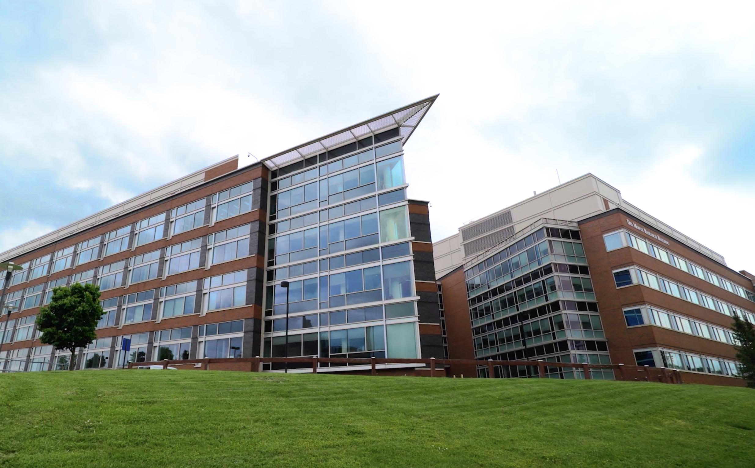
Center for Advanced Research Technologies
Center for Advanced Research Technologies
Our mission is make the world stronger, more hopeful, and ever better by working with PI’s to explore and develop next-generation treatments and technologies.
CART.urmc.edu
CART
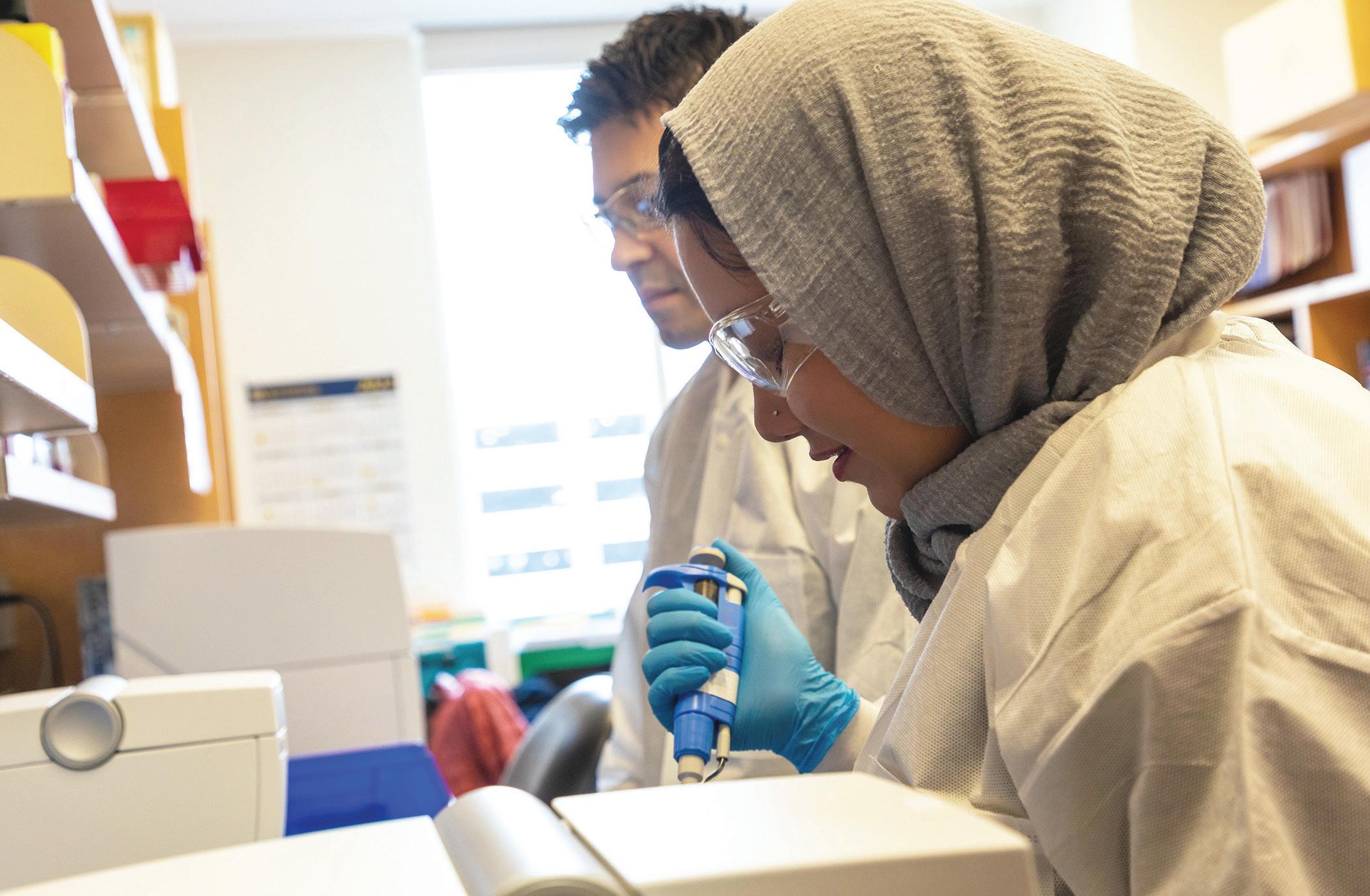
The Center for Advanced Research Technologies (CART) is a collection of core facilities within URMC that share resources and expertise with our 3,000+ researchers and 500+ labs, and with principal investigators both inside and outside of the University of Rochester Medical Center.
301 GRANTS SUPPORTED LAST YEAR 415 PRINCIPAL INVESTIGATOR COLLABORATIONS LAST YEAR 1,622 TOTAL PUBLICATIONS SUPPORTED & AUTHORED 43 STAFF MEMBERS TO SUPPORT RESEARCH
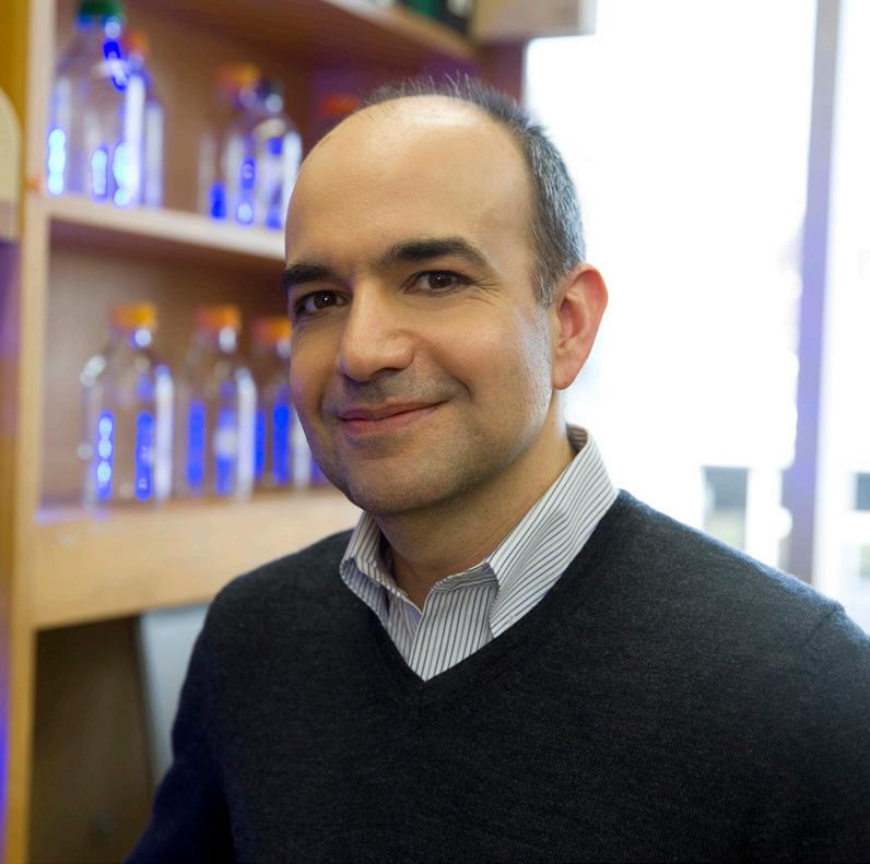
Note from Our Director
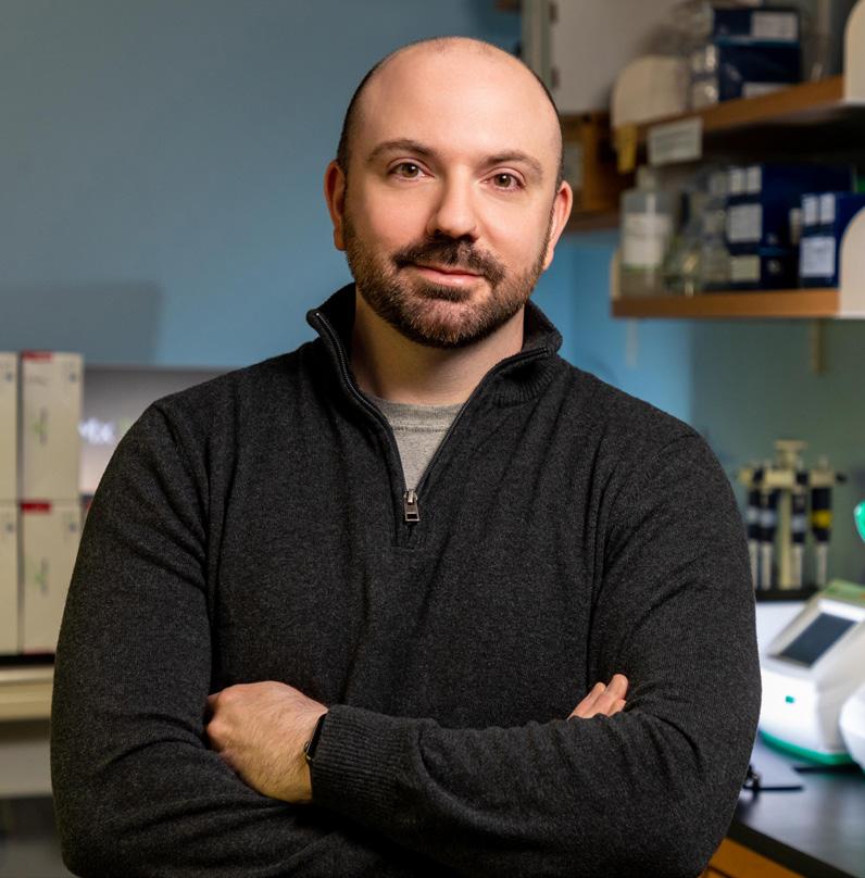
Timothy Bushnell, PhD., MBA Director, CART Associate Professor, Pediatrics
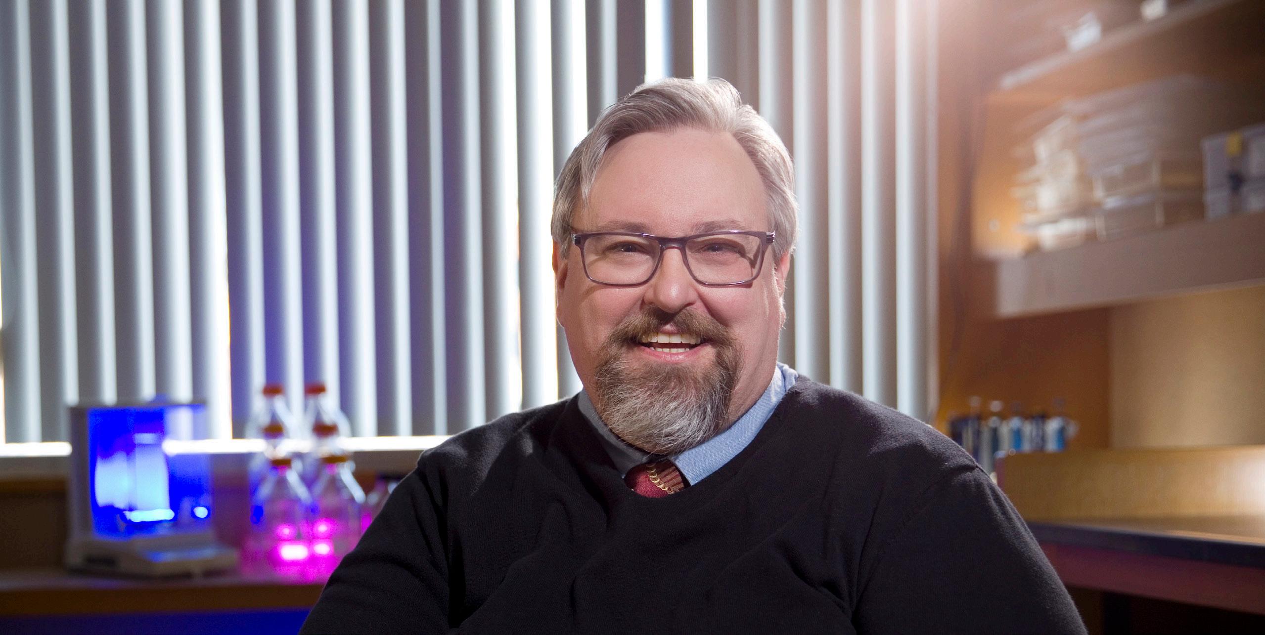
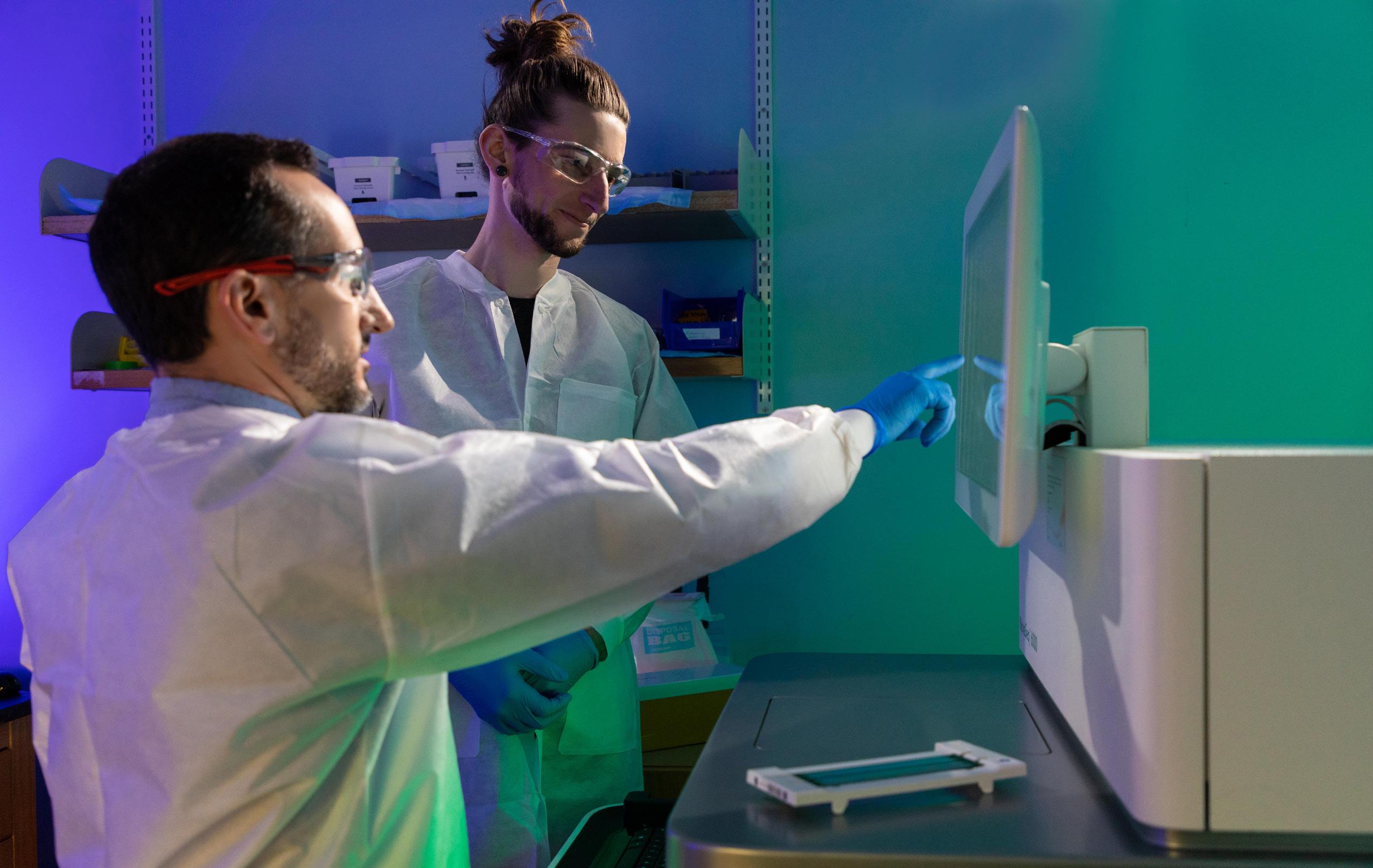
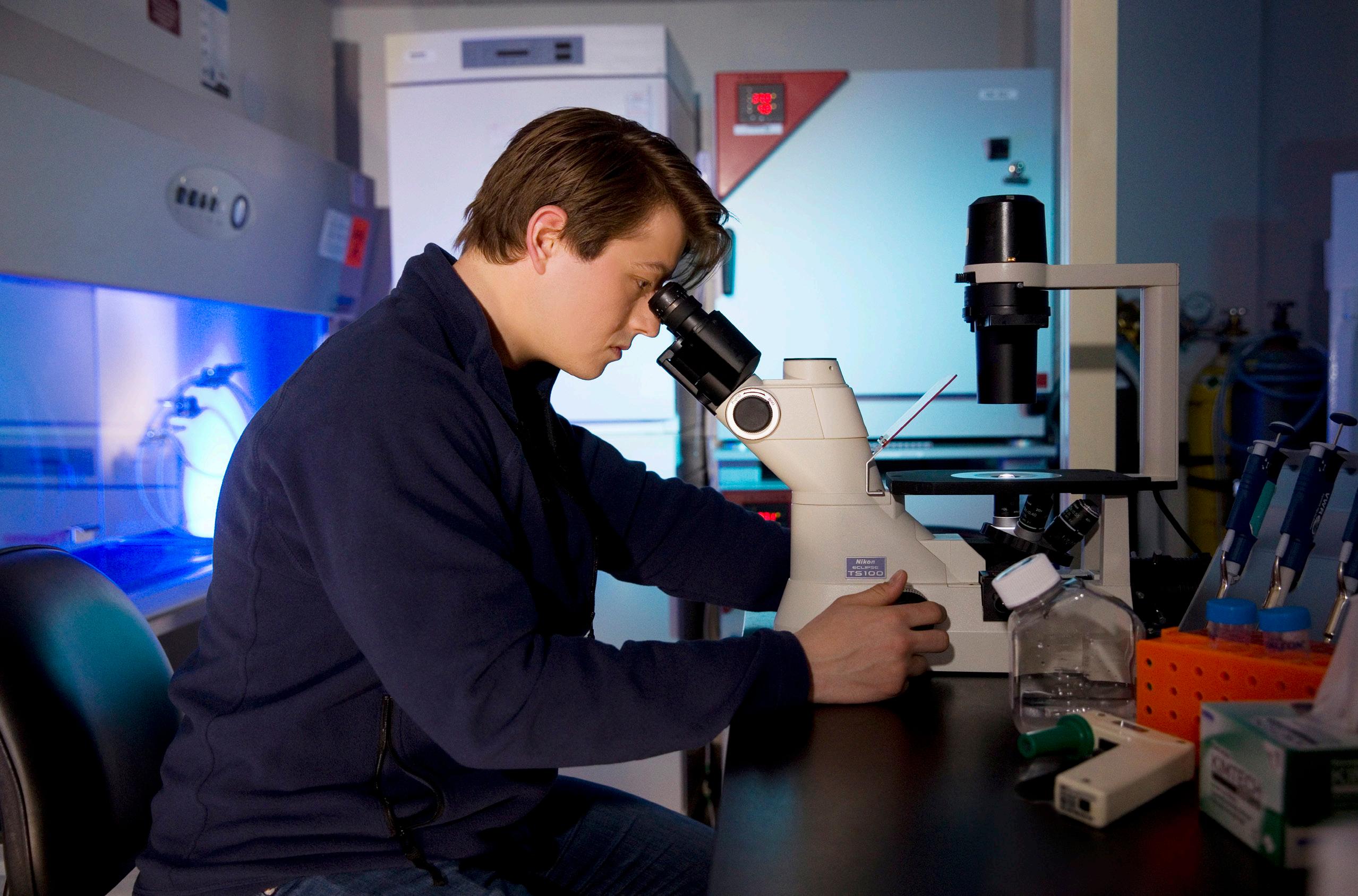
The Center for Advanced Research Technologies (CART) is a collection of scientific resources necessary for performing successful biomedical research in today’s highly competitive environment. We work constantly to maintain state-of-the-art technology within our facilities in areas including microscopy and genomics, cytometry and mass spectrometry.
But the technology is only one component that CART provides.
The strength of CART is in our team of well-trained and highly-dedicated researchers whose job it is to focus on their chosen technology, so you don’t have to. When you meet with a member of CART, you get an expert who knows how their technology can help advance your research goals. You have a partner, every step of the way.
CART is there with you when it comes to reproducibility and rigor in biomedical research. We work hard to ensure that the data generated in our facilities is of the highest quality and meets the highest standards.
No matter where you are in your career, the CART team is here to support you, from experimental design to analysis of your data, leaving you to focus on your research goals. We look forward to meeting you and supporting your research.
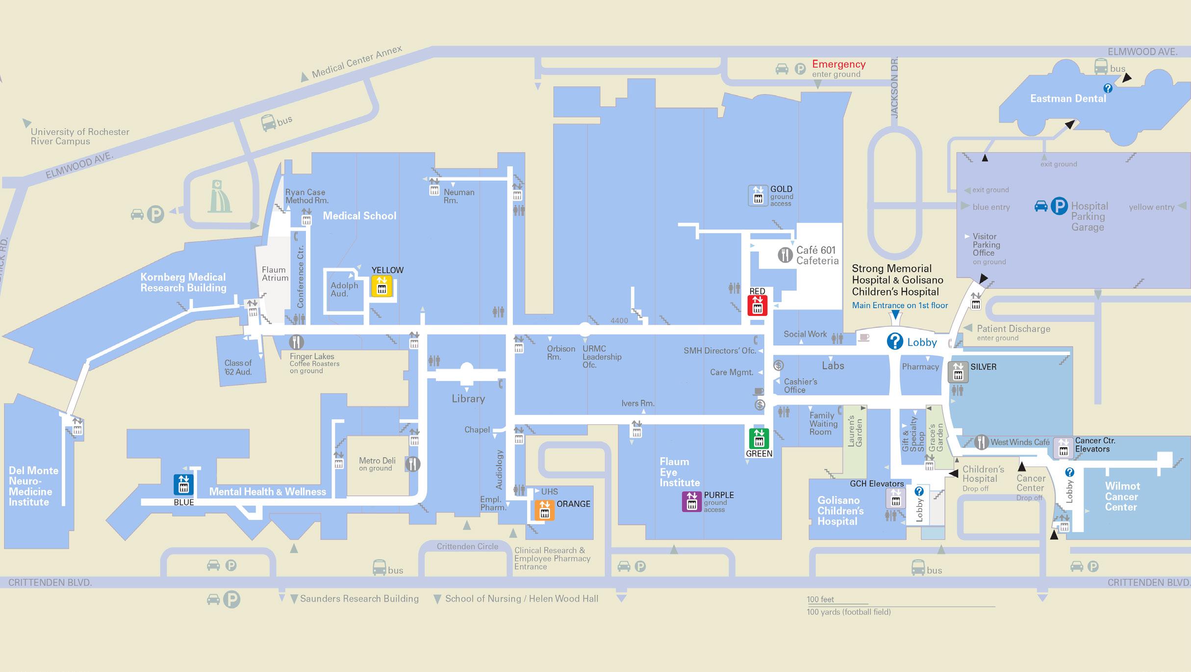
FACILITY LOCATIONS BSL-3 - BioSafety Level 3 / call for details CALMN - Center for Advanced Microscopy & Nanoscopy CSC - Cold Storage Core EMR - Electron Microscopy Resource FCR - Flow Cytometry Resource GRC - Genomics Research Center MSRL - Mass Spectrometry Resource Laboratory Metabolomics - coming fall 2022 GRC MSRL FCR EMR CALMN CSC
Genomics Research Center LOCATION: James P. Wilmot Cancer Institute / 3-0771 CONTACT US: URgenomics@urmc.rochester.edu / 585-275-7250 The Genomics Research Center (GRC) is the premier genomics shared resource in the Rochester region, providing consultation, technical expertise, and data analytical support for investigators performing cutting-edge, high-throughput sequencing and screening experimentation. • Transcriptomics • Whole Genome/Exome • Epigenomics • Metagenomics • Single-Cell ‘Omics • Spatial Transcriptomics • Custom Approaches • Data Analysis Support* *most services offered by GRC include comprehensive integrative analysis as a deliverable ABOUTSERVICES WEB:
CUTTING-EDGE
John M. Ashton, PhD., MBA Director, Genomics Research Center Assistant Professor, Biomedical Genetics
John Ashton has over 20 years of experience in genomics/genetics. Under his leadership, and with the operational support of Elizabeth Pritchett, PhD and the technical support of Jeff Malik, PhD, the Genomics Research Center is an integral facet of the URMC research community. John is passionate about advancing scientific discoveries and finding innovative solutions to complex research problems.
PUBLISHED
Our goal is to maximize
with principal investigators include:
• Platelet olfactory receptor activation limits
reactivity and growth of aortic aneurysms | PMID: 35324479
• Epigenetic aging of the demographically nonaging naked mole-rat | PMID: 35039495
• Coordination of endothelial cell positioning and fate specification by the epicardium | PMID: 34230480
• Modulation of the Human Pancreatic Ductal Adenocarcinoma Immune Microenvironment by Stereotactic Body Radiotherapy | PMID: 34862242
$2.1 MILLION IN GRANTS SUPPORTED LAST YEAR 129 PRINCIPAL INVESTIGATOR COLLABORATIONS LAST YEAR 418 PUBLICATIONS SUPPORTED OR AUTHORED 15 STAFF MEMBERS TO SUPPORT RESEARCH
your research impact. Our work
platelet
EDUCATION & TRAINING • NovaSeq 6000 DNA Sequencer • Nanostring GeoMX Digital Spatial Profiler • 10X Genomics Chromium Controller The GRC has over 20 instruments to support your research. A complete list is available online. The GRC strives to impart knowledge to the research community, through graduate school curriculum lecturing, independent practical “hands-on” workshops, informational seminars, internship opportunities, and publishing key information on our website.
INSTRUMENTATION
The
Flow Cytometry
Resource LOCATION: University of Rochester Medical Center / 3-4100 CONTACT US: Matthew_Cochran@URMC.Rochester.edu / 585-273-4020 The mission of the Flow Cytometry Resource (FCR) is to provide investigators with state-of-the-art instrumentation along with the human expertise to support all that is possible now, while pushing the limits of what can be done with flow cytometry.
Flow Cytometry Resource is a fullservice facility providing flow cytometry education and instrumentation for the University of Rochester Medical Center and River Campus, and to outside institutions. The FCR provides free consultation, experiment design help, and instrument training for University of Rochester researchers. We maintain and support a wide variety of instruments, which have robust QA/QC protocols in order to provide the highest levels of performance for our investigators. ABOUTSERVICES WEB:
Matthew Cochran, M.S. Technical Director, Flow Cytometry Resource Associate, Pediatrics
Our facility is fortunate to have access to a broad range of sophisticated technology that we can make available to University investigators, but getting the most out of that equipment can be a difficult task. In the FCR we’ve set up efficient and robust practices so we can provide high functionality and stability of operations. This allows our team to spend more time assisting our colleagues on advancing their science using flow cytometry. Whether a “simple” cell cycle assay, or a multi-year longitudinal study with numerous targets, our goal is to work with you to generate the best quality data possible.
PUBLISHED
Our published work includes:
• Development of an Orthotopic Murine Model of Rectal Cancer in Conjunction With Targeted ShortCourse Radiation Therapy | Advances in Radiation Oncology 2022 Mar-Apr; 7(2): 100867
• Lung megakaryocytes are immune modulatory cells | J Clinical Investigation 2021 Jan 4; 131(1): e137377
• In situ neutrophil efferocytosis shapes T
immunity to influenza infection | Nature IImmunology 2020 Sep;21(9):1046-1057
EQUIPMENT
$305k IN GRANTS SUPPORTED LAST YEAR 158 PRINCIPAL INVESTIGATOR COLLABORATIONS LAST YEAR 574 PUBLICATIONS SUPPORTED & AUTHORED 12 STAFF MEMBERS TO SUPPORT RESEARCH
cell
EDUCATION & TRAINING • Luminex Image Stream • Nexcelom Celigo S • Malvern NS300 • Agilent Seahorse CELL SORTING • BD FACSAria II • BioRad S3e ANALYTICAL CYTOMETRY • BD C6+ • BD LSRII • BD LSR-Fortessa • BD Symphony A1 • Cytek Aurora • Fluidigm Helios • BioRad Bioplex200 The Flow Cytometry Resource provides educational opportunities in a number of forms beginning with a bi-weekly introductory lecture. Hands-on instrument training is available for almost all equipment, and we also consult with investigators on a wide variety of topics. Advanced lectures are scheduled regularly for classes, large groups or lab meetings, so please contact us if you have topics you’d like to see. The FCR has a wide selection of flow cytometry equipment that is rarely matched, including:
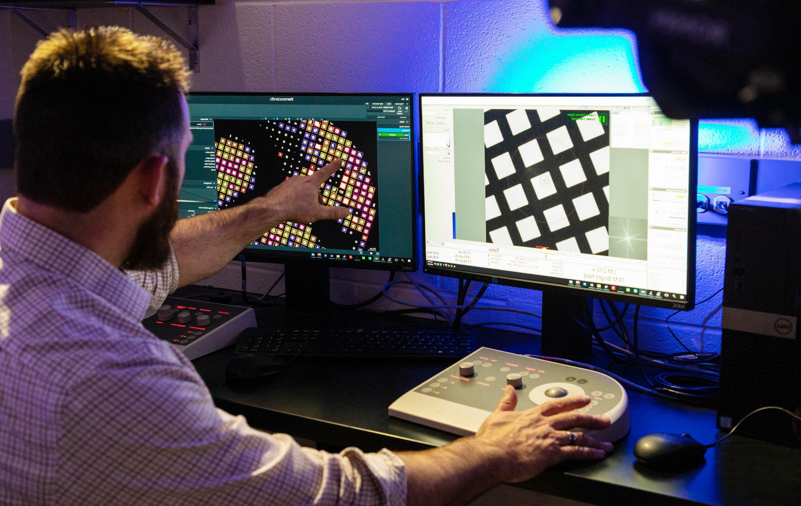
Electron Microscopy Resource LOCATION: University of Rochester Medical Center / B.7806 (yellow elevators) CONTACT US: Karen_Bentley@URMC.Rochester.edu / 585-275-1954 The goal of the Electron Microscopy Resource (EMR) is to support investigators in their pursuit of studies involving electron microscopy and Cryo-EM at the lowest possible hourly fee. • Routine TEM and SEM • Biofilm Analysis using SEM and TEM Imaging • Detection of antigen/antibody reactions with Immunoelectron Microscopy • Organelle Subcellular Fraction • Morphometric Analysis • Nanoparticle Identification Using Darkfield STEM • Elemental Analysis Using EDS • Cell Culture “Pop-off” Technique • Viral Negative Staining • Color Hi-Res Plastic Sections • Negative stain of macromolecules • Vitrification of samples for Cryo-EM • Screening of vitrified samples ABOUT SERVICES WEB:
• FEI
• Gatan
• Transmission
• Ultramicrotomes
• Critical Point
• Glow Discharge
• Plunge Freezer,
• Light
• Stereo
Karen Bentley, M.S. Director, Electron Microscopy Resource Senior Associate, Pathology & Laboratory
Welcome to the Electron Microscopy Resource (EMR). As a faculty member in the Department of Pathology, researchers can benefit from my clinical EM background in the interpretation of their research specimens and to correlate their EM imaging study to light microscopy, immunofluorescence or confocal results. We invite you to email us for a Zoom consultation to discuss how Transmission-EM, Scanning-EM or Cryo-EM can advance your thesis project or grant- funded research.
Recent publications include:
• Periarteriolar spaces modulate cerebrospinal fluid transport into brain and demonstrate altered morphology in aging and Alzheimer’s disease | Nature Communications 2022
• Evidence of Staphylococcus Aureus Deformation, Proliferation, and Migration in Canaliculi of Live Cortical Bone in Murine Models of Osteomyelitis. J Bone Miner Res. 2017
EQUIPMENT PUBLISHED $65k IN GRANTS SUPPORTED LAST YEAR 36 PRINCIPAL INVESTIGATOR COLLABORATIONS 196 PUBLICATIONS SUPPORTED & AUTHORED 3 STAFF MEMBERS TO SUPPORT RESEARCH
EDUCATION & TRAINING
Talos 120C electron microscope
Elsa 698 Cryo Arm for FEI Talos 120C
Electron Microscope Hitachi 7650
(2) Leica UC-7 & RMC Powertome XL
Dryer, Tousimis 931 Series, CPD
Cleaning System, PELCO easiGlow 91000
Leica EMGP2
Microscope, Olympus BX-40, Double Headed
Microscopes, Olympus SZ61 and Bausch & Lomb • Diamond knives (5), Diatome The Electron Microscopy Resource offers no-fee consultations while supporting the latest technologies to keep researchers at the forefront of imaging technologies.
Mass Spectrometry Resource LOCATION: Kornberg Medical Research Building / G-9828 CONTACT US: Kevin_Welle@URMC.Rochester.edu / 585-276-6804 The Mass Spectrometry Resource Laboratory (MSRL) provides instrumentation and technical expertise to local researchers seeking to conduct MSbased protein or small molecule assays. • Protein and peptide identification in purified or complex samples • Protein and peptide quantitation (including label-free, metabolic labeling and isobaric tags) • Interactome analyses • Identification of post-translational modifications (PTMs) • Targeted small molecule quantitation • Sample clean-up and processing • Qualitative and quantitative data analysis • Project design and planning • Assistance with data interpretation, grant writing and publication ABOUT SERVICES WEB:
Sina Ghaemmaghami, PhD Scientific Director, Mass Spectrometry Resource Associate Professor, Biology
The University of Rochester Medical Center’s Mass Spectrometry Research Laboratory (MSRL) is an established core facility that has provided a comprehensive suite of mass spectrometry services to investigators in Western New York for over 25 years. The core supports investigators with the planning, execution, analysis, and interpretation of proteomic and small molecule projects. We provide our
access to high-end instrumentation and complete consultation and analysis services to meet their specific project needs.
PUBLISHED
Our work with principal investigators include:
• Lin H, Tang M, Ji C, Girardi P, Cvetojevic G, Chen D, Koren SA, Johnson GVW. BAG3 Regulation of RAB35 Mediates the Endosomal Sorting Complexes Required for Transport/Endolysosome Pathway and Tau Clearance. Biol Psychiatry. 2022 Jul 1;92(1):10-24.
• Broly M, Polevoda BV, Al-Mamari W, Lim SC, Pais LS, Brown N, Riazuddin S, Bézieau S, Fu D, Isidor B, Cogné B, O’Connell MR. THUMPD1 biallelic variants cause loss of tRNA acetylation and a syndromic neurodevelopmental disorder. Am J Hum Genet. 2022 Apr 7;109(4):587-600.
user-base
EQUIPMENT
60 UR FACULTY UTILIZED MSRL SERVICES IN 2021 28 DEPARTMENTS WITHIN UR UTILIZED MSRL IN 2021 235 PROJECTS CONDUCTED IN MSRL IN 2021 4 MSRL STAFF MEMBERS TO SUPPORT YOUR RESEARCH
EDUCATION & TRAINING The Mass Spectrometry Resource offers one-on-one consultation on mass spectrometrybased approaches to keep our researchers at the forefront of proteomics research. We also are available for classroom presentations and speaking engagements. • Fusion Lumos Tribrid Orbitrap • Thermo Q Exactive Plus Hybrid Quadrupole-Orbitrap
Center for Advanced Microscopy & Nanoscopy LOCATION: University of Rochester Medical Center / 4.7500 CONTACT US: Kaye_Thomas@URMC.Rochester.edu / 585-275-1317 The mission of the Center for Advanced Light Microscopy & Nanoscopy (CALMN) is to provide state-of-the-art microscopy imaging and image analysis capabilities to further biomedical and bio-optical research. The CALMN lab provides access to high-end confocal, multi-photon and conventional fluorescence microscopy, conventional brightfield microscopy and laser capture microdissection. The CALMN provides assistance with all imaging procedures, aids in customizing staining protocols, provides UR researchers the ability and expertise to obtain high quality imaging-based data from tissue, culture, and materials-based research studies and professional, customized assistance in image analysis to support high quality quantitative imaging and publications. ABOUT SERVICES WEB:
V. Kaye Thomas, PhD Director, CALMN Assistant Professor, Pharmacology & Physiology
Welcome to the Center for Advanced Light Microscopy & Nanoscopy. In addition to providing access to high-end, state-of-the-art imaging equipment, the CALMN team endeavors to serve as a resource of imaging knowledge and experience. We provide hands-on, individual training to our researchers and can also guide scientists through experimental design, trouble-shooting and analysis phases of a project. Our aim is to form a partnership to support scientists in the research mission of the University, anticipating the future technological needs of the University community as we strive to embrace the value of Ever Better.
PUBLISHED
Recent publications include:
• Loss of function of renal Glut2 reverses hyperglycaemia and normalises body weight in mouse models of diabetes and obesity | PMID: 35290476
• CXCL10 peripheral activation niches couple preferred sites of Th1 entry with optimal APC encounter. | PMID: 34380032
• Perigenual and Subgenual Anterior Cingulate Afferents Converge on Common Pyramidal
in Amygdala Subregions of the Macaque | Journal of Neuroscience 2021
EQUIPMENT
$110k IN GRANTS SUPPORTED LAST YEAR 79 PRINCIPAL INVESTIGATOR COLLABORATIONS LAST YEAR 255 PUBLICATIONS SUPPORTED & AUTHORED 3 STAFF MEMBERS TO SUPPORT RESEARCH
Cells
EDUCATION & TRAINING The CALMN offers seminars on microscopy basics and the latest technologies to keep our researchers at the forefront of the imaging field. Abberior Stimulated Emission Depletion Microscope Nikon A1R HD Confocal LSM with TIRF Leica Stellaris 5 Confocal LSM with WLL Olympus FVMPE-RS Multi-photon Keyence BZ-X800 Microscope Zeiss Palm MicroBeam Laser Microdissection Imaris Image Visualization and Analysis Software
The BSL3 Core laboratory functions as a safe working environment for high-risk and high-security microbial pathogens. It is not a fee for service core, but functions instead as a core facility where properly trained users can study specific, Institutional Biosafety Committee-approved infectious agents under safe conditions, protecting both the user, other university personnel, and the public from harm. Users pay an hourly fee for use of the facility, which is charged out quarterly. The funds in the user account goes towards administrative fees, telecommunication charges, equipment service, and some user consumables such as certain components of personal protective equipment and safety and waste handling materials.
The Biosafety Level 3 (BSL3) core facility is available for use by any researcher whose work requires the manipulation of biological agents that may cause serious or potentially lethal disease as a result of exposure by the inhalation route.
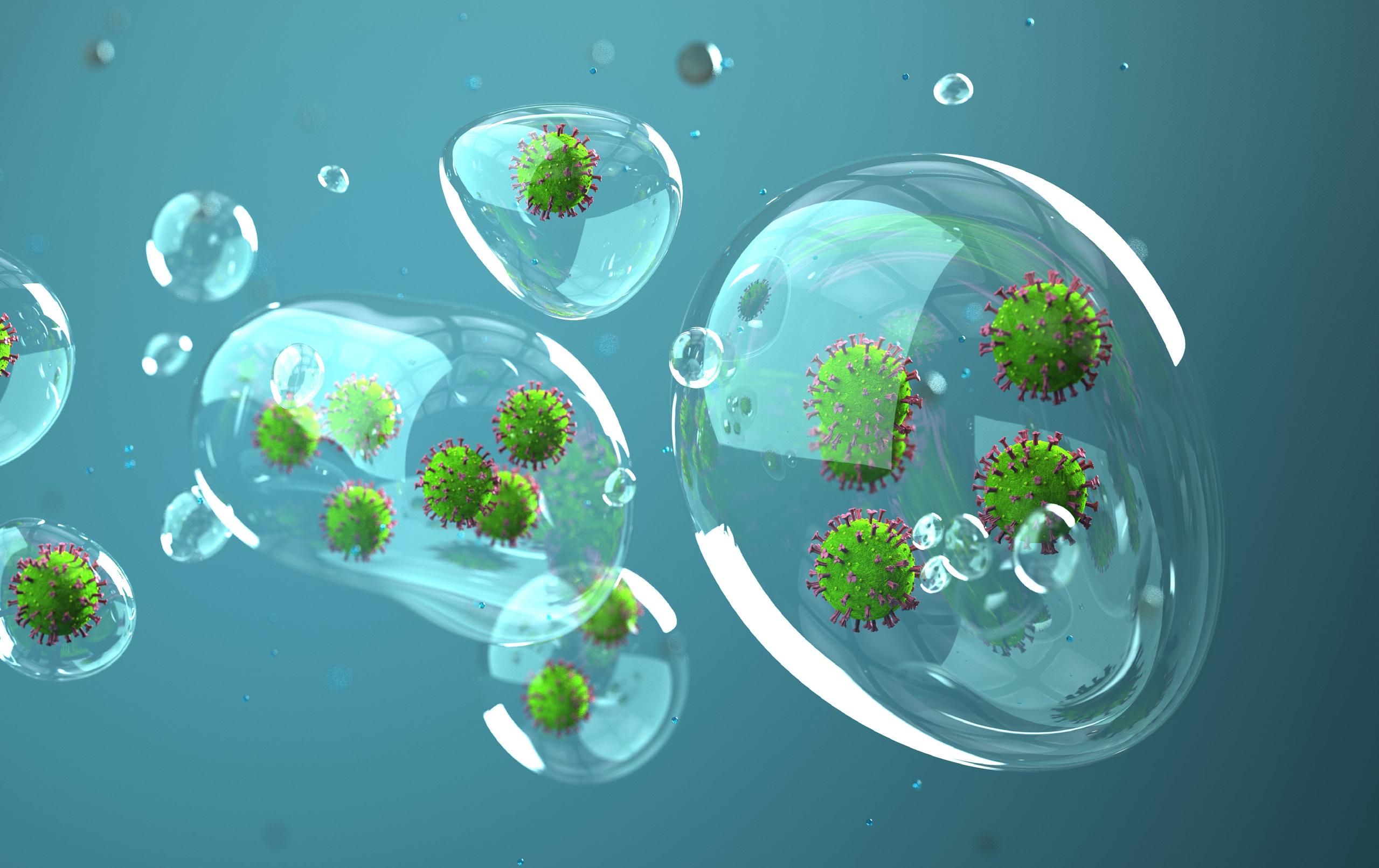
Biosafety Level 3 Facility
CONTACT:
Martin_Pavelka@URMC.Rochester.edu / 585-275-4670
ABOUT SERVICES WEB:
Martin S. Pavelka Jr., PhD Director, Biosafety Level 3 Facility Professor, Microbiology & Immunology
PUBLISHED
Our work with principal investigators include:
Raymonda MHCiesla JHMonaghan MLeach JAsantewaa GSmorodintsev-Schiller LALutz MMSchafer XLTakimoto TDewhurst SMunger JHarris IS Pharmacologic profiling reveals lapatinib as a novel antiviral against SARS-CoV-2 in vitro.; Virology; Vol 566. 2021 Nov 27.
Embong AK, Nguyen-Contant P, Wang J, Kanagaiah P, Chaves FA, Fitzgerald TF, Zhou Q, Kosoy G, Branche AR, Miller BL, Zand MS, Sangster MY, Topham DJ. “Formation
of Memory B Cells against Coronavirus in Acutely Infected COVID-19 Individuals.”
Pathogens.. 2022 Jan 29; 11(2)Epub 2022 Jan 29.
16k IN GRANTS SUPPORTED LAST YEAR 6 PRINCIPAL INVESTIGATOR COLLABORATIONS LAST YEAR 1 STAFF MEMBER TO SUPPORT RESEARCH
and Expansion
EDUCATION & TRAINING EQUIPMENT • Four class II biological safety cabinets • Pass-through autoclave • Stationary air and CO2 incubators, shaking incubator • Centrifuge with micro-, low-speed, and high-speed rotors • Electroporator • Visible light spectrophotometer • Tissue homogenizer • Sonicator • Inverted microscope • -80 freezer and liquid nitrogen storage unit • Cell lysis equipment • Plate reader • Plate imaging system The Biosafety Level 3 facility offers training to new users. For any training requests, please contact Martin.

Christopher has been a member of the University of Rochester Infectious Disease Division for 28 years.In addition to directing the Cold Storage Core, he serves as the director of the Clinical Trials Processing Laboratory, and as manager for the School Of Nursing’s Biomolecular Laboratory. Christopher Lane Director, Cold Storage Core Cold Storage Core LOCATION: University of Rochester Medical Center / 7-8802 CONTACT US: Christopher_Lane@urmc.rochester.edu / 585-273-4104 The Cold Storage Core (CSC) is an alarmed and environmentally controlled area that provides a secure on-site environment for longterm storage of research materials at the main medical center campus. We also maintain four spare -80oC freezers in the CSC for emergency use by UR investigators. • Multiple Freezer Units • Secure, Key Card Entry • 24 Hour Emergency Alarm Notification • Emergency Spare Freezers Available • Long-term Partial Unit Program ABOUTSERVICES
