Obstructive sleep apnoea: Improving a sleep service through technology
DEBATE
two of our deep dive into clinical risk management
Survey into adoption of dose-to-medium in medium reporting SPECT-CT and the potential effect it could have on imaging HISTORY RADIOTHERAPY NUCLEAR MEDICINE
SL33PAPN0E4 BIG
Part
How a world-famous philosopher became a lab assistant
Volume 33 | Issue 1 | Spring 2023 ipem.ac.uk
Since 2012, Imaging First have provided Ultrasound products and services throughout the NHS and Private Healthcare organisations. We are members of the NHS Supply Chain National Framework Agreements for new equipment, and for maintenance and servicing.


.
Our services include:


• Sales of new Edan systems • Sales of high quality refurbished systems from all major manufacturers • Both long term and short term rental of systems • Leasing and to spread the cost of equipment • Maintenance contracts, tailored to each customer's requirements from basic annual service contracts, to Fully Comprehansive contracts • Probe repairs, saving money on buying replacement probes • Sales of new and refurbished probes to suit all budgets when probes are beyond repair • Quality Assurance checks, ensuring patient safety and customer compliance at all times


In addition to our established ultrasound products and services, in 2021, we became the UK partner for iCAD, the global leaders in Intelligence, using their ProFound AI in the detection of Breast Cancer. ProFound AI offers a solution that empowers radiologists to breast cancer earlier and includes solutions for 2D mammography and tomosynthesis, ProFound AI also offers multi-vendor compatibility.


A varied issue
Usman Lula outlines the content in the latest issue of Scope, including undertaking research, mathematical modelling and service improvement, among much more.
Welcome all to the first issue of Scope for 2023. At the time of writing, another “sudden stratospheric warming event” was likely, just like the previous “Beast from the East”. Sometimes I do wonder if it is time for scientists to find better ways to predict such climate changes in terms of timing and scale. Science plays such an incredible part in such research and may one day (soon, I hope) provide improved warning signs, especially with earthquakes, which can be so devastating in all sorts of ways.

In this issue, for those employers exploring various recruitment options to increase staffing, Lauren Harrison (IPEM’s Training Development Officer) provides ways in which this can be made possible. To learn more, turn to page 14. And if you are looking at
PROFILE


undertaking research, Clara Ferreira (a Scope Editorial Advisory Board (EAB) Member) has kindly provided just the article to help you get started.


One of the ways in which we’ve been trying to improve the content and quality of Scope is by commissioning features that consider developments in areas not generally featured. One such feature, submitted by a Cardiff University team, is on combining mathematical modelling with experimental and clinical expertise to look at issues around in-vitro fertilisation. In essence, we could say science should form a foundation in the society we live in and help solve problems in every area.
A new regular feature
You may remember that we had a member profile in the third issue of Scope last year. The Scope Editorial Advisory Board, together with IPEM communications leads and


the Editor, felt that a regular profile would provide a glimpse into what our members do during their typical day, the biggest challenges they see, what they’d like to change and
what they do in their spare time. In hindsight, this may be really valuable to those seeking employment in healthcare sciences, trainees in the sector and also anyone who intends to
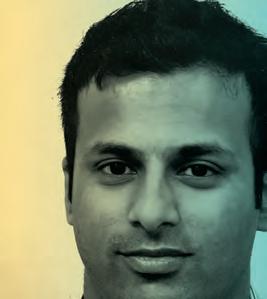
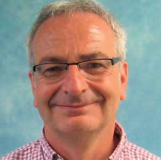
move upwards in their role. In this issue, we profile Professor Stuart Green who provides an interesting take on his role as a Director of Medical Physics.
I must say, one of my favourite features in this issue was the one on using technology in a “sleep service”. Service improvement is a key part of our role and this work aims to solve a number of problems in the sleep service, touching on auditing, software capabilities, support and maintenance, reporting, quality assurance and interactions with other systems and healthcare professionals. I have been promised a follow-up feature once the new software has been in clinical use to reflect on the changes, new system and feedback from service users. Finally, I was planning to present results from a national survey in this issue around service improvements in radiotherapy physics workflows. However, there was another (national) survey on the state of adoption of “dose-to-medium in medium” reporting that needed to be included in this issue (time sensitive!). I didn’t want two surveys in one issue, so I now plan to present my work in the summer issue of Scope Happy reading.
Usman Lula Chair of IPEM Scope EAB
I must say, one of my favourite features in this issue was the one on using technology in a “sleep service”
IPEM.AC.UK 3 SPRING 2023 UPFRONT COMMENT U F
CHAIR OF IPEM SCOPE EDITORIAL ADVISORY BOARD
Commissioning Editor Ejay Nsugbe
Commissioning Editor Clara Ferreira
Commissioning Editor Natasa Solomou
FEEDBACK
Discuss, debate, share. mycommunity.ipem.ac.uk/login
WEBSITE
News, events, support. ipem.ac.uk
ARCHIVES
Back issues of Scope online. ipem.ac.uk/scope
THE BIG DEBATE 16 / CLINICAL RISK MANAGEMENT PART 2
Commissioning
Editor: Usman I. Lula 0121 371 5056 | usman.lula@uhb.nhs.uk
Commissioning Editor: Dr Paul Doolan Medical Physicist, German Oncology Center, 1 Nikis Avenue, 4108 Agios Athanasios, Limassol, Cyprus 00357 2520 8025 | paul.doolan@goc.com.cy
Scope is published on behalf of the Institute of Physics and Engineering in Medicine (IPEM) by Redactive Publishing Ltd redactive.co.uk
In the second instalment of our deep dive into DCB0129 and DCB0160 – mandatory risk management standards for England – our panel look at the pressing issues, from alternative and complimentary approaches, to the challenges of implementation.
Publisher: Tiffany van der Sande tiffany.vandersande@redactive.co.uk | +44 (0)20 7324 2728
Editor: Rob Dabrowski
Senior designer: Gary Hill
Picture researcher: Akin Falope
Production: Aysha Miah-Edwards aysha.miah@redactive.co.uk | +44 (0)20 7880 6241
Advertising sales: scope@redactive.co.uk | +44 (0)20 7880 7556
I very quickly came to the conclusion that although at the time this was not specifically targeted for medical device systems, it was a very valid approach that could be applied to medical device IT systems.

Scope is published quarterly by the Institute of Physics and Engineering in Medicine but the views expressed are not necessarily the official views of the Institute. Authors instructions and copyright agreement can be found on the IPEM website. Articles should be sent to the appropriate member of the editorial team. By submitting to Scope, you agree to transfer copyright to IPEM. We reserve the right to edit your article. The integrity of advertising material cannot be guaranteed.
Copyright: Reproduction in whole or part by any means without written permission of IPEM is strictly forbidden. © IPEM 2023. ISSN 0964-9565
– Patrick Maw, Consultant Clinical Scientist page 16
16 32 UPFRONT 03 / CHAIR’S COMMENT 07 / NEWS 10 / IPEM NEWS 12 / POLICY UPDATE 14 / ROUTES TO REGISTRATION Scope is the quarterly magazine of the Institute of Physics and Engineering in Medicine IPEM Fairmount House, 230 Tadcaster Road,
YO24 1ES T: 01904 610821 | F:
|
of
Scope Editorial
York,
01904 612279 office@ipem.ac.uk | ipem.ac.uk
Chair
IPEM
Advisory Board: Usman I. Lula Principal Clinical Scientist, 1st Floor, Radiotherapy, Building, Medical Physics – University, Hospitals Birmingham NHS Foundation Trust, Queen Elizabeth Hospital, Queen Elizabeth Medical Centre, Birmingham, UK B15 2TH 0121 371 5056 | usman.lula@uhb.nhs.uk
IPEM SCOPE 4 SPRING 2023
CO
NTENTS
GENERAL
20
/
IMPROVING
A SLEEP SERVICE THROUGH TECHNOLOGY
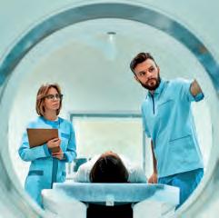
We look at the development of a new suite of software and hardware for the overnight oximetry service in Clinical Measurement at the James Cook University Hospital.
24 / GENIUS IN THE LAB
David Thwaites outlines how a leading philosopher became a physiological measurement laboratory assistant in the 1940s.
32 / RADIOTHERAPY DOSETO-MEDIUM REPORTING

ENDNOTES

54 / BOOK PITCH: IMAGINING IMAGING

28 / IN-SILICO
MEETS IN-VITRO
FERTILISATION
A Cardiff University team combine mathematical modelling with experimental and clinical expertise to investigate issues around in-vitro fertilisation.

A look at the results of a survey into the state of adoption of dose-to-medium in medium reporting among UK radiotherapy centres.
35 / QUANTIFICATION IN SPECT-CT: THE NEW LIGHT IN NUCLEAR MEDICINE?
Nuclear Medicine Technologist
Clara Ferreira looks at issues surrounding SPECT-CT and the potential effect it could have on imaging.
42 / HOW TO LAUNCH A RESEARCH PROJECT
Practical step-by-step guidance for members interested in taking part in a research project, from getting started to outlining the complex approvals process.
46 / SCIENCE IN DEVELOPMENT: A PHYSICS WORKSHOP REPORT
Chrysanthi Michailidou and Andria Hadjipanteli report back from the fifth ESTRO Physics Workshop 2022.
51 / MEMBER PROFILE: STUART GREEN
We hear from the Director of Medical Physics at University Hospital Birmingham.
52 / AN EXAMPLE FOR FUTURE LEADERS
Dr Iyobosa Uwadiae, the Secretary of the Nigerian Association of Medical Physicists, on being the first recipient of the IPEM Low- and Middle-Income Countries Award.
Dr Michael R Jackson outlines the ideas behind and the content within his new book, which explores the collision of art and science within radiology, from prehistory to the 21st Century.
28 Volume 33 | Issue 1 | Spring 2023 IPEM.AC.UK 5 SPRING 2023
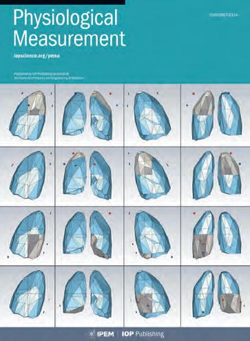
Physiological Measurement


iopscience.org/pmea


Physiological Measurement (PMEA) covers the quantitative assessment and visualization of physiological function in clinical research and practice, with an emphasis on the development of new methods of measurement and their validation.


Editor-in-Chief
Xiao Hu
To find out more about submitting, visit iopscience.org/pmea or e-mail pmea@ioppublishing.org.
IPEM SCOPE 6 SPRING 2023
JOURNAL IMPACT FACTOR
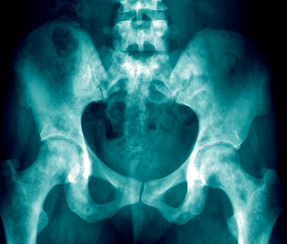
Upright radiotherapy: are there benefits?
Radiotherapy is typically delivered in supine position, however, upright positioning may have beneficial impacts, claims a new study.
The authors state that an upright position could affect organ volume, positioning and movement, compared to supine positioning.
In total, 16 patients with pelvic tumours were included in this study.
They had three setups in an upright position – an initial setup with acquisition of reference optical images and two repositioning setups.
The intra-fraction motion was assessed during two 20-minute chair rotation sessions. The patient comfort in supine and upright position was assessed with a 5-point Likert scale questionnaire.
Eight women and eight men treated on regular linacs between October 2021 and June 2022 were included.
Median age and weight were 62.5 years (35 to 81 years) and 75.1 kg (41 to 107 kg).
The findings of the pilot study have been reported in the journal Technical Innovations & Patient Support in Radiation Oncology.
Initial patient set-up took four to six minutes when performed by two radiation therapy technologists, while subsequent positionings took between two and five minutes.
Inter-fraction repositioning was achieved with below 1 mm accuracy on average and intra-fraction motion over 20 min was within 3 mm for
more than 90% of the study patients.
Most patients reported that upright positioning was as good as, and in some cases better than, the supine position that they had to maintain during their standard radiation treatment.
The positioning system is designed to place the patient in appropriate postures, depending on the cancer type that is being treated.
For prostate and pelvic treatments, patients were perched on the chair, supported by the back of the thigh and a knee rest. Patients were seated vertically for head-and-neck treatments, seated leaning slightly backward for lung and liver radiotherapy and slightly forward for breast radiotherapy.
The inter-fraction shift means were −0.5 mm (SD = 2.5), −0.4 mm (SD = 1.3) and −0.9 mm (SD = 2.7) in left–right (LR), anteroposterior (AP) and cranio-caudal (CC) directions, respectively.
The intrafraction shifts after 20 mins were 0.0 mm (SD = 1.5), 0.2 mm (SD = 1.1) and 0.0 mm (SD = 0.3) in LR, CC, and AP directions, respectively. Average global comfort was 4.1 (3 to 5) for the upright position and 3.9 (2 to 5) for the supine position.
The authors conclude: “The first study on pelvic cancer patients positioned in upright position on a chair is promising and it opens a potential new direction for the treatment of cancer patients.
“Evaluation of thoracic and head and neck tumours is ongoing and imaging with vertical CT is expected to start soon.”
bit.ly/3Ji6LKT
16 PATIENTS
1 MM Inter-fraction repositioning was achieved with below 1 mm accuracy. 20 MINS Intra-fraction motion was assessed during two 20-minute chair rotation sessions. PELVIC RADIOTHERAPY
with pelvic tumours were included in this study.
UPFRONT NEWS / TECHNOLOGY / POLICY / DEBATES FEEDBACK
debate, share my.community.ipem.ac.uk WEBSITE News, events, support ipem.ac.uk FAST FACTS IPEM.AC.UK 7 SPRING 2023
Discuss,
MAGNETIC RESONANCE IMAGING


Machine learning to predict brain tumour progression

ACanadian research team has created a computational model to predict the growth of deadly brain tumours more accurately.

Glioblastoma multiforme (GBM) is a brain cancer with an average survival rate of only one year. It is difficult to treat due to its extremely dense core, rapid growth, and location in the brain.
Estimating these tumours’ diffusivity and proliferation rate is useful for clinicians, but that information is hard to predict for an individual patient quickly and accurately.
Researchers at the University of Waterloo and the University of Toronto partnered with St Michael’s Hospital in Toronto to analyse Magnetic resonance imaging (MRI) data from multiple GBM sufferers.
They analysed two sets of MRIs from


CLINICAL ENGINEERING LOW-COST HEAVY METAL SENSOR
Heavy metals, such as lead and cadmium – present in items including batteries, cosmetics and food – are toxic when they accumulate in the human organism. They can potentially cause several health problems, but detecting them in body fluids requires expensive equipment and a controlled
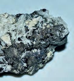

Single crystal electron diffraction
each of five anonymous patients suffering from GBM. The patients underwent extensive MRIs, waited several months, and then received a second set of MRIs.
Because these patients chose not to receive any treatment or intervention during this time, their MRIs provided the a unique opportunity to understand how GBM grows when left unchecked.
A deep learning model was used to turn the MRI data into patient-specific parameter estimates that inform a predictive model for GBM growth.
Now that the scientists have a good model of how GBM grows untreated, their next step is to expand the model to include the effect of treatment on the tumours. Then the data set would increase from a handful of MRIs to thousands.
bit.ly/3R2oDvf
laboratory environment.
Researchers at the University of São Paulo in Brazil have now developed a portable sensor made of simple materials to detect heavy metals in sweat, which is easily sampled.
The sensor is simple in terms of the materials used to make it and stages of its production.


The device is connected to a potentiostat, a portable instrument that determines the concentration of each metal by measuring differences in potential and current between electrodes. The result can be displayed on a computer or smartphone.
bit.ly/3H74mjp
The new National Electron Diffraction Facility will use electrons, instead of conventional X-ray crystallography, to investigate and determine the structure of much smaller crystals than previously possible. The new facility will feature two new XtaLAB Synergy-ED fully integrated electron diffractometers. The instruments will be housed in refurbished laboratories in Southampton and Warwick, which will also include sample preparation facilities and space for visiting researchers.
bit.ly/3wxnu5n


Influenza vaccine development
For the first time US researchers have created an atomic-level computer model of the H1N1 virus that reveals new vulnerabilities through glycoprotein “breathing” and “tilting” movements. This work suggests possible strategies for the design of future vaccines and antivirals against influenza. Distinguished Professor of Chemistry and Biochemistry at UC San Diego Rommie Amaro said: “This research could be used to develop methods of keeping the protein locked open so that it would be constantly accessible to antibodies.”
bit.ly/3iYMyz7
Electromagnetic interference
Researchers have demonstrated, for the first time, a mechanically flexible silver mesh that is visibly transparent, allows high-quality infrared wireless optical communication and efficiently shields electromagnetic interference in the X band portion of the microwave radio region. The film showed high transmission for a broad wavelength range from 400 nm to 2000 nm and sheet resistance as low as 7.12 Ω/sq. bit.ly/3R7Ivx5
NEWS IN BRIEF
IPEM SCOPE 8 SPRING 2023 UPFRONT NEWS U F
Stem cell transplant reporting
Researchers have developed a custom-built application to automate determination of engraftment – a key outcome after hematopoietic stem cell transplant (HSCT).
The application supersedes a tedious manual process and improves accuracy of reported hematopoietic cell transplant engraftments.
To improve accuracy and efficiency, an informatics team embedded within the Children’s Hospital of Philadelphia (CHOP) Cell Therapy Programme, built an
NANOMEDICINE
WHAT IS NANOMEDICINE?
application using R/Shiny, an open-source framework for developing interactive web applications that can perform complex data acquisition and manipulation tasks.
The tool extracts data and calculates engraftment dates based on the rules of the Centre for International Blood and Marrow Transplant Research.
Neutrophil engraftment
reporting was found to be incorrect in only two of 53 cases (3.8%), both of which were due to typographical errors and not the application, versus 15% of cases when engraftments were calculated manually prior to April 2021. Over the same period, platelet engraftment reporting was found to be incorrect in one of 53 cases (1.9%), also due to a typographical error, a significant decrease from the 28% error rate prior to the implementation of the tool.
bit.ly/3HuPUmF
they create and test new nanomedicines.
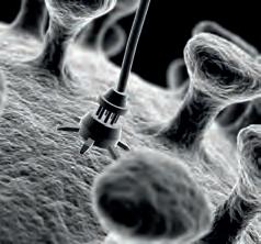
The medical application of nanotechnology. It is an area of study using nanoparticles for drug delivery, diagnoses and in vivo imaging.
HOW SMALL IS “NANO”?
“Nano” refers to particles that are only a few hundred nanometers in size, which is significantly smaller than the width of a human hair.
HAVE THEY BEEN IN THE NEWS?
Yes - there is some debate on a lack of standards when it comes to how these medicines are analysed and characterised in the laboratory. It follows a recent paper published in Nature Communications that revealed a high level of disagreement between lab results that researchers rely on as
WHAT CAN INFLUENCE THE PERFORMANCE OF A NANO MEDICINE?
Michigan State University researcher Morteza Mahmoudi, one of the men behind the paper, said: “My team and I have identified several critical but often overlooked factors that can influence the performance of a nanomedicine, such as a person’s sex, prior medical conditions and disease type.”
WHAT SHOULD BE DONE?
Taking the above factors into account when designing studies and interpreting results could enable more reliable and accurate data and lead to better nanomedicine treatments.

WHERE CAN I READ MORE?
Read the Nature Communications paper at bit.ly/3XDPAYA
ULTRASOUND WEARABLES FOR CARDIAC IMAGING
Engineers and physicians have developed a wearable ultrasound device that can assess the structure and function of the human heart.
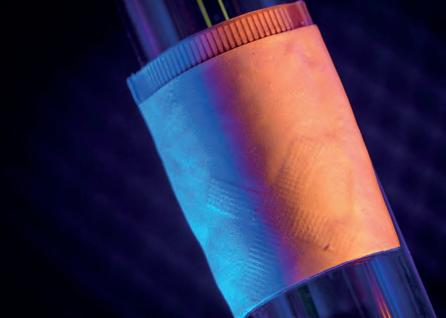
The portable device, which is roughly the size of a postage stamp, can be worn for up to 24 hours and works during strenuous exercise.
The goal is to make ultrasound more accessible to a larger population, said Sheng Xu, UC San Diego Professor of Nanoengineering.
“The technology enables anybody to use ultrasound imaging on the go,” he said.
Thanks to custom AI algorithms, the device is capable of measuring how much blood the heart is pumping. This is important because the heart not pumping enough blood is at the root of most cardiovascular diseases. And issues with heart function often manifest only when the body is in motion.
bit.ly/3R3YpbI
CLINICAL RESEARCH
IMAGES: SCIENCE PHOTO LIBRARY/ISTOCK/SHUTTERSTOCK IMAGE:
IPEM.AC.UK 9 SPRING 2023 UP CLOSE
© DAVID BAILLOT-UC SAN DIEGO JACOBS SCHOOL OF ENGINEERING
CLINICAL TECHNOLOGY
IPEM TRAINING SCHEME
The latest cohort on IPEM’s Clinical Technologist Training Scheme (CTTS) has successfully passed the course.
The CTTS has earned a strong reputation in the sector, offering a robust, externally validated education and training framework for clinical technologists, and ensuring a workforce fit to practice.
Successful completion sees graduates awarded IPEM’s Diploma in Clinical Technology and opens a route to joining the Register of Clinical Technologists (RCT).
A total of 23 trainees recently completed the course and were awarded their Diploma in Clinical Technology.
bit.ly/IPEM_CCTTS

Radiotherapy and cancer


An inquiry into the cancer crisis facing the UK has been launched by parliamentarians.
The All-Party Parliamentary Group for Radiotherapy (APPGRT) launched the inquiry into radiotherapy provision across the country and its ability to cope with urgent present and future challenges in cancer care.
PROFESSIONAL PROMOTION PRIZES AND AWARDS FOR OUTREACH
Two IPEM members have won prizes and awards for their outreach work to educate the public about medical physics and clinical engineering.

Dr Ejay Nsugbe, an independent researcher in upper-limb prosthesis control, was awarded IPEM’s Spiers’ Prize for Outreach.
He has been active in outreach for a number of years. He was recently a finalist at the annual STEM for Britain
IPEM submitted written evidence to the inquiry, highlighting the need to address workforce shortages, funding to replace ageing equipment, increased access to artificial intelligence (AI) technologies and a review of patient access to services.
Nicky Whilde, Chair of the Radiotherapy Professional Standards Panel, represented IPEM at the inquiry session.
event where he was invited to disseminate his research at the House of Commons to MPs and academics.
Elizabeth Davies, a medical physicist at University Hospitals of Leicester NHS Trust, won the Roy Ellis Patient Benefit Award for her work in supporting the provision of better quality, more consistent information on radiation risk to patients.
Dr Robert Farley, IPEM’s President, said: “Congratulations to both Ejay and Elizabeth for their dedicated work to promote and educate the public about medical physics and clinical engineering, a cornerstone of IPEM’s charitable objectives.”
PARLIAMENTARY INQUIRY
IPEM SCOPE 10 SPRING 2023 UPFRONT NEWS U F
OBITUARY
Professor John Clifton
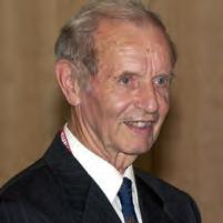
It is with great sadness that we have to report the passing of Professor John Clifton, an IPEM Fellow who made an outstanding contribution to medical physics.
Born in 1930 and with a career spanning almost 70 years, he was at the forefront of the development and advancement of physics applied to medicine in the UK.
He graduated from the University of Southampton in 1955 and started a career in medical radiation physics at the Royal South Hants Hospital.
Two years later he joined the medical physics department of University College Hospital Medical School (UCHMS) and was subsequently appointed Head of Department in 1962. Over the course of the mid-20th century, medical physicists pushed the boundaries of physics and new technologies, applying these innovations directly to medical practice. They developed and disseminated new forms of diagnosis and treatment, laying the groundwork for the sophisticated scientific medicine used today.
Joel Professor of Physics
In 1981, UCHMS became part of University College London (UCL) and John was appointed Professor of Medical Physics. Seven years later, the Middlesex Hospital Medical School merged with UCL, resulting in the creation of the academic department, UCL Medical Physics, led by John. In 1990 he was appointed the fourth Joel Professor of Physics Applied to Medicine at the University of London.
IPEM member Professor Alan Cottenden MBE, Emeritus Professor of Incontinence Technology, said: “He was Head of Department when I joined
UCL in 1984 and was very welcoming and supportive as I found my feet. He was a good and encouraging listener. Following our discussions, I often discovered that - without mentioning it to me - he had quietly done or said something somewhere that would smooth the way. A lovely man.’
EFOMP President
Professor Clifton was President of the Hospital Physicists’ Association (HPA) from 1976 to 1978 and was a founding member and the first President of the European Federation of Organisations for Medical Physics (EFOMP) from 1980 to 1983. He was also Honorary Editor of IPEM’s international peer-reviewed journal Physics in Medicine and Biology from 1979 to 1983.
He retired from UCL in 1992, becoming an Emeritus Professor of Physics as applied to Medicine.
The current Head of UCL Medical Physics and Biomedical Engineering and IPEM Fellow Professor Andrew Nisbet said: “Professor Clifton was not only instrumental in the development of Medical Physics and Biomedical Engineering at UCL and UCLH but his contributions to the field on a national and international scale are significant and it is very fitting that we have a student award named in his honour.”
The John Clifton Prize was initiated in 2011 in his honour and is awarded by UCL to the most outstanding performance from a non-final year undergraduate student.
Dr Robert Farley, IPEM President, said: “Our condolences go to John’s family and friends and he leaves behind him a significant legacy.”
OBITUARY Joe Farwell
On 9 November 2022, Joseph Farwell (Joe), husband of Ewa and father of Sylvie, passed away at age 57. Joe touched the lives of many in his long career working in various departments and through his contributions in the field of radiation protection.
Former colleagues have shared happy memories of their time spent in Joe’s company. “Eating lunch together in the garden of St Paul’s Cathedral, when Joe worked at St Barthlomew’s Hospital in 1997.” “Fond memories of working with Joe at Stoke Mandeville Hospital between 2010 and 2016. He was certainly a wonderful soul indeed.” “Having Joe pop over to watch a football match after he had put his baby daughter Sylvie to bed. He was such a devoted father. His face lit up every time Sylvie’s name was mentioned.”
In 2016, Joe and his family moved to Sticklepath on the edge of the Dartmoor National Park when he took up a role at the RD&E Hospital in Exeter. Joe continued his love of sailing after moving to Devon. He loved rural life and often said how happy he was. Just two weeks before he died he had started in a role in Taunton.
He will be sorely missed not just by his family, but also by his friends and all those that knew him. He had such an infectious cheery demeanour. There will forever be a gap in the lives of all who were privileged to be able to call him their friend.
IPEM.AC.UK 11 SPRING 2023
Increasing workforce capacity
The Westminster Health Forum
conference
According to their website, Westminster Forum Projects’ conferences are frequently the platform for major statements on policy from senior Ministers and regulators and so it was clear it was important IPEM should be represented at this one. However, I left unclear on who the target audience was or what the expected outcome of the meeting was!
It was initially chaired by the current Chair of the All Party Parliamentary Group for Cancer, Elliott Colburn, MP for Carshalton and Wallington in South London, followed by Tom Roques, Vice President of the Royal College of Radiologists. The speakers were from a wide range of professions, all with an interest in cancer, including representatives from Macmillan Cancer Support and Cancer Research UK, clinical and medical oncologists, surgeons, epidemiologists, pathologists and pharmacists. Despite the disparate group, there was one central theme from all the presentations, and one that is no surprise to any of us and that was workforce – or rather the lack of it – and the cancer backlog.
All professions highlighted the lack of supported training places, the challenges of overcoming mountains of red tape for overseas recruitment and problems with
LACK OF FUNDING FOR CLINICAL SCIENTIST TRAINING
A lack of funding for Clinical Scientist training places is putting patient safety in Scotland at risk, according to IPEM.
In a letter to Karen Reid, the Chief Executive Officer of NHS Education for Scotland (NES), Dr Robert Farley, IPEM’s President, said the lack of funding would have a negative impact on patient safety.
In his letter, Dr Farley said he understood NES was proposing funding equating to less than a
single training post in medical physics and clinical engineering in 2023, despite the Scottish Government’s Chief Healthcare Science Officer’s public acknowledgement of the importance of training.
NES responded to say while it was fully supportive of the training of the healthcare science workforce and recognised the important contribution they make to the delivery of safe patient care, there would be
retention. The lack of resilience in the workforce was raised, both in terms of the health of staff and also the practical problems of delivering a service with increasing demand.
The use of data that we already collect in the UK was also discussed and how it could be better used to both improve cancer survival rates and rationalise diagnostic and follow-up testing for those who really need it, based on risk stratification.
The National Physical Laboratory presented a tool it has been developing that can find bottlenecks in cancer pathways to support change within an NHS trust.
no additional funding available.
Responses were submitted by IPEM to inquiries launched by two House of Commons committees.
The Retained EU Law (Revocation and Reform) Bill is currently passing through Parliament and the Public Bill Committee is examining it. The Bill would completely overhaul a body of UK domestic law known as “retained EU law”, which was created by the EU (Withdrawal) Act 2018.
IPEM’s submission was based on the potential impact any changes could have on patient, public and employee safety.
Evidence was also submitted to the Science and Technology Committee inquiry on the Governance of Artificial Intelligence. The submission said in healthcare settings, healthcare workers were best placed to make the use of AI more transparent and explainable to the public and patients.
To read both responses in full, visit ipem.ac.uk/about/publicengagement/consultations
IMAGE: ISTOCK
WESTMINSTER HEALTH FORUM
held a
on “Next steps for cancer prevention, diagnosis, treatment and care in England”.
IPEM SCOPE 12 SPRING 2023 POLICY UPDATE P U
Nicky Whilde, Chair of the Radiotherapy Professional Standards Panel, went along to find out more.
There were some interesting discussions on the epidemiology of cancer and how certain social groups are still behind in survival rates. It was also pointed out that despite years of projects to support the early diagnosis and treatment of cancer, we are still lagging behind most developed countries in survival rates.
My thoughts on the meeting are that the remit was far too wide, and the attendees a disparate mix whose roles were unclear to me. However, there was one person who may have some influence – and that was Elliott Colburn MP. To quote him directly: “The big problem that is coming through loud and clear is that issue of workforce, which, sadly, is not an overnight fix. There are clearly things we can do in the short term, particularly around pensions, incentives and retention, to try and minimise the issue that we’re facing. But ultimately, this is about increasing
Dosimetry Solutions from Phoenix Dosimetry Ltd


NEW Polimaster EPD Real Time Personal Dosemeter Small (badge size), 63x50x18mm, Bluetooth enabled, Wireless charging, compatible with Radsight software.
0.1 Micro Sv/h to 1 Sv/h, 15KeV-1.5Mev
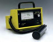
RadEye B20 measures Alpha, Beta, Gamma surface contamination with optional H*10 fi lter and B-20 then becomes a dose/doserate meter
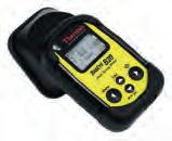
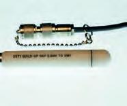
Ionisation Chambers 0.6cc NE2571/ NE2581 and NPL-2611 Secondary Standard
ALL WE CAN DO IS KEEP THE PROFILE OF OUR PROFESSIONS HIGH IN THE NATIONAL CONSCIOUSNESS
workforce capacity, overall, in our health service in the UK. I’ve heard that loud and clear.”
The optimist in me would like to think he went away to pressure the Health Secretary immediately, but as we know, there are workforce pressures across the whole of the NHS. All we can do is keep the profile of our professions high in the national consciousness through the work of IPEM and our local networks.
The UK Distributor for Thermo and specialists in Dosimetry Equipment and ‘Harshaw TLD Systems’.


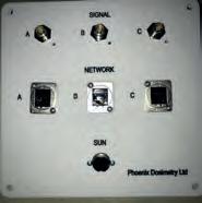
Patch Panels for bunkers improve cable management, simple to install, custom made Dosimetry Cables: Any length, any connector, now with 2 year warranty
Bart’s Solid Water and Phantoms
Fast Repair Service: typically 1-2 weeks for Mini and Legacy NE/Thermo Equipment. Free inspection service with no obligation quote
IPEM.AC.UK 13 SPRING 2023 Please visit our new website phoenix-dosimetry.co.uk
There was one central theme and that was workforce – or rather the lack of it – and the cancer backlog.
or call 01252 871990
In the autumn edition of Scope, Lauren Harrison, IPEM’s Training Development


ROUTES TO REGISTRATION
“Clinical Scientist” is a protected title within the UK and can only be used by those who are registered with the Health and Care Professions Council (HCPC). There are multiple routes to registration as a Clinical Scientist and it can be difficult to know what is right for you. There are national training schemes across the UK which provide a set training pathway to becoming registered as a Clinical Scientist, but what if you have not followed a traditional education or training route, or a training scheme is not right for you?
In the UK there are two routes that allow medical physicists and clinical engineers to demonstrate their equivalent skills and experience they have gained across their career so far – the Association of Clinical Scientists’ (ACS) Route 2 and the Academy for Healthcare Science’s (AHCS) Scientist Training Programme (STP) Equivalence.


In addition to these routes, the HCPC also operates an international application route, which allows those who have “undergone training in one of the relevant professions
outside of the UK [and] have not previously been registered with HCPC”. International applicants can apply directly to the HCPC and demonstrate the equivalent education and experience they have gained in order to become registered in the UK. The HCPC website details all of the documentation
ACADEMY FOR HEALTHCARE SCIENCE’S STP EQUIVALENCE


The AHCS STP Equivalence route is “based on individual applicants presenting periods of professional experience, qualifications and training (evidence) for assessment by a panel of assessors”. This is based on the principles of Good Scientific Practice and demonstrates equivalent knowledge and skills to the STP curriculum. Successful assessment by the AHCS awards the Certificate of Equivalence, which can be provided to the HCPC in order to register as a Clinical Scientist.
and forms required, such as demonstrating equivalence in your educational knowledge, English proficiency and identity documents.
Dr Jemimah Eve, IPEM’s Head of Workforce Intelligence and Training, said: “Whilst the training programmes on offer for becoming a Clinical Scientist are fantastic for delivering a secure pipeline into the profession, the alternative routes to registration both strengthen numbers and ensure diversity of background in the profession.”
Dr Jaap Vaarkamp, Head of Radiotherapy Physics at Betsi Cadwaladr, IPEMnominated ACS Director and ACS Chair, added: “I would make the case that the work-based training and the subsequent portfolio-based assessment offer a thorough preparation for candidates entering the profession, and for employers a flexible option to bring somebody into the workforce and develop the required skill mix. As it is open to candidates with a wide range of experience, it offers a way into the profession for those on personal career
Officer, looked at the different routes to becoming a registered Clinical Scientist. Here she explores how those routes can be used by those who have significant experience but may not have followed a traditional training route.
U F UPFRONT IPEM SCOPE 14 SPRING 2023
routes, and for employers access to a diverse workforce delivering care to our patients.” For those considering applying to any of these routes, we would recommend completing a gap analysis exercise where you review the competencies or standards required and map your skills and experience to them. Where you are unable to demonstrate a particular competency, this will allow you to see where your training needs are and can also help with showing which route is more appropriate for you.
Career progression
HCPC registration as a Clinical Scientist provides professionals with the opportunity to progress into more senior roles, such as Principal Clinical Scientist and onwards to Consultant Clinical Scientist.


ASSOCIATION OF CLINICAL SCIENTISTS’ ROUTE 2


The ACS Route 2 is for those who “have extensive experience in the relevant field sufficient to demonstrate [the ACS] competences based on a cross referenced portfolio of evidence, and (if approved) an interview”. Successful achievement of the ACS
Certificate of Attainment will make you eligible to join the HCPC register as a Clinical Scientist. The ACS competencies can be found on their website, and cover seven areas: Scientific, Clinical, Technical, Research and Development, Communication, Problem
In 2015, the AHCS opened the Higher Specialist Scientist Register (HSSR). The National School of Healthcare Science was accredited to run the Higher Specialist Scientist Training (HSST) programme, which is a five-year workplace-based training programme including a Doctoral level academic award. This supports those looking to progress into Consultant Clinical Scientist posts.
As with initial registration with the HCPC as a Clinical Scientist, there is also an equivalence route, through the AHCS, for those who gained skills and experience equivalent to this. Applicants demonstrate their experience and how this can be compared to the Standards of Proficiency. Successful candidates are awarded a Certificate of Equivalence and are eligible to join the register.

Kathryn Adamson, Principal Clinical Scientist at Guys’ and St Thomas’ NHS Foundation Trust, attended an IPEM event in 2020 on how to achieve HSST equivalence and following this has achieved registration on the HSSR.
“Applying for Higher Specialist Scientist Equivalence (HSSE) was my COVID project – some people learnt a foreign language, or how to cook flatbread during lockdown, but I did HSSE. I really applied to it to see if HSS registration was possible for someone like me, who has many years work experience in nuclear medicine, but not a PhD.
Solving and Professional
Accountability, from which you can draw on a range of experience. “Experience/ training being claimed as working in the role of a supervised pre-registered clinical scientist will require a GMC- or HCPC-registered Clinical Scientist to act as a supervisor signatory towards the application”. IPEM members are also eligible for reduced assessment fees.
“Getting on the register was a great outcome for me personally, but achieving registration isn’t something that is currently enhancing my career. I feel there is some way to go before the value of being on the HSSR (via equivalence or via the HSST) is fully recognised and considered to be an integral ‘qualification’ that will help further a career in imaging physics.
“To improve this I think we need those engaged in recruitment to make sure HSS registration is put on job descriptions and person specifications as essential, or at the very least desirable, and there should be a general expectation in the profession that HSS registration is a requirement for consultant medical physics-level posts.”
The Higher Specialist Scientist Register is a Professional Standards Authority accredited register, and it is currently not statutory for Consultant Clinical Scientists to be on the register. However, at the end of 2022 it became a requirement in Wales for new Consultant Clinical Scientists to be registered on the Higher Specialist Scientist Register, demonstrating an increasing importance in HSS registration.
Following IPEM’s event in 2020, a summary video was recorded to share information about the process of equivalence and advice given by the Consultant Clinical Scientists who attended, to support others considering applying for HSS registration through equivalence and this is freely available on the IPEM website at bit.ly/HSSTwebinar
IMAGE: GETTY IMAG E: G ETT Y
IPEM.AC.UK 15 SPRING 2023
THE BIG DEBATE Clinical risk management part 2
What is the Medical Physics and Clinical Engineering (MPCE) community’s role in the implementation of DCB0129 and DCB0160?
ANDY
Everyone should familiarise themselves of the requirements of the standards. They are very concise and easy to read. Anyone involved in the development, or procurement and use of a health IT system should confirm that there is safety case report demonstrating compliance with the appropriate standard.
ALASDAIR
There are two main areas where these standards appear to be most readily applicable, and that is firstly in the development of health IT systems (generally in-house manufactured software) and in their implementation, and secondly in the traditional model of procuring and commissioning ready-made systems. MPCE staff are well placed in this area as technical experts who also often use the systems and therefore have intimate knowledge of relevant workflows and departmentspecific configurations. They will also have risk assessment training throughout their careers through working with radiation, medical devices, or a variety of other areas.

PATRICK
When I received the training I very quickly came to the conclusion that although at the time this was not specifically targeted for medical device systems, it was
a very valid approach that could be applied to medical device IT systems. During the course it was made clear that the Clinical Safety Officer (CSO) role should be a senior clinician with appropriate training. Initially that seemed fair but as my experience developed I came to think that this is a role I could perform and in most cases knew more than clinicians about the safety concerns. It is now acknowledged that anyone with suitable professional registration can now perform that role, which I think is fair. The key thing to keep in mind is that the person carrying out the role needs both training and experience. It really is something that is learned by experience. People within the MPCE community certainly have the background to carry out a CSO role. Within my organisation I have to advise a number of senior colleagues on the need for patient/ safety cases to support the deployment of new medical device IT systems. At first they are reluctant but do eventually see the need.
Ensure that compliance with the standards are explicitly included at the procurement phase and included in any contractual arrangement.
IMAGES: ISTOCK
Q
Q Even though the standards are mandated in England, awareness and buy-in appears to be variable, both within the NHS and with manufacturers. Do you have suggestions for how this might be addressed? ANDY
IPEM SCOPE 16 SPRING 2023 UPFRONT DEBATE U F
In the second instalment of our deep dive into DCB0129 and DCB0160 – mandatory risk management standards for England – our panel looks at the pressing issues.
The standards are not written into law in Scotland, but voluntary adherence appears to be on the up with many local departments, such as the Department of Clinical Physics and Bioengineering at NHS Greater Glasgow and Clyde, ensuring best practice is met. I think there are a few areas that would improve compliance. Firstly, I would expect a number of departments will already comply to ISO 14971 for medical device development and so improving the visibility and understanding that the same principles underpin both standards will go a long way to reducing the upfront workload. Secondly, awareness of what makes good processes and good documentation will help staff translate best practice into their own work – which leads into the need for a community of staff working in this area sharing their work and offering advice and support - which would be a particularly useful resource.
Use of the DCBs by both NHS and suppliers is essential to give a sufficient level of safety assurance when deploying systems that are used in the diagnosis and treatment of patients. It is sometimes a learning process within an NHS organisation that when system deployments go wrong one of the things that would have prevented the issues would have been to follow the 129/160 process. This is an unavoidable learning step, unless of course you have a system in place that takes clinical/patient safety seriously. As for the suppliers one way to enforce the use of 129 is to make it a pre-requisite for procurement where there is a choice. It is a mixed picture with some of the bigger suppliers being quite on the ball, whereas smaller ones try to avoid due to the overhead that is involved in producing a 129-compliant safety case. It may need the availability of consultants to offer the services to the smaller companies whereas the larger companies are


 ALASDAIR
PATRICK
ALASDAIR RUTHERFORD Principal Physicist Radiotherapy Systems NHS Greater Glasgow & Clyde
PATRICK MAW Consultant Clinical Scientist Medical Physics and Biomedical Engineering University College London Hospital
ANDY CARR Clinical Safety Officer
NHS England
ALASDAIR
PATRICK
ALASDAIR RUTHERFORD Principal Physicist Radiotherapy Systems NHS Greater Glasgow & Clyde
PATRICK MAW Consultant Clinical Scientist Medical Physics and Biomedical Engineering University College London Hospital
ANDY CARR Clinical Safety Officer
NHS England
IPEM.AC.UK 17 SPRING 2023
We had invited a panel of five to debate this issue, but unfortunately two female panellists had to withdraw due to work commitments. We will aim to restore balance next time.
MEET THE PANEL
better able to absorb the cost. It is also a matter of being familiar with the process. Once you have carried it out a number of times it becomes easier and is mostly what is being done anyway with the 129 process giving the assurance that it is documented.
QWhat challenges might individuals face when trying to implement the standards?
ANDY
Working in clinical safety can initially feel quite daunting. It’s important that everyone involved in the development or implementation of a health IT system is aware of the standards and their importance, and it’s not just the responsibility of the CSO.
ALASDAIR
In my opinion, one of the clear difficulties in these standards is how the local department fits in with NHS Digital’s (now merged with NHS England) view of the overall organisation. In the guidance accompanying DCB0160, “top management” is presented as being at many different levels, and some clarity on where this sits for large MPCE departments would be useful, particularly given the complex and specialist work that occurs within and the need for that contextual understanding. Furthermore, there is difficulty in getting the time to work on documentation alongside the usually significant workload associated with developing, accepting or commissioning a new system, and this requires support from management.
PATRICK
The process does require resources in the form of time. This time resource will start with any initial training and then be the time taken to hold the required meeting and workshops to gather all the information. It is also very valuable if you can be working in an organisation that implements it already so you can benefit from the experience of existing staff. Alternatively, a network of individuals that have the required knowledge from who advice can be sought.
Q Are there alternative or complementary approaches worth considering?
ANDY
Ensure that an overview of the standards is included in training of all staff. For those working closely with development, implementation or ongoing maintenance and use of systems, it would be valuable for them to access the variety of e-learning and courses available.
ALASDAIR
An alternate approach would be to use the underlying standards, however the DCB standards are those that are accepted by the Data Coordination Board, and any deviation must be considered with due care.
PATRICK
Standards such as 14971 do offer risk-based assessment techniques that apply to medical devices but I have been involved in the 129/160 for many years and do not see why it would not be used. And one method needs to be used to ensure consistency and that is mandated as being 129/160.
ANDY
The standards were originally established to bridge the gap between products classified as medical devices and those that were un-regulated, yet could potentially contribute to patient harm. The scope of the standards was extended in 2018 to include medical devices that are implemented in such un-regulated products. The restriction does not need to be reconsidered as the medical device regulations provides a much stringent risk management regimes for these higher class medical devices.
ALASDAIR
I haven’t spent time looking at this area in depth, so would feel unconformable giving any comments.
Q Although the standards apply to Software as a Medical Device (SaMD) and medical device networks, software embedded in medical devices is out of scope, even though some of the most complex devices may fall into this category. Should this restriction be reconsidered?
U F UPFRONT DEBATE IPEM SCOPE 18 SPRING 2023
PATRICK
Well, there is 80001 that applied to medical device networks, though this is another risk-based process and again is very common sense. I think the restriction on embedded software is very sensible as that is covered by standards such as 60601 and 62304. Then 129/160 take those approved medical devices and employs them in a patient-centered workflow using a software system. Maybe in the future there could be a re-think on how the standards apply. 60601 evolved to account for software. Though the governance for the health IT systems could be seen as spanning two areas – medical physics and ICT – and 129/160 enables those areas to work together.
they?
ANDY
They would be eligible to act as a CSO if they are suitably qualified and experienced and also registered with their professional regulatory body. They must also
be knowledgeable in risk management and its application to clinical domains – training to support this is available from NHS England Clinical Risk Management Training.

ALASDAIR
Clinical Scientists and technologists with the relevant bodies should be eligible to act as CSOs, assuming the other prerequisites are met (i.e. they have had appropriate training or experience in risk management in clinical domains). I can only speak for Clinical Scientists as that is where my background is, but we are assessed against good scientific practice, which includes clinical practice –and our training and learning is all undertaken in the context of patient care.
PATRICK
I think it is very applicable to have someone from an MPCE background fulfil the role of the CSO as they will have experience of the clinical environment, which is an absolute must when dealing with the safety of systems used to treat patients. I wouldn’t see why this couldn’t be any one on the HCPC register. But they must have training and experience of the process.
QWhat role should IPEM play in the implementation of these standards?
ANDY
IPEM should help promote awareness of the standards; the responsibilities that health and social care organisations, and system suppliers have in relation to the standards and continue to promote the training available.
ALASDAIR
IPEM is well placed to facilitate the sharing of resources in clinical risk management, and to produce policy statements that support individuals working in this area (for example a position statement justifying why Clinical Scientists and technologists can act as CSOs).
PATRICK
IPEM is ideally placed to fulfil a role as being able to facilitate the expert user group for CSO working in MPCE. They are able to deal with NHSE\D\I etc to co-ordinate the initial training and then be able to setup a network that can give expert advice to those who are developing the skills to be a CSO.
QThe standards define the role of a CSO as someone who must be a suitably qualified and experienced clinician, holding current registration with an appropriate professional body. Many members of the MPCE community are either Clinical Scientists registered with HCPC, or clinical technologists registered with the RCT. Would these individuals be eligible to act as a CSO? Should
To view the standards in full, visit bit.ly/SC_DCB0129 & bit.ly/SC_DCB0160 IPEM.AC.UK 19 SPRING 2023
MAYBE IN THE FUTURE THERE COULD BE A RE-THINK ON HOW THE STANDARDS APPLY
SL33PAPN0E4
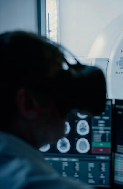
We look at the development of a new suite of software and hardware for the overnight oximetry service in Clinical Measurement at the James Cook University Hospital.
IPEM SCOPE 20 SPRING 2023 COVER FEATURE C F
IMPROVING A SLEEP SERVICE THROUGH TECHNOLOGY
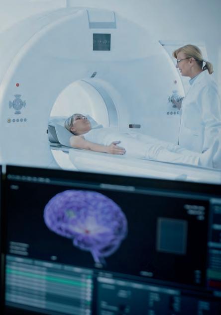
The Clinical Measurement team at the James Cook University Hospital perform overnight oximetry diagnostic testing on patients with suspected obstructive sleep apnoea (see box, right). This involves providing patients oximetry equipment that they take home with them and return after two nights of use. The results are then downloaded and reviewed by Clinical Scientists before being sent to a sleep medicine consultant. However, the existing software is outdated and at risk of becoming unmaintainable. Therefore, Andrew Simpson and Tony Alton are developing a new software solution to replace the existing software.
The existing software
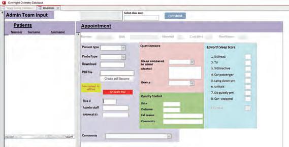
The existing system hinges on an old Microsoft Access database 1 that acquires patient demographic data from the patient administration system (CaMIS), via an open database connectivity (ODBC) data source, by using linked tables. The overnight oximeter data is downloaded using commercial software called Visi-Download and creates a PDF results file. This file is
WHAT IS OBSTRUCTIVE SLEEP APNOEA?
Obstructive sleep apnoea (OSA) is a relatively common condition where the walls of the throat relax and narrow during sleep, interrupting normal breathing.
This may lead to regularly interrupted sleep, which can have a big impact on quality of life and increases the risk of developing certain conditions. There are two types of breathing interruption characteristic of OSA:
Apnoea – where the muscles and soft tissues in the throat relax and collapse sufficiently to cause a total blockage of the airway; it’s called an apnoea when the airflow is blocked for 10 seconds or more.
Hypopnoea – a partial blockage of the airway that results in an airflow reduction of greater than 50% for 10 seconds or more.
People with OSA may experience repeated episodes of apnoea and hypopnoea throughout the night. These events may occur around once every one or two minutes in severe cases.
The term “obstructive” distinguishes OSA from rarer forms of sleep apnoea, such as central sleep apnoea, which is caused by the brain not sending signals to the breathing muscles during sleep.
stored in a folder which can be accessed by a results web application that is linked to the access database.
The problem with the existing system
The database lacks normalised tables and is difficult to audit due to free text in columns that have changed over time. The front end of the database is also inside the Microsoft Access database and features such as date picker and other user interface components are being deprecated in recent versions. The information that can be provided to the consultant is limited. The results web application only provides access to the PDF results file and whether the study has passed our quality control check. The existing solution will eventually become unsupported and increasingly more difficult to maintain. Therefore, a new system was required.
The new system
The new system, which is currently in development, consists of the following:
● Desktop software to extract patient appointments and demographic from the hospital patient administration system, CaMIS.
IMAGES: IKON
IPEM.AC.UK 21 SPRING 2023
● A web application to manage the clinic, patient appointment and issuing equipment.
● A barcode scanner to “check-in” the equipment after it has been used by the patient.
● A web application programming interface (API) to interact with the barcode scanner and the Microsoft SQL Server database.
The look and feel of the system is based on NHS Digital’s design library, using the cascading style-sheet colour scheme found at: service-manual.nhs.uk/design-system.
SleepTalk desktop application
Overnight oximetry clinics and appointments are stored within CaMIS, which also includes the patient’s demographics. CaMIS itself is not suitable for the complete management of our clinics because it does not provide features such as allocating equipment, status of quality control and capturing information from questionnaires.
SleepTalk, developed in C#, is the workhorse for the backend of the new sleep system. A healthcare science assistant uses this application to register the patient and the clinics as the application extracts data from CaMIS and loads the data into our Microsoft SQL Server database tables. The SQL server utilises user-defined table types and stored procedures to merge the data extracted from CaMIS into the normalised SQL Server Tables. After the clinic is registered, it is then available on the new SleepWeb Web Portal Application which allows all staff to manage the patient’s journey from appointment to return of equipment.
SleepWeb web portal application
SleepWeb 2 is a comprehensive clinic management system which allows the healthcare science assistant to manage appointments during their clinic, allocate equipment, record comments from the appointment and assign tasks to Clinical Scientists for review.
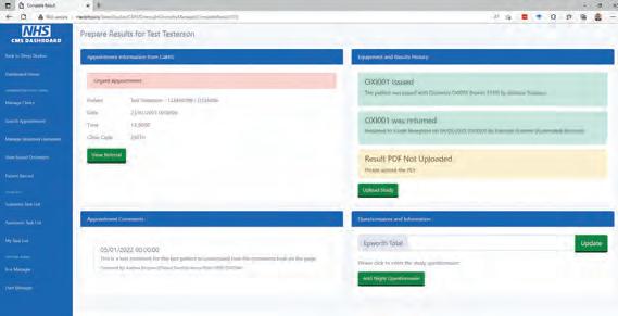
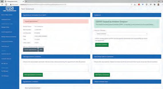
SleepWeb was developed by using C# asp.net model-view-controller (MVC) and entity framework (EF) to interact with the Microsoft SQL Server Database. An object relational mapper, such as EF, is advantageous because it automatically generates an object model of the database tables and imports the relationships between tables, thereby avoiding the need to use lengthy SQL queries and JOIN statements. These models can be passed into a “view” (the web page the user can see) either directly or via a Controller (the C# logic and code behind the web page).
In the old Access Database, the healthcare science assistant must search for the patient’s appointment date and is then presented with a list of patients for that date, once the patient has returned their equipment However, in the new software, the returned equipment is presented as a worklist irrespective of the
Figure 1 Old oximetry access database
Figure 2 Overnight oximetry clinic management page
Figure 3 Testing the new software
IPEM SCOPE 22 SPRING 2023 COVER FEATURE C F
Figure 4 Returned oximeter page
appointment date. This allows the healthcare science assistant to keep better track of equipment that is ready to be downloaded 3 4
The Clinical Scientists will then be able to perform their quality control on the results. In the old Access Database, Clinical Scientists need to search for downloads by the date they are downloaded, or the appointment date of the patient, to be able to see the results for quality checking. However, in the new system, this again will be presented in a worklist irrespective of any dates, so clinical scientists can see all outstanding results to quality check. After the quality control, the results will be released to the consultant for viewing on the new results web portal.
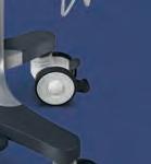
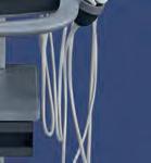
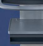
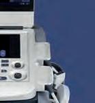
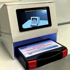
Barcode scanner
The novel part of this system was to introduce a barcode scanner 5 6, allowing patients at the hospital reception to return their equipment by scanning it. This would then automatically record the equipment as returned and add it to the healthcare science assistant’s worklist. This was important because the only way to check if equipment had been returned was to walk to the other side of the hospital and check manually. Furthermore, if equipment goes missing, we are unable to ascertain whether a patient had actually returned the equipment or whether it was lost while it was in the hospital.
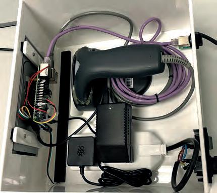
The barcode scanner uses a Datalogic barcode scanner placed inside a custom-made housing. The scanner interfaced with a Raspberry Pi and touchscreen. The software was written in python, which interacts with a newly developed API) which was written in C#.
SleepAPI
The Sleep Application Programmable Interface (API) was primarily designed to serve and receive data to and from the barcode scanner and will eventually evolve to
be used by the other sleep applications that we have developed, for example, for sending emails and updating other tables in the database.
For now, the barcode scanner utilises the API to interact with the SQL server database. For example, when equipment is returned, the scanner verifies the equipment exists in the database by using a GET method and then uses the PUT method to update the equipment status in the table to returned. The API also uses the POST method to insert data into the equipment history table which can be later used for auditing 7. The rational for using the API was to avoid the use of SQL queries on the barcode scanner and minimise the need for storing data on the scanner. All the queries are performed by the API, which can be easily amended without needing to update the barcode scanner python code.
Further work and considerations
The system is still in development. We are including more NHS Digital front end CSS as development continues. We are also working on having this system approved by our information governance team and then plan to migrate the data from the access database into the new SQL server tables. We hope to begin producing useful data that can be audited and provide better asset management of the oximetry equipment, by developing an equipment management module in the system. We also are looking into performing usability evaluations with patients for the barcode scanner. We are also using this system as a test case for complying with the DCB0129 requirements.
SQL Server Sleep API GET PUT UPDATE INSERT POST Send true or false Check that the equipment exists Mark that the equipment has been returned Add information into the equipment history Return Data SELECT Barcode Scanner
Figure 6 Working Prototype of the barcode scanner
Andrew Simpson is a Clinical Scientist in Clinical Measurement, working in the Medical Physics Department at James Cook University Hospital. Tony Alton is a clinical technologist in Clinical Measurement.
Figure 5 Inside the barcode scanner
IPEM.AC.UK 23 SPRING 2023
Figure 7 The API interacts with the SQL server database.


G F GENERAL FEATURES IPEM SCOPE 24 SPRING 2023
HE WAS SURELY THE ONLY HOSPITAL PORTER ALLOWED TO USE THE MEDICAL STAFF DINING ROOM!
GENIUS IN THE LAB


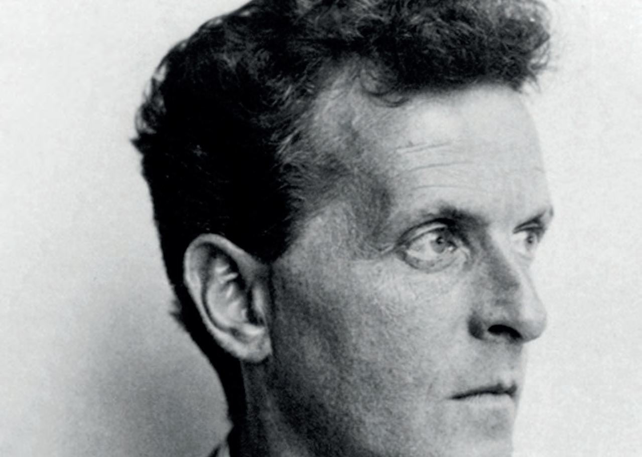
udwig Wittgenstein (1889-1951) was an Austrian-British philosopher who is widely viewed as one of the greatest thinkers of the 20th century and was often termed a genius. This includes by his supporter, Bertrand Russell (1872-1970), the British mathematician, philosopher, Nobel Literature laureate, pacifist and activist. Wittgenstein worked mainly in the areas of logic and the philosophy of mathematics, of the mind and, over all, of language, making fundamental contributions to the analytic philosophy approach emerging in the early 1900s from Russell and his Cambridge colleague, George Moore, and the German mathematician/philosopher, Gottlob Frege. Wittgenstein and Russell have each been called the greatest philosopher of the 20th century by different commentators.
Ludwig Josef Johann Wittgenstein 1 was born in Vienna, the youngest of nine siblings. His father was a steel magnate, one of the richest men in Europe and heavily

involved in Vienna’s cultural life. The children grew up surrounded by debate, art and music. However the family also experienced tragedy. Three of Ludvig’s four brothers committed suicide as young men and Ludvig suffered from depression throughout his life, including regular suicidal thoughts. He was often withdrawn and irascible, finding social relationships difficult and it has been suggested that he suffered from Asperger’s syndrome.
From engineering to maths to philosophy


Wittgenstein studied mechanical engineering in Berlin from 1906, coming to the Victoria University of Manchester in 1908 for doctoral work in aeronautics with Professor Arthur Schuster. This initially considered the behaviour of balloons and kites in the upper atmosphere for ionization and meteorology studies, with experiments conducted near Glossop, Derbyshire 2. He also studied, and patented, plane propeller designs incorporating small gas jet engines. Air and
propeller blades and were compressed by centrifugal force in combustion chambers at the blade ends and then ignited. Although experimental systems were successfully tested, this was largely theoretical work, requiring complex mathematical modelling of gas flows, combustion under high pressure and other design aspects. Wittgenstein became interested, obsessed even, with the foundations and philosophy of mathematics and logic.


In 1911, he sought advice from Frege in Jena, Germany, who recommended contacting Russell, so he simply turned up in Cambridge and asked to study mathematical logic. Russell stated that Wittgenstein absorbed all that he, Russell, knew in a year. Wittgenstein then craved solitude to work and in 1912 went to Skjolden, a small Norwegian village, to develop the ideas that became his first major work, the Tractatus Logico-Philosophicus


L
gas were forced along the centre of revolving
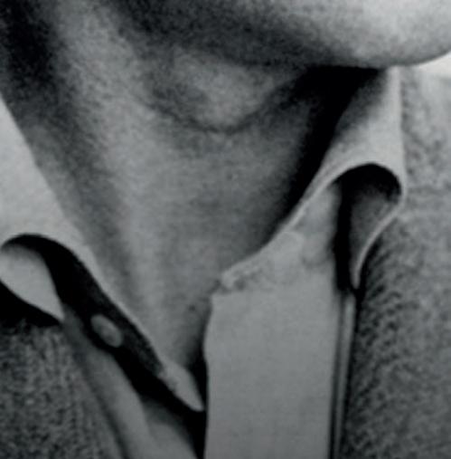
1 Left. Portrait of Wittgenstein, taken by Moritz Nähr (1930).
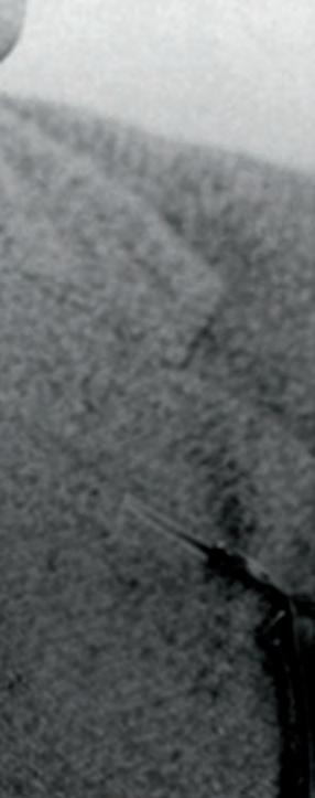
2 Below. Wittgenstein with friend William Eccles at the kite-flying station in Glossop (1911).
3 Right. Blue plaque for Wittgenstein at Guy’s, on the corner of the central quadrangle of the old buildings, bordering the colonnade.
IPEM.AC.UK 25 SPRING 2023 IMAGES: ALAMY
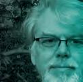
David Thwaites outlines how a leading philosopher became a physiological measurement laboratory assistant in the 1940s.
IMAGE: ©WITTGENSTEIN ARCHIVE CAMBRIDGE
(Logical-Philosophical Treatise) 4. This was written in 1914-18 while he served with distinction in the Austrian army in WW1. The unknown Wittgenstein found it difficult to get this esoteric book published at first and it didn’t appear in German until 1921, now with Russell providing an introduction, and in English in 1922.
Leaving philosophy and returning to it

Wittgenstein’s father died in 1913, leaving him a fortune, which he began to give away to support artists and authors and then finally, in 1919, to his remaining siblings, believing he should only spend what he had earned. Also at this time he felt that the Tractacus was his final word on philosophy, solving all the essential problems, so he trained as an elementary school teacher and worked until 1925 in small rural schools in Austria. By now his ideas were being widely read and discussed by philosophers, mathematicians and scientists. He realised there was more he could do and contemplated returning to philosophy. After other jobs, including a monastery gardener’s assistant, where he lived for some months in the tool-shed, he was persuaded to return to Cambridge in 1929.
One problem for working there, despite his international reputation, was that he had no degree, but he successfully submitted Tractacus as his doctoral thesis, telling his examiners that he knew they wouldn’t understand it, but not to worry! One, Moore, described it as a ‘‘work of genius”. Wittgenstein then held successive appointments in Trinity College, Cambridge, eventually becoming a professor of philosophy in 1939. Given his Jewish heritage, he took British citizenship following events in Germany and Austria and their impact on his family. For the rest of his life he worked on the ideas embodied in his second great work, Philosophical Investigations. Over this time, his views evolved and he completely discarded many statements and assumptions presented in Tractacus. Also, in his search for perfection, this second book was never

THE UNKNOWN WITTGENSTEIN FOUND IT DIFFICULT TO GET THIS ESOTERIC BOOK PUBLISHED
good enough to be finished and was not published until 1953 after his death, as were other works. He died in April 1951 from inoperable prostate cancer diagnosed two years earlier, for which he received radiotherapy, but which had metastasised to other sites. He was working on manuscripts until a couple of days before his death.
So where’s the physiological measurement?
During WW2, Wittgenstein agonised over doing something more “practical” than philosophy with a war in progress. The philosopher Gilbert Ryle introduced him to his brother, John Ryle, a Cambridge Professor of Medicine. The latter had helped Guy’s Hospital’s preparation for the Blitz and he arranged for Wittgenstein to become a pharmacy/dispensary porter there from September 1941, delivering drugs to wards and patients, reputedly often advising patients not to take them as he didn’t agree with them in principle. 3 He is also credited with being an ointment maker, including of the “finest quality zinc oxide ointment”. He had told Ryle that he would die slowly if staying in Cambridge, but would rather die quickly. This was narrowly avoided when Guy’s was damaged in bombing raids. He wished his colleagues not to know of his international academic reputation, even though he was surely the only hospital porter allowed to use the medical staff dining room!
However, he was recognised by some staff with an interest in philosophy and became friendly with one such doctor, (Ernest) Basil Reeve.
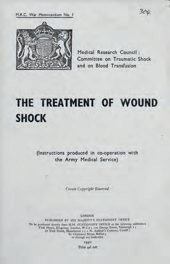
Reeve and a senior colleague, Ronald Grant, were working on radical approaches to wound shock in bombing casualties in a dedicated Medical Research Council “traumatic shock” unit. Grant initially felt there was little agreement on the symptoms defining this condition and that maybe the concept of “shock” should be abandoned. This interested Wittgenstein into discussing the semantics of the term “shock”, given his ideas on language and meaning, considering the word to cause confusion between professionals and patients/public. This led to his further interest in the work. When the bombing of London and the number of casualties had reduced, the shock unit moved to the Royal Victoria Infirmary (RVI), Newcastle-uponTyne in late 1942 to continue similar studies on road and industrial accident victims. Grant offered Wittgenstein a laboratory assistant post to support the research, paying £4 a week, and he moved into digs in West Jesmond in April 1943, close to the hospital and with the rest of the team. He found the lab work demanding and tiring and he tended to be rather withdrawn and not join in the social activity of the group. When the landlady suffered ill-health he moved to Benwell, where he lived on his own. It was said that finding a place was more difficult for him, as he looked “rather scruffy” and claimed to be a professor, which must have seemed improbable.
Understanding wound shock
The poorly defined clinical syndrome of “wound shock” had been recognised since the American Civil War and then again in WW1. Now, and arising from WW1/WW2 work, including on the role of transfusion
G F GENERAL
IPEM SCOPE 26 SPRING 2023
FEATURES
in its treatment 5, it is understood as hypovolemic shock due to rapid blood volume reduction, requiring fast action to control bleeding and restore fluids.
In Wittgenstein’s lab assistant/technician role, among other things, he improved the preparation of both frozen and paraffin histology sections and, using his engineering background, he designed and built equipment and carried out physiological measurement experiments. These were aimed at studying the characteristics of, and links between, breathing depth and rate and pulse volume and rate. He invented a new device to record pulse pressure and paradoxical pulses (abnormally large decreases in volume, systolic blood pressure and pulse wave amplitude during inspiration) in laboratory rats and humans. He tested all his devices on himself.
Wittgenstein suggested to Grant that wound sizes might be categorised by the volume of tissue damaged, using the hand or fist as a simple fast unit of measurement. Grant acknowledged Wittgenstein’s contributions as significant to the group’s work and wished he “was a physiologist, rather than a philosopher” Grant wrote that Wittgenstein had “a keenly critical mind and in discussions of medical and physiological problems (had) proved a most helpful and stimulating colleague.” Also that: “He has undertaken observations on respiratory variations of blood pressure in man, devising his own experiments and


apparatus. The results of his work so far are at variance with commonly accepted views and of considerable interest.”


In January 1944 Grant and Reeve left Newcastle to study battlefield casualties in Italy and Eric Bywaters took over the unit, finding Wittgenstein “reserved and uncommunicative, but a meticulous worker”. Both Grant and Bywaters tried to persuade Wittgenstein to stay in the unit, but he was being asked to return to Cambridge and philosophy, which he did in February 1944. His physiological measurement work was over. The research programme of Grant’s unit is described in “Medical Research Committee Reports 1939 – 1945”, but Wittgenstein is not mentioned.


Wider influence in science and medicine and a final comment
Wittgenstein profoundly impacted many areas of philosophy – primarily the philosophy of language, but also of mathematics, logic, the mind, psychology, perception, ethics, aesthetics, and science. All were approached from the viewpoint of language analysis. He is generally described as anti-scientism and he predicted that scientists would not see his work as relevant to theirs. He sought conceptual truth from a logical-linguistic-grammatical analysis of sense or non-sense, more aligned with ideas of artistic truth, as opposed to an empiricalexperimental determination of truth or falsehood. Nevertheless, while his ideas were applied mainly to the topics listed above, they can also provide insights into the conceptual foundations in other areas. There are a number of papers in the medical literature discussing Wittgenstein’s influence in fields such as neuroscience, neurology and neuropsychiatry, as well as psychology and perception. He has also influenced wider approaches or tools applicable in science and medicine, such as terminology and clarity of meaning, ontology, classification, decision-making, uses of logical truth tables and more.
As a final comment on the views of this “genius physiological measurement technician”, he stated that philosophers treat a question as an illness, wanting to find the right answer, as doctors finding a cure. In that sense, it is suggested that they might be “soul-searchers”, trying to find solutions for people’s and society’s sicknesses and a better way in life? It is notable that he considered becoming a monk and a psychiatrist at times. The restless driving force of humanity, of course, and of philosophy and of science, is that there are always more questions, with no “cure” for our insatiable curiosity.
David Thwaites is an IPEM Fellow and Emeritus Professor of Medical Physics at the University of Sydney and honorary Professor of Oncology Physics at the University of Leeds. The author acknowledges the use of multiple sources, a list of which is available from david.thwaites@sydney.edu.au
DURING WW2, WITTGENSTEIN AGONISED OVER DOING SOMETHING MORE “PRACTICAL” THAN PHILOSOPHY
4 Left. The cover of Wittgenstein’s Tractatus Logico-Philosophicus
IMAGES:
IPEM.AC.UK 27 SPRING 2023
5 Right. The Treatment of Wound Shock. Instructions prepared by the Medical Research Council Committee on Traumatic Shock and on Blood Transfusion, in co-operation with the Army Medical Service (1940).
GETTY/©CROWN COPYRIGHT, WELLCOME COLLECTION
IN-SILICO MEETS IN-VITRO FERTILISATION

Birth rates are falling globally, and in many countries, such as the US and China, fewer than two children per couple are born. Reproductive healthcare provision and optimisation is – or is bound to be – a key priority for many countries.
In-vitro fertilisation (IVF) is the primary treatment of infertility across the developed world. Eggs are fertilised in a lab, and if an embryo is developed it is transferred to the patient with the hope it leads to pregnancy. For patients, IVF is expensive, time consuming and physically taxing. Over 2.5 million IVF treatments take place annually, resulting in ~500,000 births. The IVF success rate (live birth per treatment cycle) has been ~25% for many decades, but since 2010 it has been decreasing. Meanwhile, the IVF industry is projected to grow to $37.7 billion by 2027, with an almost 10% annual growth rate. Success rates are declining partly because
of the need to culture embryos for five to six days to the blastocyst stage in order to select the “best” embryo for transfer. Moreover, single embryo transfer is mandated in many countries to avoid the risks of multiple pregnancies.
There is, therefore, a strong motivation for clinics to be able to select the best embryo as early as possible and to maximise the success rate of IVF in order to remain competitive. Selecting the best embryos is also important in frozen embryo transfers (FETs) that involve cryopreserved embryos. Research and development of novel technologies can significantly aid this important quest.


Combining mathematical modelling and experimental expertise at Cardiff University with clinical expertise from the London Women’s Clinic, we have been working on optimising IVF by tackling several challenges. We have developed image processing methods that enable noninvasive appraisal of egg and embryo health and we have worked on the logistical

IPEM SCOPE 28 SPRING 2023 G F GENERAL FEATURES
A Cardiff University team combine mathematical modelling with experimental and clinical expertise to look at issues around in-vitro fertilisation.
challenge of packing and storing frozen eggs and embryos. Moreover, we are developing a novel mathematical model of calcium (Ca2+) waves in fertilising eggs that may allow to non-invasively assess the potential for IVF success. Below we outline our work and main research results.
Frozen embryo transfer: preventing ice crystals damaging the embryo
Human embryos can undergo “vitrification” (rapid freezing) in order to be preserved so that, sometimes after years, they can be thawed and transferred to the patient. This is called a frozen embryo transfer (FET).
With 46% of IVF cycles using frozen embryos, optimising vitrification protocols in order to increase the number of successful FETs is increasingly important. Vitrification involves suspending embryos in cryoprotectant solutions, before mounting them onto a freezing device and plunging into liquid nitrogen. Minimising the freezing time prevents the formation of ice crystals, which cause irreversible damage. Hence, achieving high cooling rates is critical.
For the Cryotop cryopreservation device 1, the manufacturer protocol allows 1-4 embryos to be vitrified simultaneously. The number of embryos mounted on the device varies between clinicians and between procedures undertaken by the same clinician. Additionally, when multiple embryos are vitrified on a single device, their position and arrangement can vary. This variability led us to investigate if there is an optimum number and arrangement of embryos. Note that eggs can also be vitrified using the same device, thawed and fertilised years later. The recovery rate for eggs is only 90% in contrast to the 95% recovery rate for embryos.
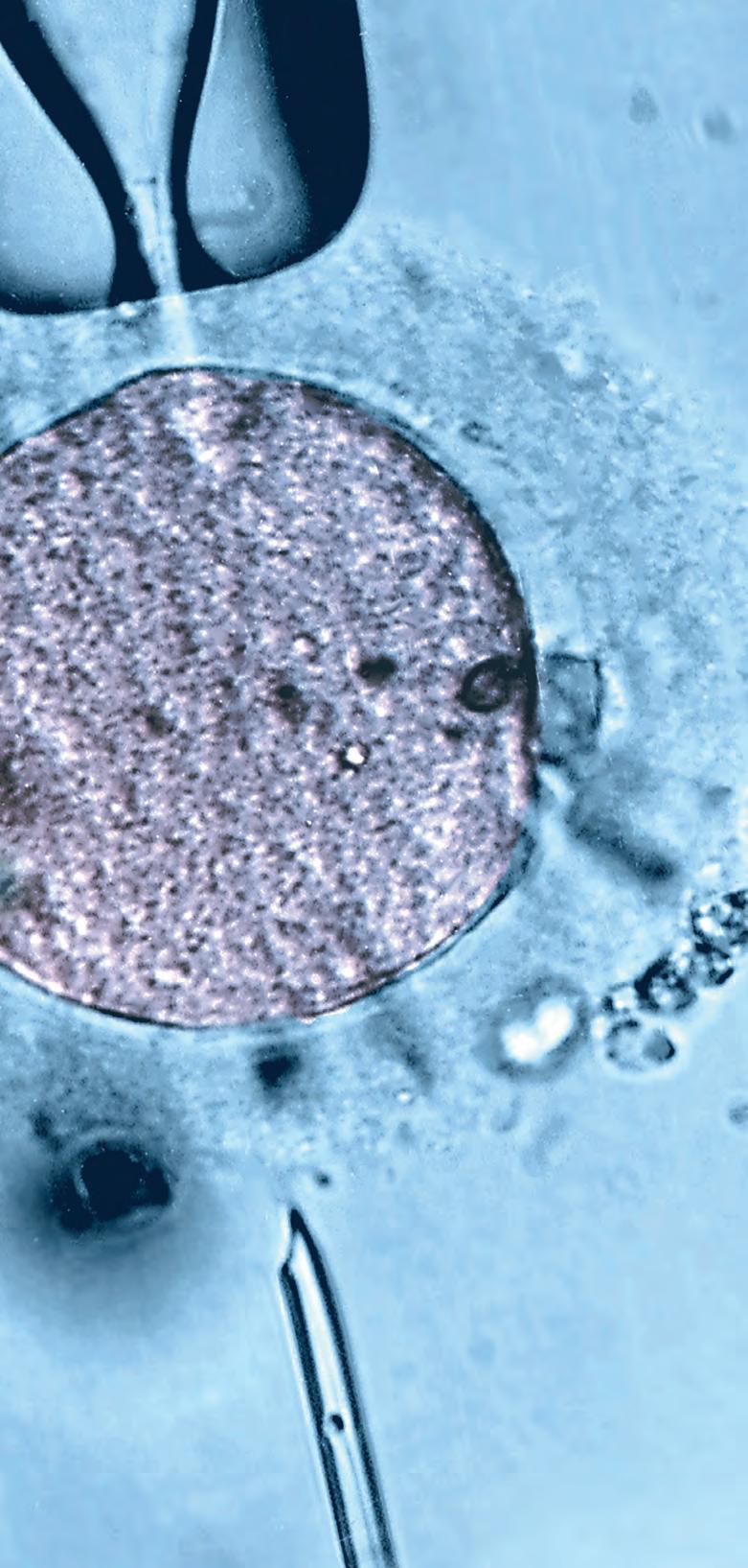
IMAGES: SCIENCE PHOTO LIBRARY
IPEM.AC.UK 29 SPRING 2023
Figure 1 An embryologist performing vitrification using the Cryotop vitrification device, which is plunged into liquid nitrogen
In this connection, we have developed a mathematical model of the vitrification process (based on the heat equation) that predicts the temperature of the mounted eggs/embryos, over time. We simulated different number and positions of eggs/ embryos and determined computationally the cooling rate. We compared all our scenarios to a “benchmark” scenario of a single egg/embryo situated in a large droplet (0.1μl); any arrangement cooling faster than this benchmark arrangement is considered to cool fast enough 2.
We found that the most important factor affecting the cooling rate is the volume of the cryoprotectant – less is better. Critically, even though the amount of cryoprotectant is limited by the manufacturer protocol, we found that within the prescribed range any arrangement of eggs/embryos is clinically viable. Our findings indicate that the current variability in clinical practice does not adversely affect IVF success rates. Our findings give more confidence to embryologists to complete vitrification quickly, as we have shown that they do not need to spend time arranging eggs/ embryos on the device in order to increase the cooling rate 3
Can the expansion rate of a frozen embryo predict pregnancy?
Embryologists usually thaw more than one embryo before a FET, in case some embryos did not survive vitrification. When there are two or more surviving embryos, embryologists must choose which embryo in the cohort should be transferred to the patient. This decision is currently based on the embryologist’s visual assessment, but unbiased, robust metrics based on data analytics can inform and automate this decision and would ensure consistency
WE DEDUCED THAT THERE IS AN OPTIMAL EXPANSION RATE
across clinicians and practices.
Embryos undergo dramatic changes during freezing and thawing. During vitrification, cells lose water and contract, and can be artificially collapsed, forming a very small, tight ball which is about 25-33% of the original size. When the embryo is thawed, the cells absorb water and eventually expand back to their original size 4
Modern time-lapse incubators generate detailed movies of the thawing process. Using machine learning image segmentation algorithms, we analysed such embryo images provided by the LWC and extracted the area of the embryo at each time point. We plotted the embryo area at time t against
area at time t+1 and compared how close to a straight line of slope 1 the data points are 5). We deduced, with statistical significance, that there is an optimal expansion rate – if the points are too far away from the straight line, that is the embryo expansion rate is either too high or too low, pregnancy is less likely. Our method can help choose embryos that are most likely to lead to pregnancy.
Choosing the best embryo in the first hours of IVF using imaging
Culturing embryos for 5-6 days is not ideal for the embryo, patients or clinics. The ideal solution would be to select the best embryo at a much earlier stage, such as on day one (zygote stage). Time lapse incubators can also be used to image eggs and zygotes in the first minutes or hours of IVF. During fertilisation there are also random motions throughout the cytoplasm described as “active diffusion” which occurs on a faster timescale than Brownian diffusion. Comparing the active diffusion rates between eggs may provide a metric of health that could help embryologists select the embryo most likely to succeed at day one. This could greatly reduce
Embryo/EggCryoprotectant
0.2mm 0.42mm 0.7mm 0.2mm 0.1mm 0.62mm 0.62mm 0.72mm 0.82mm 0.7mm Egg/ Embryo Plate
Cryoprotectant droplet
Figure 3 Various arrangements of eggs/embryos. The bottom right plot is the benchmark scenario (single egg/embryo in a large droplet).
Figure 2 The benchmark scenario of a single egg/ embryo being vitrified, inside a droplet of cryoprotectant, placed onto the plate in the Cryotop device.
IPEM SCOPE 30 SPRING 2023 G F GENERAL FEATURES
the economic cost both for clinics and patients and the psychological cost for the patients.
Techniques such as particle image velocimetry have already been used to show that cytoplasmic movement correlates to embryo development. Using additional techniques, such as differential dynamic microscopy (DDM), we can non-invasively study the motions in fertilising eggs or characterise other motions, for example the swimming of spermatozoa. We used DDM to determine the rate of diffusion in fertilising eggs. We are in the process of collecting more data to analyse so that we can finalise the metric selecting the best egg in the early hours of IVF.

Modelling Ca2+ waves could determine the best egg
One promising methodology to choose a zygote at day one of IVF is based on monitoring calcium (Ca2+) signalling and its associated movements and flows. The methodology was pioneered in UK laboratories but it has not progressed in the recent years partly due to the lack of mathematical modelling that can inform further steps. We have been working on closing this gap.
Multiple Ca2+ waves and oscillations at fertilisation cause egg activation in mammalian eggs. There is an optimal Ca2+ pattern associated with successful fertilisation and early development but
monitoring Ca2+ requires using dyes and fluorescence that damage the egg.
These Ca2+ waves cause the spasms and flows in the cytoplasm of mouse and human eggs; the movements can be detected non-invasively using low level light imaging and particle image velocimetry (PIV), which are cheap and could be readily implemented.
The timing of the spasms has been shown to predict the development of mouse embryos to the blastocyst stage (day 5-6). Hence, also this method could give embryologists a rapid (day 1) quantitative indicator of embryo viability.
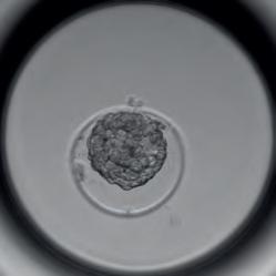
However, this technology has not been further developed, partly because of a lack of understanding of the relationship between Ca2+ waves and egg cytoplasmic movements. In this connection, we have been developing a novel mechanochemical model which describes the coupling of Ca2+ oscillations and waves to the motions in the cytoplasm.
The future of fertility
Although great progress has been made, there are still many challenges in IVF. The use of mathematical modelling has been limited while all stages of IVF could benefit from a more quantitative and less empirical approach; with our work we have taken several steps to close this gap. A crucial, unresolved challenge is that while embryo selection seems promising in in-silico studies this doesn’t translate to improvement in the clinic. This is probably because the data comparison is performed on embryos from different cohorts. Finding the “holy grail” of IVF – a tool for selecting the best embryo in a particular cohort (for one patient) – could rely on relating imaging data to quantifiable metrics derived from mathematical models of embryo development. To accurately calibrate these models, a unified database of all imaging data from many clinics would be needed. With increasingly advanced imaging techniques and larger computing power, IVF clinics are becoming able to implement more advanced methods for identifying the best embryos; they are just waiting on the right mathematical models.
Authors: Tim Ostler, Dr Katerina Kaouri and Dr Thomas Woolley (School of Mathematics, Cardiff University), Prof Karl Swann (Cardiff School of Biosciences).

Zona Pellucida Cells 0.3 0.25 0.2 0.15 0.1 0.05 0 00.050.10.150.20.250.3 Pregnancy No pregnancy Poor candidates for FET Area at time t + 1 Area at time t Embryo Area
Figure 4 A vitrified embryo, imaged using a GERI incubator, after being removed from storage and thawed. (a) Embryo undergoing re-expansion: cells begin expanding from a tight ball inside the Zona Pellucida (b) The same embryo imaged 115 minutes later - cells have absorbed water and expanded to fill out the Zona Pellucida.
Figure 5 The area of 124 embryos at time t, against their area at time t+1. The line of slope 1corresponds to embryos undergoing no expansion.
WE ARE IN THE PROCESS OF COLLECTING MORE DATA TO ANALYSE
IPEM.AC.UK 31 SPRING 2023
RADIOTHERAPY DOSE-TO-MEDIUM REPORTING
A survey of UK practice
n 2021 IPEM commissioned a dose-to-medium working party. This was as a result of a publication by the Global Harmonisation Group (GHG) for quality assurance in clinical trials, which recommended that, where possible, dose in clinical trials should be reported as dose-tomedium in medium (Dm,m) and a subsequent increase in the number of enquiries from UK centres to National Physical Laboratory (NPL) regarding validation of these algorithms. The purpose of the working party is to review the state of adoption of Dm,m reporting among UK radiotherapy centres and identify any barriers to implementation; develop guidance on treatment planning system dosimetry validation and develop a national calibration and audit service for dose-to-medium in conjunction with NPL.
Survey design
As part of this work a survey was sent to UK centres, and we are very grateful for all centres who took the
time to fill it out. This article outlines the key findings from this survey. It will not go into specifics of the relative merits or drawbacks of dose reporting modes and readers are directed to the global recommendations or the American Association of Physicists in Medicine (AAPM) TG329 report for some excellent summaries of the problems faced.
The survey was distributed to the heads of radiotherapy physics mailing list (incorporating 65 UK radiotherapy centres) using Google Forms in September to October 2022. The survey comprised 29 unique questions and included a combination of multiple choice and free text answers. Some questions were repeated allowing information regarding multiple treatment planning systems (TPS) and algorithms to be entered. Some questions were only relevant to, and answered by, those centres that are using Dm,m clinically. The purpose of the survey was to establish:
● General demographic information concerning radiotherapy hardware and software infrastructure;
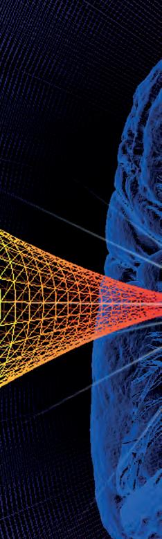
● Proportion of UK respondents currently reporting, or intending to report, Dm,m as well as algorithms used, and dose validation undertaken;
● Opinions among all respondents regarding barriers to implementation of Dm,m reporting, coupled with suggestions for useful resources and services that would aid commissioning work in future.
I
A look at the results of a survey into the state of adoption of dose-to-medium in medium reporting among UK radiotherapy centres.
WHERE POSSIBLE, DOSE IN CLINICAL TRIALS SHOULD BE REPORTED AS DOSE-TO-MEDIUM IN MEDIUM
G F GENERAL FEATURES IPEM SCOPE 32 SPRING 2023
Results
A total of 46 responses were received, representing a 71% response rate, which is comparable to other recent IPEM working party surveys, such as the 2020 survey of online treatment monitoring. All major radiotherapy manufacturers were represented, with the majority of centres using Varian linacs and software including the Eclipse treatment planning system (TPS) and AcurosXB (AXB) calculation model. The distribution of TPS and dose reporting is shown in Figure 1, while Table 1 gives information regarding the Dm,m planning algorithms used. Figure 2 shows the different treatment units in clinical use by responding centres which provides context to the work and is included here for general interest. It is worth noting some of the complex arrangements within some centres. For example, 40% of respondents had more than one TPS, and 53% had two or more algorithms in clinical use. This results in (for the 46 responses received) 89 algorithms across 72 separate planning systems. Two thirds of those with multiple algorithms (across 1 or more TPS) report dose in different ways.
Nineteen of the 46 respondents (41%) reported using Dm,m clinically in at least one algorithm or planning system. This translates to a total of 24 Dm,m treatment planning systems spread between these 19 centres. In total, 53% of these centres used Dm,m for all
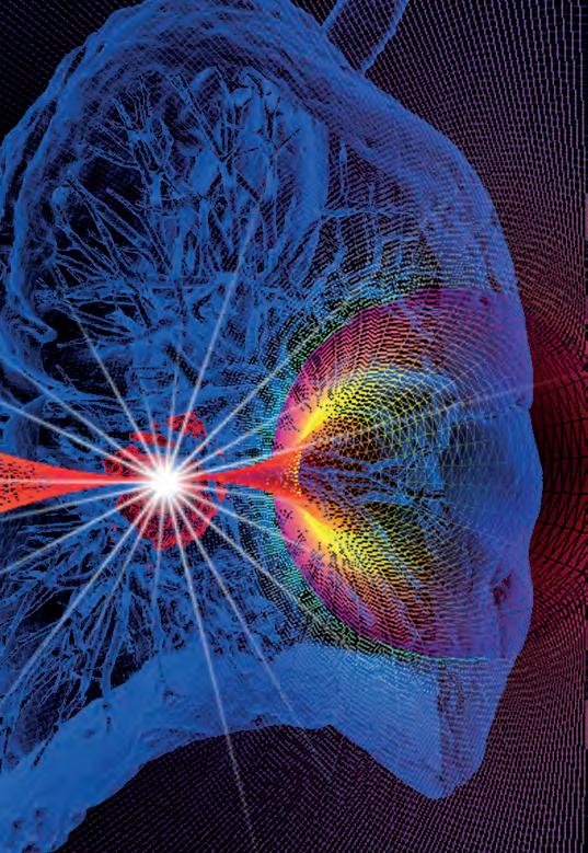
40 35 30 25 20 15 10 5 0 30 25 20 15 10 5 0 AccurayTomotherapy Eclipse Monaco Pinnacle Raystation Accuray Precision Brainlab elements Ethos Oncentra Number of responding UK centres Number of responding UK centres Varian Elekta Varian Ethos/ Halcyon Accuray Cyberknife Accuray Tomotherapy
No. Of TPSRespondentsNo. of AlgorithmsRespondents 1 27 (59%)1 21 (46%) 2 12 (26%) 2 12 (26%) 3 7 (15%) 3 8 (17%) 4 5 (11%)
Table
1 Respondents with different numbers of TPS and algorithms
Planning SystemUsersDm,m AlgorithmUsers Eclipse 30 (42.9%) Acuros 12 (40%) Raystation 11 (15.7%)Collapsed Cone/Monte Carlo3 (27.3%) Monaco 3 (4.3%)Monte Carlo 2 (66.7%) Oncentra 1 (1.4%)Collapsed Cone 1 (100%) Pinnacle 9 (12.9%)N/AAccuray Precision 7 (10%) Monte Carlo 3 (42.9%) Accuray Tomotherapy 1 (1.4%)N/A Brainlab Elements 5 (7.1%)Monte Carlo 1 (20%) Brainlab iPlan 1 (1.4%)N/A Ethos 2 (2.9%)Acuros 2 (100%)
Table 2 The proportion of TPSs used at different centres. Note that several centres reported the use of multiple planning systems.
Brainlab iPlan Dw,w Dw,m Dm,m
Figure 1 Showing the number of UK centres using each TPS and the proportion of each using different dose reporting.
Figure 2 The number of respondents with particular machines at their centre, note that the values are per centre not per machine.
IMAGE: SCIENCE PHOTO LIBRARY IPEM.AC.UK 33 SPRING 2023
anatomical sites. A further 21 centres (46%) declared an intention to commission Dm,m in the next two years. The distribution of future intent is shown in the Figure 3 below. It should be noted that there was a variation in the response from Pinnacle users as to whether Dm,m was used, so these were considered as a separate category.
The algorithm in Pinnacle takes account of both electron density and atomic composition, producing a dose close to Dm,m. In five cases, (21% of planning systems reporting Dm,m) edits were made to the materials or mass densities available including adding couch structures, bolus materials or metals.
Among those centres currently reporting Dm,m verification work consisted mainly of measurements either with film or chambers, possibly with medium set to water, or comparison with an independent TPS. Only two centres reported verifying against an independent Monte Carlo calculation. Several centres expressed difficulties in matching existing phantoms to the correct medium in the treatment planning system and there was an overwhelming majority of those reporting Dm,m (83%) who requested further guidance in validating Dm,m.
A lack of guidance was frequently cited as a barrier by centres not currently reporting Dm,m alongside resource constraints and clinical priorities. As shown in Table 2, there are complex arrangements in terms of multiple planning systems and algorithms at some centres. The challenge of consistent validation across a range of software and techniques may also be a contributing factor here. A number of other centres also express concerns regarding the lack of literature surrounding the clinical impact of doseto-medium reporting. Despite the considerable number of centres who have implemented Dm,m clinically there is still desire for UK consensus in this area. The stated barriers include clinical guidance, resourcing and other priorities.
Interest in a Dm,m chamber calibration service from NPL was indicated by 56% of respondents. The same percentage also expressed an interest in a Dm,m validation phantom, either to purchase or hire.
The percentage of Dose reporting for the treatment planning systems used clinically at responding centres.
However, it should be noted that a large number of centres were unsure of the benefits at this stage, as shown in Figures 4a and 4b. Additionally, centres reported that published stopping power and correction factors would be useful in moving forward with clinical implementation.
Summary
If you don’t already report dose to medium, are you planning on commissioning a dose to medium treatment planning system?
Would you be interested in a chamber calibration in a different medium by NPL?
Would you be interested in a dose to medium phantom, if one were to be developed?
A high response rate of 71% was achieved in this UK survey of Dm,m practice. A range of treatment planning systems are in use clinically for this purpose with the majority currently using Varian’s Acuros XB algorithm. Over a third of respondents are reporting Dm,m with many others planning on commissioning in the near future. Due to potential selection bias (with those already using Dm,m possibly more likely to complete the survey) this may be an overestimation in the proportion of UK centres that are using Dm,m. The current position is that the majority of UK centres are not able to conform to the Global Harmonisation Group recommendation of using Dm,m for clinical trials. However, there is a clear movement in this direction with a further 21 centres indicating intent within the next two years. This survey indicates a strong appetite for implementation guidance (which is currently limited and with no national consensus), and serves to motivate future work in this area, particularly considering the large numbers of centres currently using or considering Dm,m. It also shows an interest in additional services to assist with the implementation of Dm,m, such as specialist phantoms and a calibration service. The results of this survey will inform the work of this group and any literature produced. The Dm,m working party is also working on the development of matched virtual and physical phantoms for Dm,m validation calculations and measurements, with Monte Carlo-based correction factors for the materials used.
Charlie Martin,
Clark. The authors would like to thank everyone who took part in the survey. They welcome follow up comments to RTaudit@npl.co.uk.
Figure 3 a) the percentage of dose reporting methods currently in clinical use and b) the timescales of Dm,m reporting in centres who aren’t currently using.
Dm,m Dw,m Dw,w Pinnacle
In the next year In the next 1-2 years In the next 2-3 years In the next 5+ years Not in the next few years Yes Unsure No Other Yes- to hire Yes- to purchase No Unsure 15; 50% 6; 20% 5; 17% 3; 10% 1 29; 41% 9; 13% 24; 35% 8; 11%
Figure 4 The proportion of responding centres who would be interested in a) a chamber calibration service and b) a dose-to-medium phantom.
G F GENERAL FEATURES IPEM SCOPE 34 SPRING 2023
Victoria Newton, Clare Antoine, Rhydian Caines, Karen Venables, Catharine
QUANTIFICATION IN SPECT-CT
The new light in nuclear medicine?
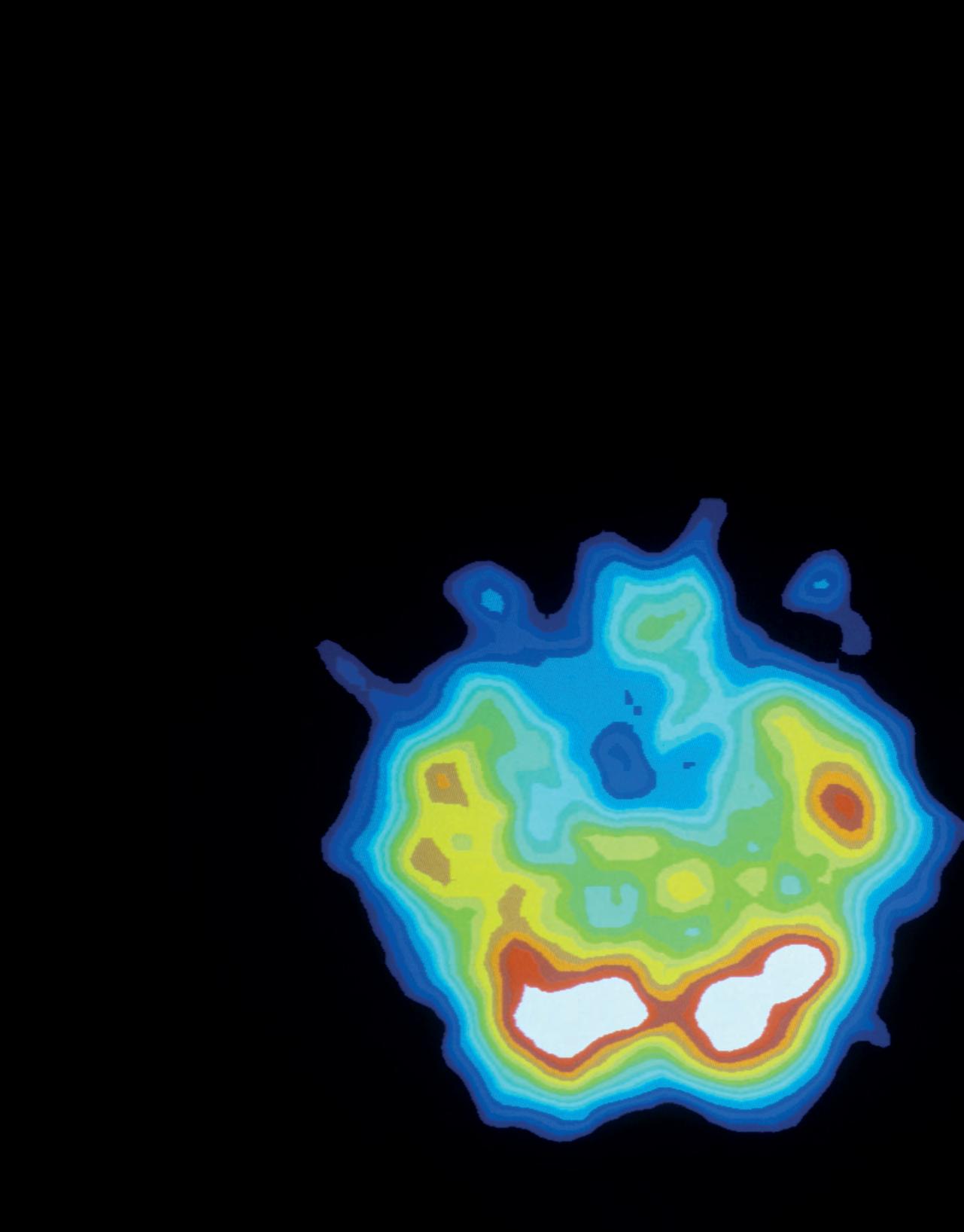
Nuclear Medicine Technologist Clara Ferreira looks at issues surrounding SPECT-CT and the potential effect it could have on imaging.
fter Henry Becquerel discovered radioactivity in 1896, there was an immediate interest in quantification. This interest is becoming more relevant in single photon emission computed tomography-computed tomography (SPECT-CT) systems now that they are gaining acceptance as an accurate quantitative imaging modality with the CT component providing not just anatomical reference, but also accurate attenuation and scatter correction in order to improve quantification in diagnostics and therapy.
Although SPECT-CT is an extremely helpful, widely used clinical imaging modality, it will not achieve its full potential when it is used in a purely qualitative manner because it depends on the relative tracer uptake.
On the other hand, the quantitative SPECT-CT (qSPECT-CT) imaging allows the direct measurement of the activity distribution within a given region and it will have an enormous effect in the practice of nuclear medicine (NM) due to greater objectivity in the interpretation.
Some other semi-quantitative metrics have also been popularised, of which the standardised uptake value
A
G F GENERAL FEATURES IPEM.AC.UK 35 SPRING 2023
(SUV) has been the most popular. The SUV has several variants, but, in a general way, it corresponds to the normalisation of the activity (decay corrected to the same time point) in the region-of-interest (ROI) by dividing the injected activity by some patient metric of distribution volume, such as the body mass.
The value SUV is very vulnerable, but it continues to be used due to dynamic imaging and blood samples not being needed in the positron emission tomography-CT (PET-CT) imaging. There is a wide range of radionuclides in which qSPECT-CT is used – 99mTc, 111In, 131I, 177Lu, 186Re and 201Tl.
Physics
The main features for a reconstruction algorithm are described in Table 1
The reconstruction algorithm of choice today is based in the statistical iterative methods – with many using ordered-subset maximum-likelihood expectation maximisation algorithm (OSEM) – several
publications have reported higher quantitative accuracy in iterative reconstructions than of non-iterative methods. The use of the algorithm has several advantages, such as, the ability to model physical characteristics of the acquisition in the reconstruction with the objective to enhance image quality and accuracy and better control of signal-tonoise characteristics in the final image.
If the data is reconstructed in kBq/cm3, there is the change of applying SUV, considering that the administered activity and the delay between the injection and the acquisition is known, as well as the patient weight/height (depending on the type of SUV equation used).
There are also some factors that may influence the quantitative accuracy of reconstructed SPECT-CT data and they are directly related with the subsequent absorbed dose calculations – Table 2
In order to obtain optimised calculations, an accurate evaluation of the different uncertainties is needed, which is especially important in clinical trials.
Main features for a reconstruction algorithm
Needs to behave in a linear fashion regarding the reconstructed activity concentration
Algorithm to compensate for photon absorption within the body
Algorithm to remove scattered radiation
Ability to calibrate the reconstructed data (for example, in kBq/cm 3).
To this day, most of the SPECT images are performed without attenuation or scatter correction. Although, it is considered a necessity for qSPECT-CT. The attenuation correction can be performed after reconstruction using the attenuation map from the CT data or the correction can be directly integrated in the iterative reconstruction. Thus, CT scan does not provide just anatomical information, but also information about the density of the body tissues or attenuation photons within the body, enhancing the diagnostic accuracy and patient throughput.
it is necessary to achieve a compromise between collimator efficiency and image quality – thicker collimator septa reduce the septal penetration, but they also reduce its efficiency. In such manner, a certain amount of septal penetration is allowed, which needs to be corrected in the reconstruction part.
Factors influencing the accuracy of qSPECT-CT
Partial-volume effect
Dead-Time
Radioactivity decay during the acquisition
Corrections and normalisations for spatial and temporal variations in detector response
Photon Scatter
Photon Attenuation
VOI delineation
Varying detector trajectory
The major routine application of attenuation correction in SPECT is the removal of attenuation artifacts in myocardial perfusion imaging (MPI), but the emphasis has not been on quantitative assessment.
The most used scatter correction method is called transmission-dependent scatter correction and it gives accurate results in a range of different imaging. The simpler energy-based method, known as the triple-energy-window approach, has also demonstrated accurate quantification and it is offered by several vendors.
It is important to keep in mind that
Most of the reconstruction software includes an option for scatter, attenuation correction and point-spread function correction. The recent developments in collimator modelling and 3D reconstruction also allow improvements in the reconstruction techniques, thereby enabling absolute qSPECT. The use of contrast recovery, background variability and recovery activity percentage can also contribute to the optimisation of the image.
Quality control
The future of qSPECT-CT quality control will mirror the one used in PET-CT systems
 Table 2 List of factors that influence the accuracy of qSPECT-CT (Bailey & Willowson, 2013)
Table 1 Indication of the main features of a reconstruction algorithm in SPECT-CT (Bailey & Willowson, 2013).
Table 2 List of factors that influence the accuracy of qSPECT-CT (Bailey & Willowson, 2013)
Table 1 Indication of the main features of a reconstruction algorithm in SPECT-CT (Bailey & Willowson, 2013).
G F GENERAL FEATURES IPEM SCOPE 36 SPRING 2023
nowadays – where it is possible to check the consensus between the dose calibrator and the reconstructed SPECT-CT values for radioactivity.
It is important to highlight that the big difference between SPECT-CT and PET-CT cross-calibration is the photon energy associated with the different radionuclides. For example, in PET, all the radionuclides have the same photon energy – 2 photons of 511 keV –; in SPECT-CT, different radionuclides are used and all of them emit photons with different energy peaks. The different photon energies lead to the
use of different collimators and pulse height analyser energy windows, which require separate calibration. Many of the required parameters (such as scatter correction) can be predefined to a specific radionuclide, collimator, but regular validation is essential.
The biggest application of qSPECT-CT happens in the theranostic area – especially after the introduction of 177Lu for dosimetry. The qSPECT-CT can provide diagnostic information beyond just the absence or presence of the disease, but its use is still limited in clinical practice.
Several authors agree that manufacturers should supply techniques for calibration of the systems, operational limits and routine validation as part of the regular SPECT quality control programme.
In the clinical environment, an increase in the quantification variability between the different SPECT-CT systems is expected due to, for example, patient positioning and patient’s body mass index. According to one of the researchers, patients with high BMI (higher than 47 kg/m2) quantifying a lesion will be a more challenging process, which can be explained by the increased attenuation, decreased signal-to-noise ratio and decreased spatial resolution considering the high distance source-detector. The use of more iterations in the image reconstruction is a possibility because it might improve convergence, the resolution and prevent artifacts; otherwise, the increased attenuation can be cancelled by an increase in the scan time or in the patient dose.
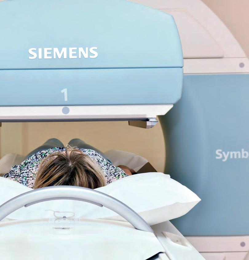
Standardisation of protocols is needed in such way that quantitative results can be reliably compared between systems considering that the accuracy of quantification has been shown to vary widely between centres.
The standardisation of dosimetry methodology is an essential component of any multi-centre trial aiming to provide a robust evidence-based for absorbed dose-response thresholds, which needs to have the different makes and models in consideration.
According to the authors of the clinical trial SEL-I-METRY, the calibration factors vary between the various makes and models of SPECT-CT systems, but there was sufficient similarity between them to allow the use of global specific calibration factors.
Some authors asked several nuclear medicine departments about why they do not use quantitative analysis in their scans – most of them mentioned its use is questionable and the transferability between different centres and platforms. Thus, the several challenges include the clinical validation and transferability.
The “Metrology for Clinical Implementation of Dosimetry in Molecular Radiotherapy” (MRTDosimetry) project was a joint research project within the
IMAGE: SCIENCE PHOTO LIBRARY
IPEM.AC.UK 37 SPRING 2023
THE QSPECT-CT CAN PROVIDE DIAGNOSTIC INFORMATION BEYOND JUST THE ABSENCE OR PRESENCE OF THE DISEASE
European Metrology Programme for Innovation and Research (EMPIR), which ran for three years finishing in 2019. This project dealt with the problem of assessing the absorbed dose of individual patients who were undergoing molecular radiotherapy leading to the development of a protocol for commissioning and quality control of 177Lu qSPECT-CT imaging and testing in a multicentre by comparing the differences between the different partners. The experimental protocol included: determination of an appropriate image calibration factor, correction of partial volume effects and validation of quantification imaging using a 3D phantom.

Bone
In some diagnostic applications, the increased tracer uptake is correlated with the presence or severity of the disease. Hand and wrist pain is one of the most frequent
THE REMOVAL OF A HIGH NUMBER OF SLNS LEADS TO ADVERSE EFFECTS
symptoms and reason for admission in rheumatology; occasionally, it is difficult to identify the cause of the pain because of the extensive differential diagnosis and similar clinical manifestations.
In a 2021 study, the researchers compared joint SUVs and pain symptoms in 87 patients with rheumatic disease
manifesting as hand and wrist pain. The patients underwent bone SPECT-CT of the hand and wrist region in a Symbia Intevo 16 hybrid SPECT-CT (Siemens Healthineers, Erlangen, Germany) three hours after the administration of 740 MBq of 99mTc-MDP. The image analysis was performed using MIM version 6.6, MIM Software Inc., OH, WA. Figure 1 shows the results of this research.
The qSPECT-CT skills showed the potential for differentiating articular problems from pain perception in rheumatic patients with hand and wrist pain because the SUVmax measured directly represents inflammation and/or damage in peripheral structures rather than just using the pain perception. Painful hands and wrists are frequently associated with relatively high joint SUVmax; and it was also possible to understand that the distribution and intensity in joint SUVmax
G F GENERAL FEATURES IPEM SCOPE 38 SPRING 2023
differed from pain according to the specific rheumatic diseases. Rheumatoid arthritis is represented by high SUVmax, while fibromyalgia was found to have lower SUVmax which is thought to be associated with less arthritic activity and more pain sensitive.
In these cases, the SUVmax of joints provides more objective information for arthritic activity related to peripheral joint damage, independently of pain symptoms.
Thyroid
Grave’s disease is the most common cause of hyperthyroidism, which accounts for nearly 90% of the total number of cases, states a 2019 study.
The radiopharmaceutical 99mTcPertechnetate can be used to assess thyroid function by thyroid uptake system and it is considered useful for the differential diagnosis between conditions characterised by extremely altered thyroid function.
In this 2019 study, the researchers included 190 patients (116 patients with Grave’s disease and 74 healthy volunteers), who were injected with 185 MBq of 99mTc-Pertechnetate and scanned in a SPECT-CT scanner (NM/CT 670; GE Healthcare, Pittsburgh, PA, USA) at 30 minutes post-administration.
The SUVmax and SUVmean values by qSPECT-CT were significantly different in the two groups (both p<0.01, MannWhitney U test).
The researchers also compared the results of the qSPECT-CT and the thyroid hormone levels. In this case, the SUVmean and SUVmax had positive correlations with the T3 and T4 levels, respectively.
Breast
The incidence of breast cancer is the most common in women. The axillary lymph node status is a crucial prognostic factor in breast cancer patients; this way, an accurate lymph node staging is essential to guide treatment that affects their overall survival, states a 2022 study.
Sentinel lymph nodes (SLNs) in breast cancer are detected using a combination of radiopharmaceutical and blue dye.
The pre-operative SLN of breast (SLNB) SPECT-CT imaging provides an accurate anatomical information of SLNs with
lymphatic drainage into the axilla and extra-axillary regions, distinguishing false-positive results caused by nuclide contamination and lymphatic tortuosity and reducing the false-negative caused by the patient obesity. It also compensates for the limitation of the gamma probe in determining the exact anatomy of SLNs.
The removal of a high number of SLNs leads to adverse effects and, some of them, might be healthy; this way, a group of
researchers developed a technique to distinguish between metastatic and healthy lymph nodes by using 99mTc-sulfur colloid (SC) and qSPECT-CT.
When the qSPECT-CT is analysed, a radioactivity threshold could give access to a selective removal by keeping it at 30% of the hottest node; this way, no metastatic SLNs were missed and, as a result, 30% of identified lymph nodes do not require resection.
SUV Pain (-) Pain (+) DIP PIP MCP Wrist 6 4 2 0
Figure 1 Box-whisker plots of joint SUV (SUVmax) at each level joint. DIP – distal interphalangeal; PIP – proximal interphalangeal; MCP – metacarpophalangeal; *P<0.05 (Lim et al, 2022).
SUVmax (g/ml) 1000 800 600 400 200 0 Graves group (a)Control
SUVmean (g/ml) 500 400 300 200 100 0 Control group Graves group (b) IPEM.AC.UK 39 SPRING 2023
Figure 2 Comparison of SUVmax (a) and SUVmean (b) values between the two groups (Dong et al, 2019).
group
The authors consider that the 99mTc-SC qSPECT-CT has the ability to indicate the nature of the lymph nodes in breast cancer, assisting surgeons where anatomic location may be difficult for biopsy.
Lung
In some patients, when a lobectomy is required, a V/Q scan with quantification is required in order to find out how much each part of the lung is contributing to the overall function in order to calculate the percentage of loss of lung function. The pre-operative lung perfusion quantification should include attenuation and scatteredcorrected tomography data; also the patient’s individual information from CT should be used for segmentation, according to a 2018 study.
The prevalence of lung malignant processes is high, with a drastic increase over the past decades. According to existing guidelines, resection is the therapy of choice, if possible.
In the study, SPECT-CT was used to analyse pulmonary function based on 3D perfusion data by using patients’ individual anatomical information from CT segmentation. A total of 39 patients were included in the study for pre-operative quantification of lobar lung function, exclusion of acute pulmonary embolism (PE) and exclusion of chronic thromboembolic pulmonary hypertension.
The tool Q.Lung was used by assisting the VOI-definition semi-automatically and accelerates the evaluation workflow noticeably; considering that the manual version is very time consuming.
This study was able to demonstrate the effect of evaluating planar and SPECT-CT data for estimation of lobar perfusion,
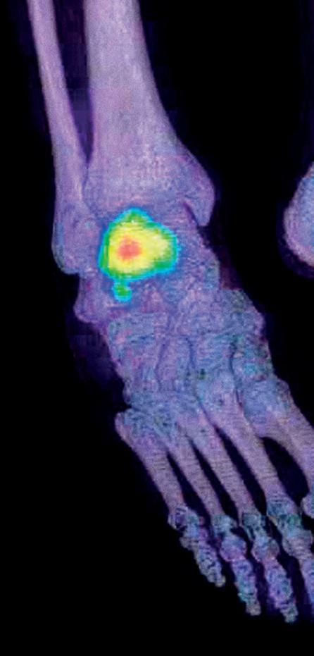
which results in under-estimation of the lobes in the planar images.
Renal
Glomerular filtration rate (GFR) is described as a flow rate of blood plasma that is filtered through glomerulus, which is an indicator of renal function. Thus, the GFR is estimated using a radiopharmaceutical that is completely filtered through glomerulus, not reabsorbed and not excreted in renal tubules, according to a 2019 study.
The most used radiopharmaceutical to evaluate renal function is 99mTc-DTPA and blood samples after the administration is the method to directly measure renal clearance, but they are laborious and time-consuming. Therefore, a 99mTc-DTPA scintigraphy is another option with the use of Gate’s method that utilises the counts measured for one minute, from two minutes to three minutes after the radiopharmaceutical administration.
For this reason, SPECT-CT with 99mTcDTPA is a promising method for the measurement of GFR because it allows the assess to quantification and reliable measuring of the renal clearance in comparison with planar imaging. The VOI is drawn on each kidney for the quantification of absolute radioactivity; this is also a reproducible and accurate method in healthy volunteers, patients with renal tumours with post-partial nephrectomy and for the analysis of disease severity in patients with kidney stones.
A quantitative 99mTc-DTPA kidney SPECT-CT study included 393 patients, retrospectively in order to develop an automated GFR quantification method based on deep-learning approach and
compare with the manual VOI. The time required for the segmentation was only a few seconds per patient; on the other hand, the manual segmentation takes around 15 minutes per scan, so it prevents the large use of this approach considering the time-consuming task.
Formulas such as modification of diet in renal disease (MDRD) or chronic kidney disease epidemiology collaboration (CKD-EPI) equations are frequently used in clinic to derive GFR from the serum creatinine level; although creatinine is not an ideal substance for measuring renal clearance.
There was no difference between manually driven GFR and automatic-driven GFR (Table 3) in all groups of patients and kidneys. Thus, it is possible to conclude that the two segmentation methods work well to represent the functional deterioration by obstructing urinary stones and subsequent improvement
Healthy Patients (n=25) Kidney Stones Patients (n=63) P-value Manual VOI 120.39 ± 19.26115.65 ± 16.91NS Deep-learning-generates VOI119.25 ±18.35115.02 ± 17.71NS P-value NS NS G F GENERAL FEATURES IPEM SCOPE 40 SPRING 2023
Table 3 – Total GFR (mL/min/1.73 m2) by manual and deep-learning-generated-based segmentation in healthy and urolithiasis patients presented in mean ± SD. NS – non-significant (Park et al, 2019).
after stone removal procedures.
The combination of CT-based automatic segmentation by deep learning approach and novel qSPECT technology may pave the way for precision nuclear medicine regarding measurements of GFR.
Therapy
Accurate absolute quantification of a radiopharmaceutical distribution is essential for dosimetry aimed at personalised radionuclide therapy and may improve prediction of therapy response, prevention of toxicity effects and treatment follow-up by the calculation of absorbed doses to organs, tissues or tumour of interest.
For example, in prostate, thyroid and neuroendocrine tumours, it is expected that dosimetry will play a pivotal role for reliable determination of dose response relationships. The establishment of a dose-effect relationship for a given radionuclide therapy allows for a
personalised therapy plan more closely resembling external beam radiotherapy; also, the dosimetry calculation became a legal requirement under the Basic Safety Standard.
Peptide Receptor Radionuclide Therapy (PRRT) for gastroenteropancreatic neuroendocrine tumours with 177LuDOTATATE has been increasing in the last few years after the NETTER-1 trial showed that results in longer progression-free survival and higher response rate than to treatment with somatostatin analogues. Therefore, red marrow, healthy liver and kidneys are identified as organs at risk. Also, there is increasing evidence that treatment outcome correlated with the absorbed dose delivered to tumours and healthy organs makes personalised dosimetry even more important.
Currently, a fixed activity of 7.4 GBq per cycle is administered, but the personalised dosimetry is needed to ensure safety and
evaluate the absorbed dose to the tumour and assess the dose-response relationship. Yet, for an accurate estimation of the activity in several organs at several time points, calibration and following the right steps in the quantification is crucial.
The following images were acquired in a SPECT-CT Discovery NM/CT 670 system (GE Healthcare) with MEGP collimator, and a 20% energy window centred on the 208 keV photopeak and a 10% scatter correction window centred on 177 keV. A 128×128 matrix was used. The patient images were acquired at 4-, 24-, 72- and 192-hours post-administration.
The Dosimetry Toolkit (GE Healthcare) was used to analyse the images and the OLINDA/EXM V1.0 software was used to calculate the organ absorbed and effective doses.
It is important to analyse the variation inter-patients, but also intra-patient. It is also important to verify that performing patient dosimetry in the different therapies is possible in nuclear medicine department.
Conclusion
As demonstrated by the clinical trial SEL-I-METRY, it is possible to perform accurate, standardised quantitative imaging between different centres participating in diagnostic and molecular radiotherapy treatments in a multi-centre and multi-company setting. At the moment, the calibration of qSPECT-CT is highly centre-dependent, even among the one who use the same systems and parameters, and few efforts have been made in order to establish traceability of activity quantification across sites.
Several authors agree that diagnostic applications of quantification can illustrate its further progress and increase the value of the images, taking into consideration that quantification can be a biomarker for disease severity, patient outcome or a predictor of treatment response.
In recent years, several SPECT-CT and software suppliers have developed tools in order to respond to the qSPECT-CT needed by offering packages for different radionuclides, such as, 99mTc, 111In, 131I, 90Y and 177Lu.
The truth is that the time for the clinical implementation of qSPECT has come.
IPEM.AC.UK 41 SPRING 2023
THE TRUTH IS THAT THE TIME FOR THE CLINICAL IMPLEMENTATION OF QSPECT HAS COME
HOW TO Launch a research project
Research is a very broad term. In a simple way, it can be defined as “finding out something new”. It is beneficial for people and patients, enabling earlier diagnosis, more effective treatments, prevention of ill health, better outcomes and a faster return to everyday life. The hospitals that are “research active” have lower mortality rates than those that are not, but all NHS hospitals are involved in research in some degree.
COVID-19 has put health research in the spotlight and shown how important it is in our lives.
Research generates evidence to refute, support or develop a hypothesis, attempting to find out what happens when we manipulate clinical practice in some way. It may require only observations and it may be prospective or retrospective. It may be qualitative or quantitative. In the end, the results will be generalised beyond the sample upon which the research was based.
The National Institute of Health and Research (NIHR) is the research arm of the NHS, providing funding for studies, academic training, facilities, career development and research capability development. It is also important that the relevant R&D department and the local research team are involved in the study set-up.

Getting started
If you have a research idea or want to collaborate with another study which is already set-up, you should get involved. Firstly, it is important to know if your idea is classified as “research” – see Table 1. Medical research and development are a lengthy process (especially for drug development). Therefore, some skills to include in the research are: planning, organising, teamwork, numerical and statistical analysis and communication.
Approval process
If you answer was “research” in the previous question, there are some approvals to seek before starting:
● HRA approval: is the permission to seek if you want your study to go ahead in the NHS in England.
It comprises an assessment of study compliance with applicable regulations and standards and includes a review of the NHS Research Ethics Committee (REC).
● Confirmation of capability and capacity: you should have this confirmation from every participating trust to indicate that they can deliver the study in their organisation by meeting all governance requirements and performance targets. This document signals that the study can start in a specific NHS trust.
In order to apply for these, it is mandatory to have a sponsor – an organisation that will be legally responsible for the organisation and management of the study (such as, an NHS trust, a university or even a commercial company). Normally, the minimum that you will need to produce is:
● A protocol;
● A patient information leaflet (PIL);
● A consent form.
The research protocol should describe the research in as much detail as possible. It should always be kept up to date as the research evolves. Making a protocol publicly available can be done in the interests of transparency – many publishers offer a protocol publication service.
G F GENERAL FEATURES IPEM SCOPE 42 SPRING 2023
If you have a proposal for a research study that involves a drug you need to follow the Clinical Trial Pathway – Clinical Trials of Investigational Medicine Product (CTIMP). CTIMPs are governed by law and inspected by the UK regulator, the Medicines and Healthcare products Regulatory Agency (MHRA).

The R&D team and the university will help you design your trial paperwork to ensure it is ethical and legal. It is vital to keep the necessary training up to date; ideally, all the training should be done in the last three years.
In a clinical trial, one treatment is compared with another involving patients, healthy people and even both. One treatment can be a dummy treatment (normally a placebo) or a standard treatment in use.
Within clinical trials, it is possible to set-up one or request to be part of one which is already set-up; in the latter, the use of World Health Organization’s International Clinical Trials Registry Platform provides access to clinical trials all around the world.
The Table 2 indicates the different phases of clinical trials. In every phase, it is important the researchers keep patients in both groups who are similar in age,
Is it definitely “research”?
Research
Audit
It is designed to generate knowledge. It may involve the participation of patients and it involved an attempt to test a new hypothesis.
It is designed to test whether an established service or technique is meeting a defined standard.
similar proportion of men and women and with similar health overall.
In most trials, a computer will be used to randomly decide which group each patient will be allocated to. In many trials, nobody knows who has been allocated to receive each treatment (blinding), which helps to reduce the bias. A patient can leave the trial at any point without giving a reason and without affecting the treatment that he/she/they are receiving.
At the end of the trial, researchers should publish the results. All the results need to be sent to MHRA, which decides whether to allow the company making the medicine to market it for a specific use.
If you are planning to apply for a grant, there are also some specific documents to prepare:
● Organisational Information Document (OID);
● Schedule of Events (SoE) or Schedule of Events
Costing Attribution Template (SoECAT).
The SoECAT is a requirement for NIHR and NIHR non-commercial partner research funders where the study may involve participants under an NHS or health and social care duty of care.
If you are planning to be involved in a commercial study, the sponsor of your study will have to prepare a commercial OID – a model draft commercial research agreement using the appropriate national template agreement – and the NIHR Costing Template, which needs to be completed online.
The review of Confidentiality and Research Agreements will usually be undertaken by R&D team which will liaise with the sponsor in order to finalise the agreement. After all is signed, a Confirmation of Capability and Capacity will be issued. The cost of the study and subsequent negotiations can start as soon as the draft protocol is available and should include:
● The NHS, through NIHR;
● The Medical Research Council (MRC);
RESEARCH GENERATES EVIDENCE TO REFUTE, SUPPORT OR DEVELOP A HYPOTHESIS
Table 1 Difference between research and audit.
IMAGES: ISTOCK IPEM.AC.UK 43 SPRING 2023
IN A CLINICAL TRIAL, ONE TREATMENT IS COMPARED WITH ANOTHER
● The Department of Health and Social Care/other government departments;
● Medical research and hospital charities;
● Pharmaceutical and other healthcare companies.
If NIHR is one of the options, the NIHR costing template should be used. When costing a study, there are a number of things to consider:
● The interventions;
● Time taken for each intervention and frequency of visits;
● Number of participants;
● Type of resource required (nurses, doctors, research assistants);
● Requirement for scans, data entry, CRF completion and coordination;
● Use of clinical research facility;
● GCP monitoring;
● Pharmacy and R&D set-up costs;
● Code breaking services;
● Travel costs and participant refreshments;
● Archiving costs;
● CTA application fee. Everyone involved in the conduct of clinical research must have training in Good Clinical Practice (GCP) to ensure they are best prepared to carry out their duties.
Health Research Authority (HRA)
The HRA opened a process for applying for approval for all project-based research in the NHS led from England. This system combines NHS REC approval and NHS R&D permissions. In the application is possible to find the assessment for governance and legal compliance undertaken by HRA staff, with the independent NHS REC opinion provided through the UK research ethics
service, where required. This replaces the need for local checks of legal compliance and related matters by each participating organisation in England, allowing the participating organisations to focus their resources on assessing, arranging and confirming their capability and capacity to deliver the study. The HRA offers e-learning modules in the following areas:
● Medical devices;
● Use of HRA schedule of events;
● Research involving participants lacking mental health capacity;
● Research involving exposure to ionising radiation;
● Research involving human tissue;
● Confidentiality and information governance considerations in research;
● HRA approval: training for commercial and noncommercial studies;
● Review the Research Design of Clinical Trials. The training is free, but a HRA training account must be created in to receive the certification of completion.
Integrated Research Application System (IRAS)
Applications to the HRA are made through the IRAS web portal, which generates all the forms needed for the relevant bodies to make sure that the research is “NHS ready”. The IRAS application form includes the NHS ethics approval, NHS permissions and additional relevant approvals for the following review bodies:
● Administration of Radioactive Substances Advisory Committee (ARSAC);
● Gene Therapy Advisory Committee (GTAC);
● MHRA;
● Ministry of Justice;
● National Offender Management Service (NOMS) – if the study involves prisoners;
● Confidentiality Advisory Group (CAG) – if the study involves identifiable patient data without consent;
Phases of clinical trials
Phase 1 ● Medicine given to a small number of people who are volunteers for the first time
● Researchers test for side-effects and calculate the right dose needed for treatment
● The treatment starts with small doses and the dose increase only happens if the patient does not feel any or only minor side-effects.
Phase 2
Phase 3
Phase 4
● Medicine is tested in a larger group of people who are ill
● This phase allows a better understanding of the effects in short-term.
● Medicine tested in a large number of people who are ill against an existing treatment or placebo to see if it is better in practice and verify the side-effects
● It normally involved thousands of patients.
● The safety, side-effects and effectiveness continue to be studied while the treatment is used in practice
● Only carried out when the results in the previous phases are positive and have been given marketing licences.
● Social Care Research Ethics Committee. Selecting one of the options above it will generate additional questions relating to these types of approval. An Organisational Information Document (OID) and Schedule of Events (SoE) should be completed for participating NHS organisations in England to be able to assess and confirm their capacity and capability to deliver the research. This is a requirement for all HRA-approved projects where the sites are NHS organisations – if some sites are acting as a participant identification centre (PIC) and other as full research sites, then more than one OID and SoE will be required. The HRA advice is that for non-commercial, noninterventional studies, the OID should be the only agreement between sponsor(s) and the site. For projects where the NHS sites are only identifying participants on behalf of the study, it can be used as an agreement.
Table 2 The different phases of clinical trials and its description.
G F GENERAL FEATURES IPEM SCOPE 44 SPRING 2023
Universities, like most other higher education institutions, are not covered by the same insurance and indemnity arrangement as NHS trusts so for some non-interventional research projects where the site is undertaking research activities on behalf of the university, an additional letter might be required.
For interventional studies, a non-commercial agreement will need to be used as the site agreement where the university is sole or co-sponsor – this document is required in addition to the SoE. For research studies including non-NHS sites, such as a private clinic or an external laboratory, a site-specific assessment is required within the IRAS process. The template version should be uploaded to HRA.
Research sponsorship
The IRAS application must be electronically signed off by the research sponsor before it is submitted to HRA. All applications for research sponsorship will be reviewed by the university’s Ethics Committee.
Once approved, the university’s R&D team will issue a letter confirming sponsorship, which needs to be uploaded to the IRAS, along with copies of the university’s insurance certificates.
Review of HRA applications
Once sponsorship is approved, all the required documents and electronic signatures are in place, the researcher should phone the Central Booking Service (CBS) to book the application in for review.
After the submission
The applicants should be ready to answer any questions
or requests for information from the NHS REC or HRA.
The REC will issue a confirmation that the application is valid, and the researcher will be invited to attend a meeting where the study will be reviewed. The researcher will then receive a letter saying whether the application has been awarded a favourable opinion, provisional opinion or unfavourable opinion. The letter will also indicate the next steps. In parallel, the HRA will undertake a legal compliance and governance assessment of the study. Once the initial assessment is completed, the HRA will issue an “Initial Assessment Letter” which will flag issues to be resolved.
Once all issues have been resolved and approvals collected, the HRA will issue a letter of HRA approval. This should be kept with the study documentation.
If the researcher(s) decide(s) to start the research activities before appropriate approvals are in place, he/ she/they could be subject to research misconduct.
UK Clinical Research Network (UKCLRN) Portfolio Eligibility and Support
If the study is eligible for adoption by the UKCLRN portfolio, the Comprehensive Local Research Network (CLRN) may be able to assist you with the HRA process. It can provide support no matter the stage of the research – idea stage, grant application stage or beyond – in both primary and secondary studies.
NHS R&D Trust Permission
University researchers who wish to conduct studies within the NHS need to obtain NHS permission from each trust that is involved in the study. A research passport and/or honorary contract might also be needed. This issue should be clarified with the R&D team of each trust.
The research passport is a validated pack of documents which can be used by researchers external to the NHS to developed research in the NHS. In order to have access to the pack, it will be given to you a Research Honorary Contract or a Letter of Access. On the other hand, if you have a substantive contract with the NHS or if you are a student supervised by an NHS employee, a Research Passport is not needed.
There are two types of Research Passports: (1) specific to the project you are working in or (2) cover for multiple projects lasting up to three years.
Student Research
The UK Policy Framework for Health and Social Care Research outlines that there are special arrangements for certain student research projects in the NHS. These do not necessarily need HRA approval but can instead be locally approved by the NHS organisation.


IPEM.AC.UK 45 SPRING 2023
SCIENCE IN DEVELOPMENT
A physics workshop report


Chrysanthi Michailidou and Andria Hadjipanteli report back from the fifth ESTRO Physics Workshop 2022: Science in Development, which took place in October in Lisbon.


E N ENDNOTES IPEM SCOPE 46 SPRING 2023
The fifth ESTRO Physics Workshop 2022 “Science in Development” was organised by the ESTRO Physics Committee. The aim of the workshop is to give the opportunity to physicists interested in pre-selected, popular and challenging topics to have in-depth discussions with colleagues from around Europe and beyond. In previous years, outcomes from the workshop included international collaborations and guidance publications. It is important to emphasise that the workshop is not a teaching event, instead, its role is to promote scientific discussions and create networking opportunities within physicists. For this year’s topics, see box. Partly funded by IPEM, we were given the great opportunity to attend two of the topics (1 and 3), for which we are sharing our experiences below.
The workshop commenced with a fascinating talk by Dr Ben Heijmen on “Computers – lifesavers for medical physics and medical physicists?!?” to all the attendees from all five workshops, following the warm welcome by the overall chair of the workshop, Dr Catharine Clark.
THE TOPICS AT THE ESTRO PHYSICS WORKSHOP 2022


1. Re-irradiation: improving dose summation for plan optimisation, evaluation, and outcomes analysis

2. Joint DREAM (Dose Response, Experiment, Analysis, Modelling): a physics & radiobiology workshop


3. Justification and optimisation of kilo voltage (kV) imaging in image-guided radiation therapy (IGRT)
4. Particle Arc Therapy: from concept to clinical reality

5. Next generation MR-guided radiotherapy: AI applications for planning and image guidance
Each group of around 20-30 people then worked separately for two whole days on one of the five topics. At the end of day two, there was time for inter-group discussion and at the end, each group presented to all the attendees a list of future actions on their topic. Lots of future work with common interest between the attendees is now planned.
Chrysanthi Michailidou, Medical Physicist, German Oncology Center, Limassol, Cyprus
TOPIC: RE-IRRADIATION: IMPROVING DOSE SUMMATION FOR PLAN OPTIMISATION, EVALUATION, AND OUTCOMES ANALYSIS

The use of re-irradiation has increased for recurrent and metastatic cancers and new primary tumours. However, there are a lot of challenges associated with re-irradiation, including significant anatomical changes that can make image registration and cumulative doses to organ at risk (OAR) difficult to assess. The lack of common guidelines in this area, makes re-irradiation in the clinic even trickier.
The aim of this workshop was to discuss the clinical challenges of re-irradiation for dose optimisation and assessment, including cutting edge of re-irradiation
IMAGES: ISTOCK
IPEM.AC.UK 47 SPRING 2023
research and clinical implementation. Some of the planned outcomes included formulating a consensus statement on the best practice for dose accumulation in re-irradiation, as well as making recommendations on which tools and functionalities are most urgently needed.
Many thanks to the chairs Dr Ane Appelt (University of Leeds/Leeds Teaching Hospitals NHS Trust) and Dr Eliana Vasquez Osorio (University of Manchester), and co-chairs Dr Chuck Mayo (University of Michigan) and Dr Andrew Jackson (Memorial Sloan Kettering Cancer Centre) who did a fantastic job organising the workshop – it was clear that participants were very excited to be part of this group.
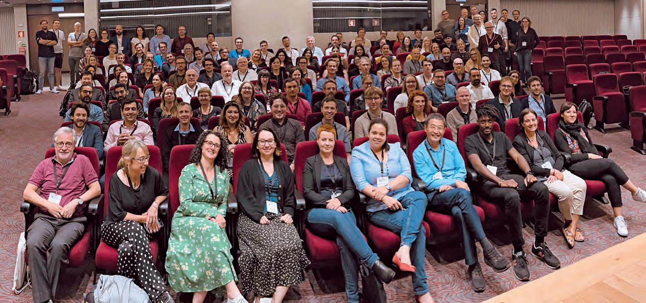
Before we all met in Lisbon, we had an online pre-workshop meeting. A welcoming introduction by the chairs with the motivations and planned outcomes was followed by talks from Dr Louise Murray (Leeds University/The Leeds Teaching Hospitals NHS Trust) and Dr Pauline Dupuis (Centre Léon Bérard) setting the scene. All participants introduced themselves and
A STANDARDISED WORKFLOW COULD IMPROVE CUMULATIVE DOSE ASSESSMENT
Dr Nick Hardcastle (Peter MacCallum Cancer Centre) introduced a dose accumulation task and survey. For this task, participants followed their clinical standard practice to assess the cumulative (physical and EQD2) doses to the different OAR, for a lung and a head and neck case that we were provided with. Our chairs also organised an interactive online mural, on which participants could add ideas for discussion, potential outcomes, as well as post-workshop outputs.
Once in Lisbon, we started the discussions with live polls related to re-irradiation – a nice icebreaker that created a positive group atmosphere. The analysis of the results of the dose accumulation task were presented at the workshop by Dr Nick Hardcastle. It showed variations in practice with regards to reviewing, summing, and
assessing cumulative re-irradiation doses, which correspond to outcome uncertainties. This study demonstrated that a more standardised workflow, using spatially registered doses, could potentially help improve consistency in cumulative dose assessment.
The first invited speaker, Dr Nicolaus Andratschke (University Hospital of Zurich), presented the European DELPHI consensus on how to define and report re-irradiation. The aim of this consensus is to provide guidelines and a standardised procedure on how to treat and report re-irradiation cases. Dr Andratschke stressed the importance of radiobiological considerations and described the clinical scenarios and considerations of reirradiation. In the DELPHI consensus, re-irradiation is defined as “a new course of radiotherapy either to a previously
E N ENDNOTES IPEM SCOPE 48 SPRING 2023
irradiated volume (irrespective of concerns of toxicity) or where the cumulative dose raises concerns of toxicity.”
Image registration techniques for dose accumulation were discussed by Dr Eliana Vasquez Osorio. The role of image registration in re-irradiation was noted whilst studies that showed organ-wise registration can improve results for large anatomical differences were introduced. Dr Vasquez then continued with registration uncertainties and how to quantify and account for them. Dr Ane Appelt talked about deformable image registration and EQD2 dose summation for pelvic re-irradiation. Examples of deformation in the pelvis were illustrated and a workflow integrating DIR and EQD2 dose scaling for evaluation of pelvic SABR re-irradiation was presented. A method to sum plans was also introduced, based on dose mapping using deformable image registration, considering organ / tissue specific alpha/beta and recovery factors.
The AAPM/ASTRO ontology and standards for data sharing in re-irradiation was also presented by Dr Charles Mayo. The work of the re-irradiation Special Medical Physics Consult (ReRT-SMPC) at the University of Michigan was introduced, a committee formed to develop a consensus-based approach and a standardised procedure for re-irradiation. Dr Mayo explained the importance of multi-institutional standardisation in creating representative databases and introduced the AAPM Operational Ontology for Radiation Oncology (OORO) (aapmbdsc.azurewebsites.net), formed to address additional gaps in data standards and provide basis for comprehensive multi-institutional registries. Dr Andrew Jackson’s talk about modelling reirradiation tolerances and discount factors using registry data focused on introducing a re-irradiation assessment model to determine the discount factor based on the time between irradiations. This model also consists of creating a “binary DVH” for each organ of interest using LQ-corrected doses for each treatment, discount the dose and create a plan sum, and finally use a fitted dose-volume model to assess the plan.
During the workshop, we held numerous constructive discussions, both between

presentations as well during the dedicated group discussions slots. Everyone had the opportunity to share their thoughts and opinions and the overall experience felt very inclusive. It was also quite useful to have vendors from different companies with us, as we had the opportunity to discuss currently available tools and share our thoughts on which software tools are more urgently needed. Bringing the workshop to a conclusion, we have addressed most of the objectives we set pre-workshop, whilst many ideas were laid on the table concerning re-irradiation aspects that need to be further investigated/ developed. Participants displayed great enthusiasm and willingness to further continue working on the re-irradiation aspects outlined in the workshop. This has led to the formation of working groups, with the aim to contribute towards standardising best practice in re-irradiation by organising educational events, gathering clinical constraints and workflow from different centres, producing guidelines on clinical workflow and best practice for dose accumulation in the re-irradiation setting as well as reviewing relevant papers.
This workshop has been a blast and I am very excited to continue working on standardising re-irradiation in the clinic. The chairs were very welcoming and worked hard to get as many people as possible involved. Any ideas or interest participants contributed to this journey were made very welcomed.
Andria Hadjipanteli, Medical Physicist, German Oncology Center, Limassol, Cyprus TOPIC: JUSTIFICATION AND OPTIMISATION OF KV IMAGING IN IGRT
Image-guided radiotherapy (IGRT) is the incorporation of imaging at the beginning of each radiotherapy treatment session. Its benefits are huge: (i) improving the geometric accuracy of patient positioning for radiation delivery; (ii) enabling highly conformal treatments; (iii) monitoring treatment target changes for potential adapted treatment. However, there are risks too: (i) secondary cancer; (ii) organ dose tolerance; (iii) precisely treating the wrong target. One of the main aims of this workshop was to discuss methods of optimising our imaging protocols so that we increase the benefits’ weight and lower the risks’ weight. In addition, there was the need to discuss the challenges of dose justification and recording, and how the imaging doses should be monitored and recorded in a radiotherapy department. The current status in the field was also presented and discussed.
The workshop was greatly organised and co-ordinated by the chairs Dr Nuria Jornet (Spain) and Georgios Ntentas (UK). Our group was formed by attendees from Australia, Cyprus, France, Germany, Holland, Spain, UK and the US. Most were radiotherapy physicists, but also radiology physicists, radiographers, researchers and industrial developers (BrainLab, Elekta, SunCheck, Varian) were present. During the two days all attendees were given the opportunity to give a pitch in order to share their work, thoughts, current status, issues on the topic and to raise questions to the developers.
The workshop kicked off with an invited talk by Dr Tomas Kron (Peter MacCallum Cancer Center, Australia). The talk stressed how imaging is becoming more and more frequent due to smaller and smaller planning target volumes (PTVs) and the importance of local optimisation in image-guided radiotherapy, as it is not
IPEM.AC.UK 49 SPRING 2023
EVERYONE HAD THE OPPORTUNITY TO SHARE THEIR THOUGHTS AND OPINIONS. IT FELT VERY INCLUSIVE
provided by manufacturers. Different typical imaging acquisition procedures available for image-guided radiotherapy, including electronic portal imaging devices (EPIDs), CT-on-Rails, kV-CBCT (linac integrated), MV-CBCT (linac integrated) were discussed by Prof George Din (Vanderbilt University School of Medicine, USA). During the workshop a nice explanation was given of how imaging dose (normally around 1-3% of therapeutic dose) is distributed differently in radiotherapy than in imaging, due to the beam energy difference. The usage of Monte Carlo for calculating the CBCT dose to one patient and incorporating it into his/her therapeutic dose were presented, even though its application is not yet commercially available. Also, techniques that can be used to reduce imaging dose, including the effect of combining different imaging protocols and techniques, reducing the imaged region of interest and optimising the imaging geometry, were presented.
Through several presentations and discussions that followed it was shown that there is great interest from many hospitals in adjusting the default CBCT protocols supplied by the manufacturer in order to decrease the dose but maintain the same image quality. This was shown to be possible. George Ntentas and Marina Khan from Guy’s and St Thomas’ NHS Trust (UK) presented their recently published work on the implementation of a comprehensive set of optimised CBCT protocols and the validation through imaging quality and audit. This study presents an efficient methodology developed and applied by physicists and radiographers, to optimise CBCT protocols by varying CBCT imaging
parameters (including patient size, tube current, exposure time, frames, gantry speed, trajectory, number of projections) and on-line and off-line image evaluation. As a side note it is worth remarking that this work also showed how a multidisciplinary approach can lead to effective methodologies.
Some talks focused on the optimisation of imaging protocols in radiotherapy specifically for paediatrics. We also discussed how we can create CBCT protocols for different sized patients (small, medium and large), in order to move a step closer to patient-specific requirements and personalised medicine. In addition, physicists from some hospitals made efforts to create protocols for achieving higher image quality than normal, in order to resolve small lesions, sacrificing dose in this case. But at what point does IGRT need to be incorporated into the treatment planning process? In AAPM TG-180 it was recommended that 5% of the therapeutic target dose should act as a limit, based on evidence from published
data. However, as it was discussed in the workshop, one might need to be careful with the absolute value of this dose and how it is estimated.
Dr Tim Wood (Hull University Hospital, UK) presented the first audits of CT imaging for radiotherapy treatment planning dose to patient in radiotherapy centres across the UK (an update by the IPEM working party), the outcome of which is available. National CBCT audits are also nearing completion and the results are due.
During the attendee’s presentations many questions or requests to the industrial representatives were raised. These mainly involved increasing the freedom available to the physicists (imaging parameters) in order to be able to fully optimise protocols. Some manufacturers already allow amendments to imaging parameters. The good news is that there are new versions of CBCT systems, software and automatic exposure control conditions are to be released by 2023–24 that will allow further optimisation by physicists and hopefully a more standardised approach amongst centres.
The concerns and interests, such as optimisation techniques, dosimetry techniques and future developments in the field, were common between the attendees. Therefore, going forward there will be collaboration to standardise methodologies and produce guidelines.
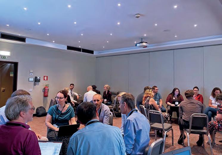
Closing remarks for the workshop
One can only gain by attending such workshops due to the small-group character they have, which encourages bravely speaking up, even for more junior scientists, as well as good coordination. The original aims of the workshop were certainly achieved. In both workshops we attended there was scientific excitement in the atmosphere and an ongoing lively discussion for the relevant topics shown from all attendees. Through the long discussions we managed to collect a lot of useful knowledge that will benefit our centre, patients in Cyprus and further afield through the guidance that will be developed. We recommend running and joining workshops like this. We are very appreciative to IPEM that we could attend such a motivating workshop.
E N ENDNOTES IPEM SCOPE 50 SPRING 2023
THERE WAS SCIENTIFIC EXCITEMENT IN THE ATMOSPHERE AND AN ONGOING LIVELY DISCUSSION
Member profile: Stuart Green


and development of practitioners in medical physics (and of other staff that contribute to our service areas, such as radiographers) so that the Clinical Scientists are able to split their role between tasks that are necessary for the service today, and developments to shape the service for tomorrow. Having time for developmental activities critically depends on having a strong and skilled workforce of practitioners to carry much of the day-to-day service load – and indeed to also play their part in driving developments.
What modern skillsets do you think are required in your role for a successful career?

I think everyone coming into a career now has to have a decent understanding of genomics and machine learning, but we must always remember that we are physicists, so those skills are the most important.
What accomplishment have you been most proud of in your career?

The period that I was President of the British Institute of Radiology was very rewarding. It came at a very difficult time for the Institute, but we made some good decisions and now, 12 years on, the Institute is thriving. But mostly I am proud of the department here in Birmingham and of each and every one of the staff that go above and beyond to do the best for our patients.
What is your typical day?


I get to work at around 8:30am and tackle my emails before making the first cuppa. Usually there are meetings (typically many meetings and these days mostly on Teams). In recent years I have tended to spend more time in discussions related to nuclear medicine than the other main service areas. Our focus has been to develop this service – which is already one of the largest in the country – and make it one of the very best. I try to leave work soon after 5pm but it doesn’t always work.
Which elements of your job do you like the most?
Working with people to drive our services forward so that we do better for patients, and through that to give opportunities for good experiences and general development of the ~150 staff in medical physics at UHB.


I have managed to build and maintain links also with some great colleagues in the University of Birmingham, and I very much enjoy collaborating with them to advance lots of areas of physics applied to medicine, but mostly with a focus on advanced radiotherapy techniques and technologies.


What are the biggest challenges – either for yourself or the sector?


Staffing. And that’s for our services in medical physics and for the NHS as a whole. The level of pay is an important factor if we are to continue to employ the excellent staff that we need.


If you could change one thing about the profession or area of specialty, what would it be and why?

We need to focus much more on the training


What are your predictions about the future of your profession and your area of specialty?


Medical physics is a strong and growing profession because we are uniquely placed to help hospitals deal with some of their greatest challenges. In radiotherapy I think we will find new ways of delivering dose to tumour cells, and better ways to combine with immunotherapy agents. Together I think these will change patient outcomes significantly.
What do you do in your free time?
I have an allotment – which is a great source of exercise and stress relief, and a 13-year-old daughter who I love doing stuff with. Recently, we have had solar panels and a home battery installed, and we are busy insulating our home to be ready for a heat-pump (as we will all have to do quite soon). It seems to take a lot of time!

IMAGE: ALAMY
My current role is Director of Medical Physics at University Hospital Birmingham (UHB).
E N ENDNOTES IPEM.AC.UK 51 SPRING 2023
I applied for the job after working closely with Prof Alun Beddoe during his time as Director. I had seen the great job that Alun did and I wanted to emulate him and to maximise the impact that I could have on the way we image and treat patients.
AN EXAMPLE FOR FUTURE LEADERS
LMIC award winner
Dr Iyobosa Uwadiae is a medical physicist in Nigeria and Secretary of the Nigerian Association of Medical Physicists. In 2020 she was the first recipient of the IPEM Low- and Middle-Income Countries (LMIC) Award, which recognises the challenges medical physicists and clinical engineers can face.

When I saw the call for applications for the LMIC Award, I was sceptical about applying, but was encouraged after reading about it and making clarifications on what it entailed. It turned out to be a very good decision. The aim of the award was to empower the recipient to develop professional activities in their country with the support of IPEM. At that time, there were several challenges facing medical physicists in Nigeria. The gap between what medical physics was, compared to what it could be, was very wide and there was an urgent
need to bridge it. After deliberating on a few project proposals, the decision to collaborate on the Harmattan School project was a no-brainer because we saw that it could be a way of solving the problem of the shortage of qualified medical physicists in the country by inspiring young physicists and engineers to pursue a career in medical physics.
The Harmattan School
The reception for the Harmattan School was beyond what we had hoped for. There was a genuine desire to know what medical physics was all about and how physics was applied in medicine. The NAMP-IPEM Harmattan School for Medical Physics kicked off in 2021 as a five-year project.
Due to the pandemic, the first two editions of the school were held online and this helped to extend the reach of the school beyond the shores of Nigeria.
I believe that the next great innovation in physics and medicine will come from scientists in the African region. The Harmattan School is one initiative that has sparked the fire. One of the major objectives of the school was to showcase the different applications of physics and engineering in medicine. The facilitators of the school were

E N ENDNOTES IPEM SCOPE 52 SPRING 2023
both from IPEM and NAMP and in 2022 we had two members from the American Association of Physicists in Medicine volunteer. We wanted participants to see where we were as a profession and where we would like to be, like IPEM. We also wanted to correct the impression most students have about not being able to get quality medical physics education or practice good clinical physics in the country, and finding good medical physicists to look up to. This is quite a common narrative in the region and
I WANT TO EXTEND OUR HEARTFELT APPRECIATION TO THE LEADERSHIP, STAFF AND MEMBERS OF IPEM
medical physics activities in their countries but lack the opportunity and resources. An initiative such as this award can go a long way to helping them. The impact of these ideas, no matter how small they may appear to medical physicists in the developed world, must not be underestimated.
It has been a wonderful two years spent under the mentorship of Professor Dan Clark OBE, who initiated the award. Through him I got to meet some of the most amazing medical physicists and clinical engineers, some of whom volunteered on the programme.
Yes, the award led to the creation of the Harmattan School and a collaboration between NAMP and IPEM but beyond that, it has set an example for future leaders and has sparked a fire in the hearts of the next generation of medical physicists here.

I want to once again, on behalf of NAMP, extend our heartfelt appreciation to the leadership, staff and members of IPEM who made this project a reality. NAMP looks forward to future collaborations with IPEM.
can be seen in the massive brain drain. I can say that there are many good physicists with brilliant minds who only lack the platform and opportunity to shine brightly.
Do not underestimate
The facilitators from NAMP were mainly those in their early careers that could serve as examples to students who needed to see a different side of the narrative. I know that there are others like me who are very passionate about promoting and developing
I also want to take this opportunity to challenge other similar medical physics organisations to take a cue from IPEM by collaborating with medical physics organisations in developing countries to build stronger professional bodies and empower future leaders. What you may count as “little”, can make a big difference somewhere. To the next recipient of this award, I say congratulations.
Applications for the next LMIC Award open at the Science, Technology and Engineering Forum in Glasgow.
IMAGE: © MATTHEW OMOJOLA DREAMSTIME.COM IPEM.AC.UK 53 SPRING 2023
Imagining Imaging R


eaders of Scope will not need reminding of the fundamental importance of physics as the bedrock on which all medical imaging modalities rely. However, as a working radiologist I have always been fascinated by the influence of innovations originating within the artistic realm on the construction and interpretation of medical images.
My book explores the collision of art and science within radiology from prehistory to the 21st Century.
Key radiographic techniques can be traced back to earliest human history; projection-based stencils in cave art, orthogonal views in ancient Egyptian portrayals of the body and tonal image inversion demonstrated by red-figure and black-figure Greek pottery in antiquity. The concept of a quantised, pixelated image utilised in cross-sectional imaging can likewise be traced back many centuries to Roman mosaics and in fabric weaving from
MY BOOK EXPLORES THE COLLISION OF ART AND SCIENCE
cultures across the globe. More recent developments including linear perspective during the renaissance and the use of multi-focal perspective by Cezanne and other post-impressionist painters have also been of profound importance in shaping medical imaging techniques. I also delve into the psychology and neurophysiology of image interpretation, including a detailed account of the human visual system. As someone whose livelihood is reliant on the accurate interpretation of images, it is sobering to realise the majority of what we “see” is generated by the visual cortex rather than the raw data entering the retina. The extent to which medical images can be regarded as objective anatomical truth is also scrutinised. What may be discarded as artefact in one examination may provide useful diagnostic information in another; image-smoothing algorithms employed in cross-sectional studies can legitimately be labelled “artistic licence”, and a host of conventions in image acquisition and display strongly influence how the body is framed. The business of what belongs in an image, and the angle
chosen to view the action, is not as straightforward as we might suppose.
Since the discovery of X-rays, artists and moviemakers have been inspired by radiographic concepts, and I examine this impact on visual culture in the final chapters of the book, including tropes from popular culture, such as the cartoon electric shock motif and X-ray glasses. Medical imaging has, in turn, continued to benefit from methods and technology developed by artists, film-makers and videogame creators, and I argue that the process of cross-pollination between the scientific and artistic realms must continue in the era of artificial intelligence.
Drawing upon a wealth of diverse visual references including astronomy, botany and cartography, Imagining Imaging is academically rigorous yet delivered in a readable and humorous style, and has drawn praise from contemporary artists and film-makers alongside radiologists and radiographers. I hope IPEM members will also enjoy this unique and personal take on medical imaging.
Dr Michael R. Jackson is a Consultant Paediatric Radiologist at the Royal Hospital for Children and Young People, Edinburgh. IPEM Members can get 20% off the price of the book on the Routledge website by using the code FLR40.

BOOK PITCH
Dr Michael R Jackson, Consultant Paediatric Radiologist, outlines the ideas behind and the content within his new book.
Imagining Imaging is published by CRC Press
IPEM SCOPE 54 SPRING 2023 ENDNOTES E N
BOOK PITCH


myQA® iON for Radiation Therapy Arrange a demo of myQA iON enquiry@osl.uk.com +44(0) 1743 462694 enquiry@osl.uk.com www.osl.uk.com myQA iON is a unique Patient QA software environment featuring an automated Analyse your results using advanced 3D tools such as DVH’s and clinical goals


RETURN ADDRESS: IPEM Fairmount House 230 Tadcaster Road York YO24 1ES UK
Physics in Medicine & Biology
development
and experimental
find out more about submitting, visit iopscience.org/pmb or e-mail pmb@ioppublishing.org. JOURNAL IMPACT FACTOR
iopscience.org/pmb Physics in Medicine & Biology (PMB) covers the
and application of theoretical, computational
physics to medicine, physiology and biology. Editor-in-Chief To
