THE FUTURE OF CT


MAGAZINE ADVANCING
PROFESSIONALS
IMAGING
PAGE 32
PAGE 26 AUGUST 2023 | VOLUME 7 | ISSUE 8 THEICECOMMUNITY.COM
PRODUCT FOCUS - AI


To learn more visit theimagingacademy.com or call 210.570.1452. WE ARE BUILDING THE NEXT GENERATION. As a student, you deserve more. THE WORLD MOVES FAST. WE MOVE FASTER.™ + powered by For the class schedule visit us at theimagingacademy.com. New Classes Begin February 13th Enroll Today! More versatile More valuable More job satisfied More upwardly mobile At Imaging Academy, we give you more.
1
Why choose Technical Prospects as your preferred parts provider?
Prompt Response, Friendly Service: When you reach out to us, you’ll speak with our team of experts that are here to help, with a smile!
3
2
Quality Parts: Our parts are tested on-site before they are sent to the customer, quality is paramount in all we do!
Expertise and Knowledge: Our skilled technicians possess extensive expertise across a wide range of equipment. They have the knowledge and experience to tackle even the most complex technical issues, ensuring precise diagnostics and effective resolutions.
5
4
Customized Solutions, Personalized Care: We understand that each facility has unique requirements. That’s why we take a personalized approach, tailoring our solutions to fit your specific needs. Our team will work closely with you to address your challenges and objectives.
Staying Ahead with Technology: We stay at the forefront of technological advancements. Our technicians leverage cutting-edge tools and resources to swiftly identify and address equipment issues, optimizing performance and minimizing downtime.
6
Exceptional Customer Care: Your satisfaction is our top priority. Our friendly staff will go the extra mile to ensure your needs are met and your questions are answered. We’re dedicated to building long-term relationships with our customers through exceptional service.
TECHNICAL PROSPECTS
877.604.6583 / 920.757.6583 | sales@technicalprospects.com | technicalprospects.com
Experts in Siemens Medical Imaging
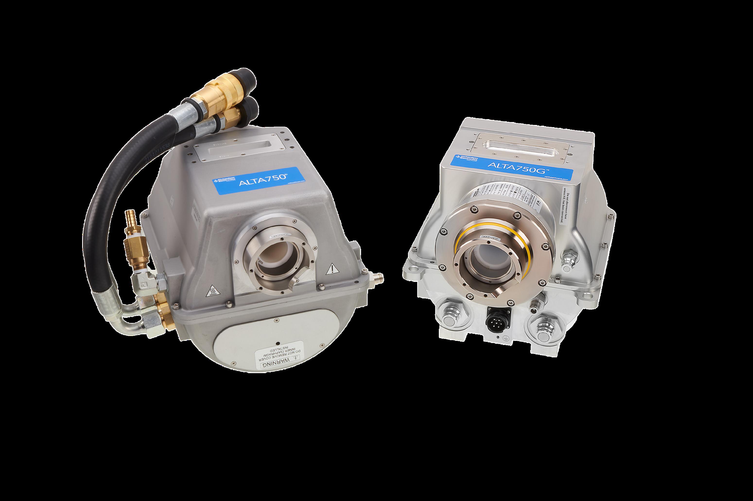
THE ALTA750 SERIES ®
OEM-QUALITY CT REPLACEMENT TUBES FOR LESS
®
Richardson Healthcare engineers form-fit-function CT replacement tubes to enable service providers to improve efficiency and lower the overall cost of healthcare delivery. With the launch of the ALTA750G and our legacy product, the ALTA750 , we proudly expand our CT tube replacement solutions for Toshiba/Canon* Aquilion Scanners ®
LOWER COSTS & MAXIMIZE CT UPTIME - CONTACT US FOR A QUOTE!

ALTA750 compatible on:
Aquilion 4*
Aquilion 8/16*
Aquilion 32/64*
Aquilion 16 Large Bore*
Aquilion CXL*
Aquilion RXL*
Aquilion VeloCT*
Aquilion Prime Gen 1*
ALTA750G compatible on:
Aquilion Prime S*
Aquilion Prime SP*
product and company names are trademarks™ or registered® trademarks of
respective holders Use of the trademarks is solely for identification purposes and does not imply any affiliation with or endorsement by the trademark holders
*All
their
® ®
R E L L H E A L T H C A R E . C O M H E A L T H C A R E @ R E L L . C O M 7 0 4 . 7 3 9 . 3 5 9 7
FEATURES
DIRECTOR’S CUT
We have reached the end of an era. Dr. Threasa Frouge, MD, has served as Banner Imaging’s chief medical officer since 2019 and will retire this month.

COVER STORY
Computed tomography (CT) imaging is an invaluable diagnostic tool and continues to grow along with technological advances.

RISING STAR
Chad Behrens, R.T.(R)(CT), is a rising star within Banner Imaging and the overall imaging field.
14 36 32 ADVANCING THE IMAGING PROFESSIONAL 6 ICEMAGAZINE | AUGUST 2023
IMAGING NEWS
Catch up on the latest news from around the diagnostic imaging world.
PRODUCT FOCUS
A look at some of the latest AI solutions for diagnostic imaging.


EMOTIONAL INTELLIGENCE
A good leader recognizes the fundamental areas of responsibility that should be overseen, and what must be done in each of those areas.

AUGUST 2023
18 26
42 ICEMAGAZINE 7 WWW.THEICECOMMUNITY.COM
CONTENTS SPOTLIGHT 10 In Focus Kennith Crump 12 Rad Idea Communication Is Key 14 Rising Star Chad Behrens 16 Off the Clock Traci L. Foster NEWS 18 Imaging News A Look at What’s Changing in the Imaging Industry 24 Webinars Webinar Shares Benefits of a 3D Lab PRODUCTS 25 Market Report 26 Product Focus Artificial Intelligence INSIGHTS 36 Director’s Cut End of an Era 38 PACS/IT The Time Has Come for a New Ambassador for AI 40 Coding/Billing Are You Appropriately Capturing All Your Visits? 42 Emotional Intelligence Essential Leadership Responsibilities 45 Roman Review Expert Body Language 49 ICE Break 50 Index ICE Magazine (Vol. 7, Issue #8) August 2023 is published by MD Publishing, 1015 Tyrone Rd., Ste. 120, Tyrone, GA 30290. For subscription information visit www. theicecommunity.com. The information and opinions expressed in the articles and advertisements herein are those of the writer and/or advertiser, and not necessarily those of the publisher. Reproduction in whole or in part without written permission is prohibited. © 2023 MD Publishing 1015 Tyrone Rd. Ste. 120 Tyrone, GA 30290 Phone: 800-906-3373 President John M. Krieg john@mdpublishing.com Vice President Kristin Leavoy kristin@mdpublishing.com Vice President of Sales Jayme McKelvey jayme@mdpublishing.com Group Publisher Megan Cabot megan@mdpublishing.com Editorial John Wallace Editorial Board Jason C. Theadore Nicole Dhanraj Melody W Mulaik Verlon E. Salley Rachel Thiesse-Yount Traci Foster Sales Emily Hise Art Department Karlee Gower Taylor Hayes Kameryn Johnson Events Kristin Leavoy Webinars Linda Hasluem Digital Department Cindy Galindo Kennedy Krieg Haley Wells Accounting Diane Costea 8 ICEMAGAZINE | AUGUST 2023 ADVANCING THE IMAGING PROFESSIONAL


626 acquisitions to expand our services to our customers To learn more visit WeAreISS.com or call 888.667.1062 The world moves fast. We move faster. Call our contrast injector specialists today to find out how we can save you money! • On-Site Repair • Depot Repair • Parts Sales and Parts Identification • Loaner Availability • Technical Support • Training • Injector System Sales • Preventative Maintenance Tools
KENNITH CRUMP
FOCUS IN
BY JOHN WALLACE
Connally Memorial Medical Center (CMMC) Imaging Director Kennith Crump, MS, CNMT, RT(R), always seemed to know he was destined to work in health care. It is a rewarding career, and he looks forward to each day.
Crump joined CMMC in January of this year after more than six years at Longview Regional Medical Center where, as the director of operations, he managed 58 full-time employees and directed daily department operations including fiscal management and technology evaluation for equipment acquisition and service. He maintained regulatory and
accreditation compliance for radiology, cardiac cath and vascular lab. He also prepared the department for state, ACR and Joint Commission regulations and inspections.
Prior to his tenure at Longview Regional Medical Center, Crump served as the operations manager at BE Smith within Southern Illinois Healthcare. He was responsible for managing radiology departments at three locations: Carbondale Hospital, Herrin Hospital and St. Joes Hospital in Carbondale, Illinois.
During his time at InSight Health Services Corp. in Fort Worth, Texas, from September 2005 to December 2011, he made significant contributions to the organization’s success.
He served in the U.S. Army National Guard from 1988 to
SPOTLIGHT
Imaging Director Kennith Crump joined Connally Memorial Medical Center earlier this year.
10 ICEMAGAZINE | AUGUST 2023
1994, participating in Operation Desert Storm and earning the prestigious Gulf War Medal. He is a Certified American Registry of Radiologic Technologist (ARRT) and a Certified Nuclear Medicine Technologist (CNMT), highlighting his specialized expertise in the field.
Crump’s unique educational background, diverse experience and remarkable achievements position him as a highly skilled and dedicated radiologic and nuclear medicine technology professional. His contributions to health care organizations have significantly impacted patient care, and his commitment to excellence and continuous professional development set him apart in his field.
The health care field was a must and once he discovered imaging he knew where his future would take him.
“From a young age, I desired a career in health care. Imaging science genuinely sparked my interest,” Crump explained.
KENNITH CRUMP
MS, CNMT, RT(R)
1. Favorite movie? “The Godfather”
“CMMC is made up of an exceptional team of health care professionals. In imaging, every day is a diverse experience, keeping it interesting,” he added when asked why he loves his job.
As a leader, Crump is focused on doing tasks the right way and modeling behaviors and a work ethic for others to replicate.
“In this field, leading by example is highly efficient,” he said.
He, after all, learned from those who came before him including mentor Brandon McDowell.
“Our CMMC Chief Operating Officer Brandon McDowell has significantly assisted my transition into the CMMC Imaging Director role,” Crump said. “I am a new addition to the CMMC team, so I was quickly astonished by how incredible this rural health care system is!”
“I’m very grateful for this opportunity,” he added. •
2. What is something most of your coworkers don’t know about you? In my youth, I had a severe speech delay that five years of speech therapy corrected.
3. What is one thing you do every morning to start your day? Coffee, of course.
4. Best advice you ever received? Temporary setbacks are blessings in disguise.
5. Who has had the biggest influence on your life? My father is the most honest person I have known.
6. What would your superpower be? Mind reading, but the ability to turn it on and off.
7. What are your hobbies? Playing golf and following LSU Tiger sports. Geaux Tigers!
8. What is your perfect meal? Being from Louisiana, I must have my red beans and rice with cornbread.
ICEMAGAZINE 11 WWW.THEICECOMMUNITY.COM
Rad idea
BY AMY RUTLEDGE, MBA, RDMS
COMMUNICATION IS KEY
The cost analysis of retaining employees’ verses recruiting new ones has been well documented . Simply put, it is financially advantageous for institutions to retain their employees rather than to recruit new ones. So, what then are some strategies to engage and retain employees? As rudimentary as it sounds, communicate! Meaningful and transparent communication should be the bedrock of any retention strategy. One tool in the leadership toolkit should be leadership rounding. Leadership rounding is the consistent practice of asking open-ended, targeted questions to key stakeholders – to obtain actionable information.
Leadership rounding focuses on:
• Establishing Relationships – “What did you think about the game last week?”
• Cultivate Victories – “What is working well in your department?”
• Identify Opportunities for Improvement – “Do you have any ideas to improve patient safety?”
• Repair/Replace systems – “Do you have the tools you need to do your job?”
• Building a Culture of Recognition – “Is there anyone I should recognize?”

Relationship-building questions during rounding help open channels of communication while also demonstrating to employees that their leaders value them as a complete individual. Additionally, 1:1 rounding allows the perceptive leader to gauge the employees’ level of stress by not only listening but also watching for
non-verbal cues. This may give the leader some actionable items to follow up on.
Many health care employees tend to notice and speak out when something is not working well, which is without a doubt necessary for quality patient outcomes. As leaders, we want to encourage that as a way identify opportunities for improvement and find systems that may need repair. At the same time, we also need to load balance and create a culture where our employees recognize what is working well in the department. We want to overcome the obstacle of always focusing on the negatives and cultivate some victories with open ended questions like “Who has been really helpful lately?” or “What is working well”?
Rounding also builds a culture of recognition. Employee recognition is always more meaningful when it is timely and tied to a specific accomplishment. Rounding allows the leader to gain that employee-level information that they may not otherwise have and recognize that employee in an authentic way.
Lastly, rounding is to assist leaders in gaining actionable information. Once the information is obtained, it is imperative that we are committed to assisting staff resolve issues and are willing to provide follow-up. Closing the communication loop or tracking the actionable item allows the staff to see how their concerns are addressed. This, in turn, will improve employee retention and engagement, which drives better service, improved financials and growth.
– Amy Rutledge, MBA, RDMS, is manager of the UHCMC Department of Radiology. •
Share your RAD IDEA via an email to editor@mdpublishing.com.
SPOTLIGHT
ADVANCING THE IMAGING PROFESSIONAL 12 ICEMAGAZINE | AUGUST 2023
Helping students advance their careers through knowledge, education and hands-on, technical learning.
“Having completed Phase I: Principles to Servicing Diagnostic X-Ray Systems at RSTI was a large factor in getting selected for my current position with Mayo Clinic in the Medical Imaging Department.”
“RSTI teaches you how to work on a modality, not single machines…learning the theory then the process of how the machine works and how to service it gives you a good foundation and makes you ready for any model.”
COMPLETE SUPPORT PARTNERFOR OUR SERVICE CUSTOMER:

Complete list of classes that we offer can be found at rsti-training.com
ISO 9001:2015 CERTIFIED
CERTIFICATE NO. Q-1158) STATE OF OHIO REG. NO. 93-09-1377T
(IQC
• Training
Imaging
Service
Technical
Equipment
(440) 349-4700 parts@rsti-exchange.com registration@rsti-training.com CONTACT US:
LEARNING
TRAINING THE INDUSTRY SINCE 1985 – ALMOST 40 YEARS! Visit us at these shows! Education & Exhibiting: CABMET August 3-5 | NCBA August 15-17
•
Parts •
Support •
Support •
Options
ENGINEERED FOR
™
– Tim Z., RSTI Alumnus
– Alex P., RSTI Alumnus
RISING STAR CHAD BEHRENS
Chad Behrens, R.T.(R)(CT), holds an associate degree in applied science and serves as the site manager for Banner Imaging Skyline/McKee Breast Center. He is a rising star within Banner Imaging and the overall imaging field. ICE magazine recently found out more about Behrens and his imaging journey.

Q: WHERE DID YOU GROW UP?
A: I grow up in a small town called Merino in northeastern Colorado
Q: WHERE DID YOU RECEIVE YOUR IMAGING TRAINING/EDUCATION?
A: I attended McCook Community College in Nebraska for my pre-requisite classes and then transferred to Southeast Community College to complete the radiology technologist program. I graduated with an Associates of Applied Science. I started working the day after graduating at Haxtun Hospital in Colorado where I received training in computer tomography and received my Colorado computer tomography operator’s license. After a couple of years, I became a certified CT technologist.
SPOTLIGHT
14 ICEMAGAZINE | AUGUST 2023 ADVANCING THE IMAGING PROFESSIONAL
Chad Behrens, R.T.(R)(CT), serves as the site manager for Banner Imaging Skyline/ McKee Breast Center.
Q: HOW DID YOU FIRST DECIDE TO START WORKING IN IMAGING?
A: I was an accident-prone child and spent more time in the ER than most kids, this allowed me firsthand experience in the imaging world. This definitely helped pique my interest in the medical field. On a job shadowing event in the ER, I got to shadow the imaging department and that sealed the deal for me.
Q: WHY DID YOU CHOOSE TO GET INTO THIS FIELD?
A: I have always enjoyed helping people and I like to be part of the action. On my job shadowing event, I was there to be an ER nurse but after the trauma and chaos was over there was just charting and typing. At that time, I would say my typing skills were nonexistent, so charting for multiple hours didn’t seem the best fit. I noticed the imaging department was involved in every trauma event I encountered that day. I asked if I could shadow them for a little while. After a couple of hours shadowing the imaging department, I knew this was the direction I wanted to take.
Q: WHAT DO YOU LIKE MOST ABOUT YOUR POSITION?
A: I would say I love my team and the fact that everyday there is a new situation or issues that arise that need a solution. I love being part of the solution and thinking outside of the box to figure it out. This allows me to grow and learn something new daily.
Q: WHAT INTERESTS YOU THE MOST ABOUT THE IMAGING FIELD?
A: The fast pace and the constant change, whether with each individual patient or with our technology constantly advancing.
Q: WHAT HAS BEEN YOUR GREATEST ACCOMPLISHMENT IN YOUR FIELD THUS FAR?
A: My greatest accomplishment has been my ability to have fellow staff grow in the imaging field. Over the course of my management career, I have had the pleasure of doing this in many different settings; coordinating and planning out trainings for staff to be crossed trained into different modalities, creating succession planning and training for our new leads and future managers.
Q: WHAT GOALS DO YOU HAVE FOR YOURSELF IN THE NEXT 5 YEARS?
A: I plan on receiving my CRA and continue to grow as a leader with Banner Imaging and eventually become an associate director for the Colorado locations. I want to help Banner Imaging expand in Colorado with additional locations and be a key member in the startup/expansion. •
FUN FACTS
FAVORITE HOBBY: Spending time with my family playing cornhole and horseshoes.
FAVORITE SHOW: I don’t watch a lot of shows besides little kid cartoons with my family, but anything that has Adam Sandler in it I’m going to watch.
FAVORITE FOOD: Cheeseburgers
FAVORITE VACATION SPOT: Anywhere tropical
SOMETHING ON YOUR BUCKET LIST: Skydive and travel to Australia
SOMETHING YOUR CO-WORKERS DON’T KNOW ABOUT YOU: Three-time state runner-up in wrestling. I was in a car wreck my sophomore year in high school and I don’t remember much prior to that.
ICEMAGAZINE 15 WWW.THEICECOMMUNITY.COM
Clock Off THE
TRACI L. FOSTER
 BY MATT SKOUFALOS
Traci Foster’s path to a medical imaging career wasn’t a direct one, but it’s ended up getting her exactly where she wanted to be. After completing an undergraduate degree in elementary education, Foster returned to her hometown of Rochester, Minnesota, to attend radiology school at the Mayo Clinic. She graduated, worked at the Mayo Clinic for a year, and then relocated to Texas, where today, she is the director of radiology and radiation oncology at the Houston-based St. Joseph Medical Center.
“I certainly didn’t have a plan for what I wanted to do when I grew up,” Foster said. “It just kind of fell that way. I do it for the patients. I do it for the staff. I love working with people.”
In comparison, Foster’s career as a live performer was far more considered, and no less accomplished. At eight, she picked up the violin — despite wanting to learn the viola, her hands were too small for the instrument when she started — and never looked back.
By high school, Foster’s musical career had taken her through a variety of performance opportunities, including those with the Rochester, Minnesota Symphony, with which she and a classmate were just the second and third high-schoolers to play in the history of the orchestra.
“That was a huge learning experience for me,” Foster said. “In my previous experience in high school, you get music and you have several months to learn these pieces before you perform them. In symphony, it’s a lot of sight-reading, and we’re performing it
next week. It’s a lot more pressure, and an amazing experience to play along with other professionals and learn from them.”
In college, Foster stepped up to join the Fargo-Moorhead Symphony Orchestra, a community orchestra comprising players from Fargo, North Dakota, and from Moorhead, Minnesota. In her experiences there, she had the opportunity to play in an opera company pit orchestra as well, which was a new and distinct challenge.
“It’s completely different than being in a symphony,” Foster said. “If you make a mistake, it’s a little more obvious.”
In high school, Foster toured Europe with her performance group; in college, she toured Canada. But after she graduated, Foster also enjoyed a distinct experience through the Fargo-Moorhead Symphony: playing back-up to 1960s majestic rock act, The Moody Blues.
The British band frequently performs with the backing of an orchestra, but in lieu of touring America with one, they would pick up players in every town along the way, rehearse the repertoire, and then run the performance live. Each of the orchestra players’ instruments were individually amplified — which meant, nerve-rackingly, that any mistakes would be as well.
For as much as her musical career had offered her to that point, Foster hadn’t experienced anything like it before.
“It was an amazing experience,” she said. “The rehearsals, you can really tell, they jelled. They can communicate with body language; just a look at each other, and they’re going to do that refrain again, or ‘Let’s keep going because the audience is really liking this.’ ”
“Watching that happen, and the talent that they
ADVANCING THE IMAGING PROFESSIONAL 16 ICEMAGAZINE | AUGUST 2023
SPOTLIGHT
have, it was an experience I will never forget,” Foster said. “It was a big undertaking for them. For me, it was a once-in-a-lifetime opportunity to be able to see how such a successful band comes together, and can put on this fabulous show with a bunch of strangers getting together a few hours beforehand. I carry my backstage pass in my violin case still.”
While participating in the Fargo-Moorhead Symphony, Foster and a few of her fellow players created a quartet based around a violist who wrote original compositions and arranged other well-known radio hits for a chamber ensemble. The group booked all manner of events, from weddings to parties, and Foster herself also taught private violin lessons to about 30 students, of all ages, from her own studio.


“We’d give recitals,” she remembered; “I really enjoyed that, and being able to see them progress, which gave me a lot of pleasure.”
Today, Foster has become a student of music again, taking lessons on the piano, and having fun amid the effort of training her brain to think in terms of a different instrument altogether. Playing the piano involves reading musical notation on two staffs — bass and treble clef — whereas playing the violin involves only one of these at once. Now, when she joins a recital, it’s as a beginner among others. Her aims are to reach a proficiency that will allow her to simply sit down and play in public, be it classical or popular music.
“It’s so interesting to sit back and be a student again,” she said. “Musically, my goal is to become as comfortable on piano as I was on violin. I just need more time and experience with it to really feel comfortable sharing it with strangers. I love my teacher. He’s very well-versed, and I’m learning much more of a variety of styles.”
When she’s not working or honing her chops, Foster is also an avid runner who’s completed more than 20 marathons, several half-marathons, and an Ironman
triathlon, which involves running, cycling and swimming. She said she’s “gotten the races out of [her] system, and the medals,” and now enjoys simply discovering new and fun places to enjoy running outside.
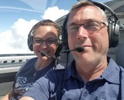
For fun, Foster and her husband, Darryl, enjoy taking trips in their Van’s Aircraft RV-7, a two-seat, single-engine airplane that Darryl built and maintains himself. In it, the couple has the flexibility to travel across the country for sightseeing, date nights or to visit any of the children in their blended family.
She still plays violin for special services at her community church, and, occasionally, for audiences at the hospital. Although Foster isn’t involved with a symphony at the moment, her work in radiology keeps her close to patients, which she said allows her to “make the best difference and the biggest difference.” •
 Traci Foster performs duets at Texas Children’s Hospital.
Traci Foster and her husband, Darryl, are seen flying in a homemade RV-7.
Traci Foster performs duets at Texas Children’s Hospital.
Traci Foster and her husband, Darryl, are seen flying in a homemade RV-7.
ICEMAGAZINE 17 WWW.THEICECOMMUNITY.COM
Traci Foster is seen with children (from left to right) Ian, Traci, Elexis and Adam.
Imaging News
A LOOK AT WHAT’S CHANGING IN THE IMAGING INDUSTRY
GE HEALTHCARE ADVANCES PET/MR CAPABILITIES
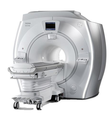
GE HealthCare unveiled SIGNA PET/MR AIR, at the Society of Nuclear Medicine and Molecular Imaging (SNMMI) 2023 annual meeting. The company showcased the integration of its advanced AIR technologies with the SIGNA PET/MR AIR system, to enhance diagnostic precision, simplify treatment evaluation, while elevating patient comfort.
Recent FDA approvals of new PET radiotracers and therapeutic approaches for high-prevalence diseases, such as prostate cancer and Alzheimer’s Disease, emphasize the need for reliable and comprehensive imaging solutions throughout the patient care journey. The unique AIR technologies from GE HealthCare incorporated into SIGNA PET/MR AIR address the evolving demands of these patient populations. These technologies include AIR Coils, which enhance and improve patient comfort, AIR Recon DL, which improves MR image quality and enables scan time reduction, and MotionFree Brain, which mitigates motion-related PET image degradation.
Prostate cancer is the second most prevalent cancer in the world. Recently, prostate cancer care has undergone a transformation with the introduction of prostate-specific membrane antigen (PSMA)-targeted radiotracers for imaging diagnostics and theranostics, which combines diagnosis and therapy. The integration of PSMA-targeted PET radiotracers with GE HealthCare’s high sensitivity PET and the anatomical precision of MRI in SIGNA PET/MR AIR holds the potential for precise diagnosis and staging/re-staging of prostate cancer. Additionally, it can aid in the selection of patients for subsequent PSMA-targeted radioligand therapy.
SIGNA PET/MR AIR delivers deep learning based MR image reconstruction with AIR Recon DL, helping to ensure both improved image quality and up to 50% in reduction of scan time. GE HealthCare’s AIR Coil technology effortlessly conforms to the shape of the human body, enhancing the quality of MR imaging while enhancing patient comfort. This technology offers a thinner, lighter solution, allowing for more accurate PET quantitation in contrast to conventional rigid RF coils.
In other news, GE HealthCare has entered into a distribution agreement with DePuy Synthes, the orthopaedics company of Johnson & Johnson, to bring GE HealthCare’s OEC 3D Imaging System, in conjunction with DePuy Synthes’ extensive product portfolio, to more surgeons and patients across the United States. This new collaboration demonstrates GE HealthCare’s ongoing commitment to bring imaging technologies to clinicians providing patient care through the practice of some of health care’s most complex spine procedures. •
NEWS
ADVANCING THE IMAGING PROFESSIONAL 18 ICEMAGAZINE | AUGUST 2023
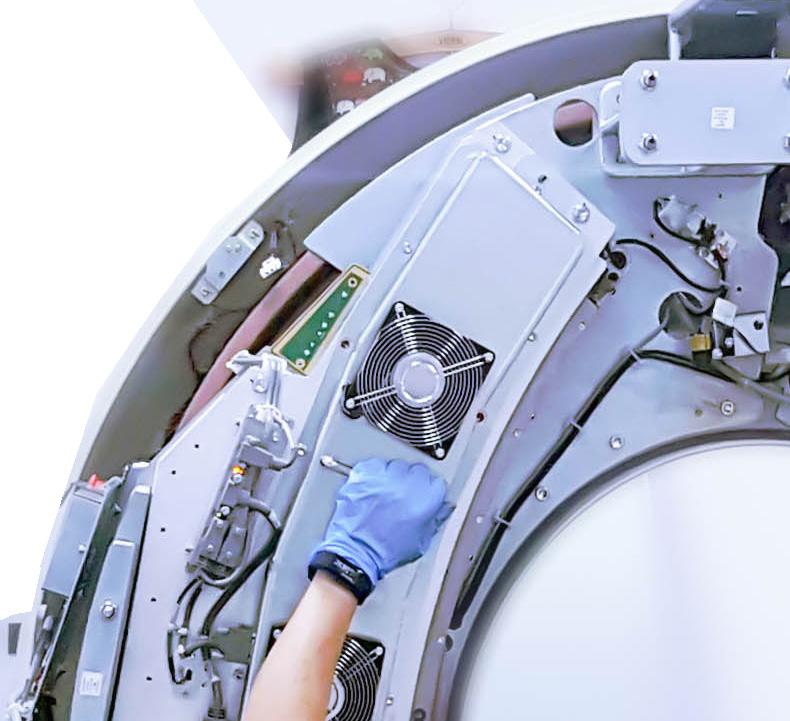
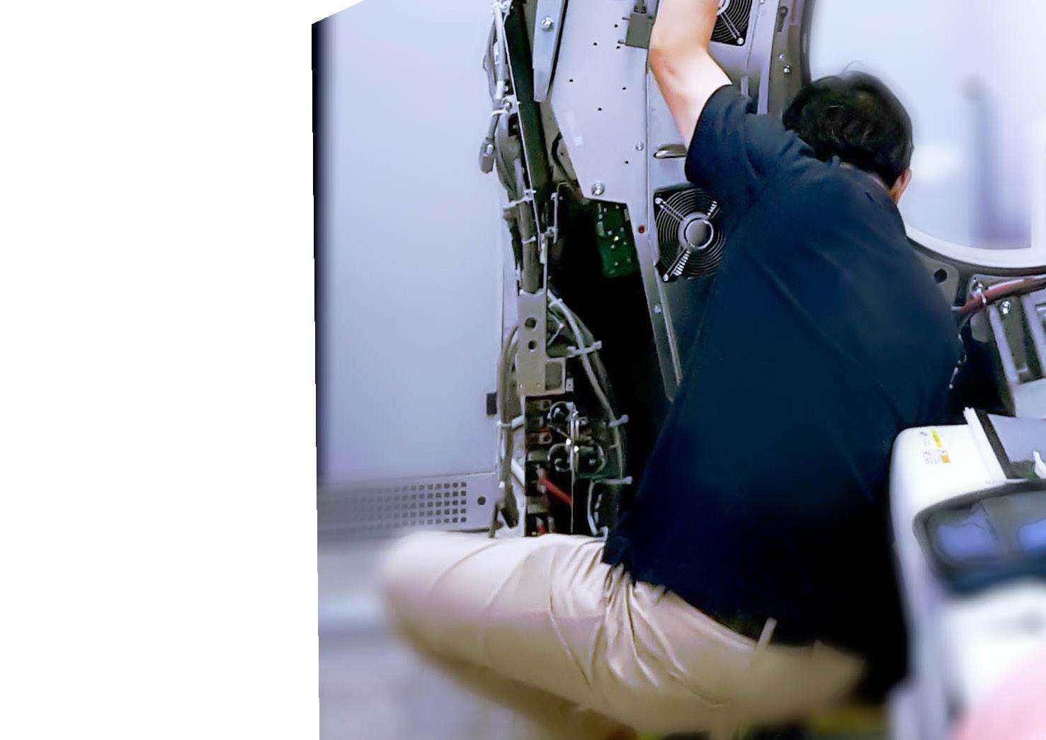
RSTI CELEBRATES TRAINING MILESTONE
Radiological Service Training Institute (RSTI) recently celebrated a record-breaking enrollment milestone! It is said that a picture is worth a thousand words and the video crew, photographers and drone footage captured that and more. Surpassing enrollment numbers since their doors opened almost 40 years ago, was a special moment to capture
“We appreciate the opportunity to support the training needs of the imaging service industry,” RSTI CEO Todd Boyland, CRES, CPSM, said. “It was equally as fun getting to share this with the RSTI team and the group of students in classes that week too.”
Since 1985, RSTI has provided diagnostic imaging training. Offering a wide selection of over 60 courses each year, RSTI has trained more than 15,000 service professionals from over 50 countries in radiology, mammography, MR, CT, ultrasound, networking, PACS and DICOM. •
BANNER IMAGING IS HIRING


Come experience everything Arizona and Colorado have to offer. Life doesn’t get much better than 300 days of sunshine, hiking, biking, lakes, rivers, and so much more. We invite you to our out patient imaging centers where your feedback and life work balance are valued.


apply today at bannerhealth.com/careers

ICEMAGAZINE 19 WWW.THEICECOMMUNITY.COM
SIEMENS HEALTHINEERS DEBUTS BIOGRAPH VISION.X
At the 2023 Annual Meeting of the Society of Nuclear Medicine & Molecular Imaging (SNMMI), Siemens Healthineers debuted the Biograph Vision.X, a next-generation positron emission tomography/computed tomography (PET/CT) scanner that builds on the established performance of the Biograph Vision and delivers a time of flight (TOF) of 178 picoseconds (ps) — the industry’s fastest TOF.
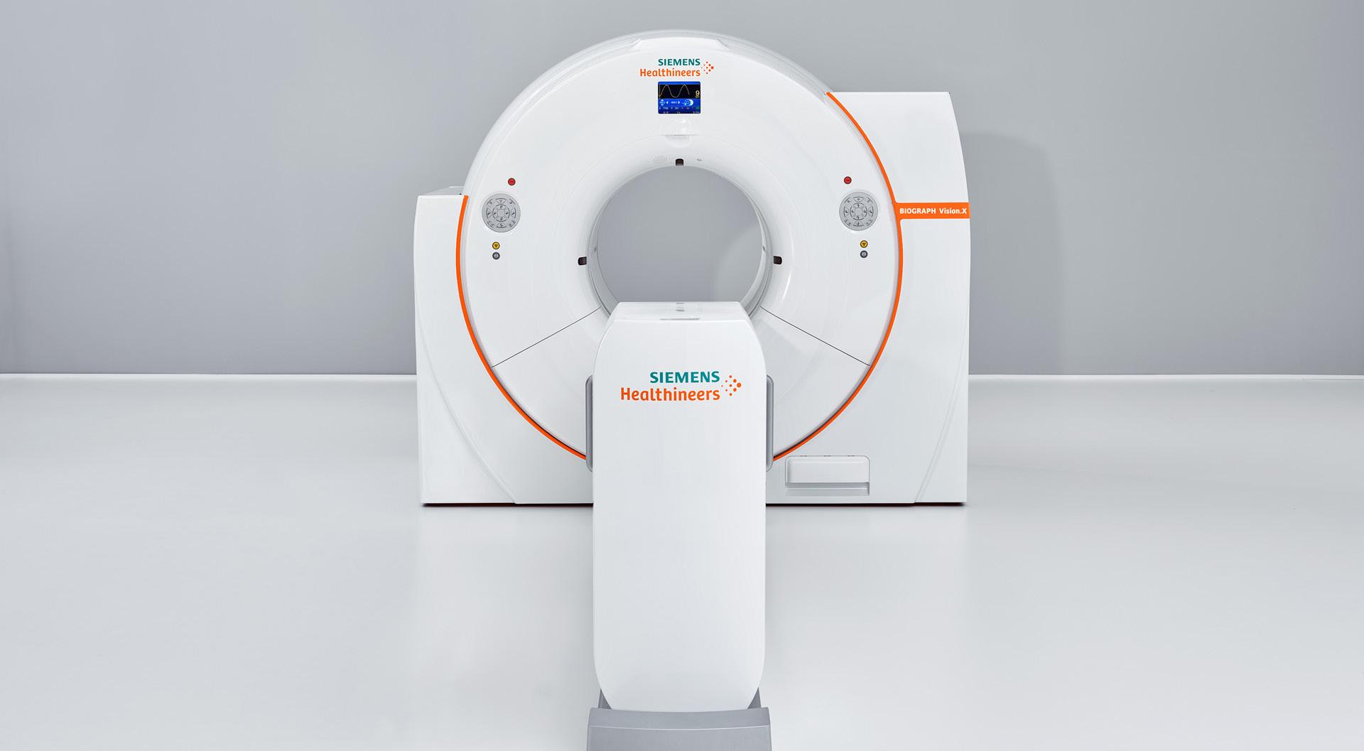
The Optiso Ultra Dynamic Range (UDR) detector technology of the Biograph Vision.X is based on silicon photomultipliers (SiPMs), which enables the use of small 3.2 mm x 3.2 mm lutetium oxyorthosilicate (LSO) crystal elements to deliver higher spatial resolution than larger crystals. Leveraging these extremely small LSO crystals that are 100 percent covered by SiPMs, the scanner delivers high 48-mm volumetric resolution and its industry-best temporal resolution of 178 ps. For these reasons, the Biograph Vision.X can deliver a 20 percent performance improvement3 that can lead to improved patient throughput as well as reduced patient radiation exposure and radiotracer cost.
“The Biograph Vision.X PET/CT scanner builds on the strong foundation of our Biograph Vision system and demonstrates our ongoing commitment to being the industry leader in time of flight for improved lesion detection and increased anatomical detail,” said James Williams, Ph.D., head of Siemens Healthiners Molecular Imaging.
“This speed translates into true increased sensitivity for better imaging performance. This level of precision is more important than ever, as more physicians now use PET/CT imaging not only for diagnosis but throughout the patient care cycle – from diagnosis to therapy monitoring to post-therapy follow up. And such precision in PET/CT is invaluable in theranostics, where diagnostic and therapeutic capabilities are combined to help treat post-recurrent prostate cancer patients.”
The AIDAN intelligent imaging platform of the Biograph Vision.X leverages artificial intelligence (AI) to increase operational efficiency and create an accelerated patient workflow for a more precise diagnosis. AIDAN’s unique methodology is built on more than 700 machine- and deep-learning patents from Siemens Healthineers, which was the first company to bring AI-powered applications to PET/CT scanners.
The large 78-cm bore of the Biograph Vision.X can reduce patient anxiety and enable easier positioning for radiotherapy devices or bariatric patients. The scanner fits into any room that houses a PET/CT scanner from the Biograph Vision family.
The Biograph Vision.X will be available as an in-field upgrade to current users of the Biograph Vision, allowing them to extend the performance of their current system. •
NEWS
ADVANCING THE IMAGING PROFESSIONAL 20 ICEMAGAZINE | AUGUST 2023
ALL-TOUCHSCREEN SONOSITE ST POCUS SYSTEM ANNOUNCED

A world leader in point-of-care ultrasound (POCUS) solutions has added to its portfolio of systems with the launch of Sonosite ST. Designed with the busy proceduralist in mind, Sonosite ST offers a new 21-inch all-touchscreen interface, a large 10” by 7.5” image area, automated setting optimization for each exam type, Auto Steep Needle Profiling (SNP) and more. Sonosite ST also shares the same family of transducers with Sonosite PX and Sonosite LX to support compatibility across systems.
“As the company that invented POCUS we’ve spent the last 25 years working with clinician customers to innovate and advance the use of ultrasound at the patient bedside,” said FUJIFILM Sonosite President and CEO Rich Fabian. “That collaboration has once again delivered results in the form of a system designed for proceduralists, something engineered to help them save time, increase confidence and improve outcomes.”
“At a time when hospitals are facing unprecedented clinical staffing challenges, we believe Sonosite ST has the potential to improve both the clinician and patient experience,
while at the same time enabling increased throughput,” Fabian said. “I’m very confident the Sonosite ST will become an industry workhorse for procedure-focused clinicians.”
Sonosite ST features optimized exam types for the POCUS-guided procedures used most often, including nerve block administration in regional anesthesia, vascular access in emergency medicine and joint injections in musculoskeletal care. This enables proceduralists to rapidly access the appropriate exam types for their procedures at the touch of a button, allowing them to surface the most needed optimizations, annotations and calipers to simplify their workflow
“The value of POCUS has been clearly demonstrated in my care delivery and I’m thrilled to see it becoming standard practice for needle-based interventions across a variety of health care specialties including anesthesia, vascular and musculoskeletal care and beyond,” says anesthesiologist, Dr. David Auyong. “I look forward to Fujifilm’s new Sonosite ST, which will take the value of POCUS-guidance a step further for proceduralists by prioritizing and streamlining access to critical exam types with amazing image clarity.” •
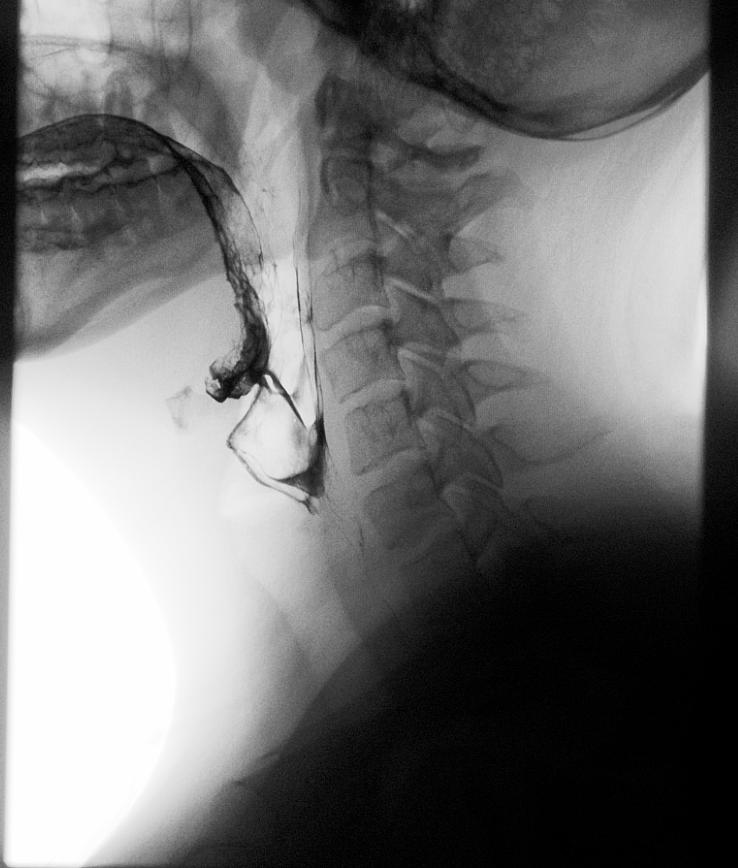
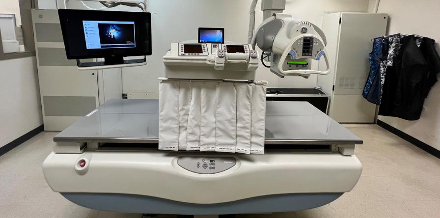
- New Windows 10 Platform with Dynamic Digital DR Panel - Eliminates the OEM Saturn Windows 95 Platform - Dose Reduction - HIPAA Compliant, 510k approved. - Fluoroscopy digital recording loops for MBS studies - Upgrade Provided to your Proven R/F System in 4 days. - Complete Refurbished Systems Available Imaging Chain issues with your GE Legacy or Precision-500 R/F systems? Upgrade system with Insight DRF Enhanced BRIAN.CARLOCK@PREMIERIMS.COM | 800.722.1991 RADONMEDICALIMAGING.COM
ICEMAGAZINE 21 WWW.THEICECOMMUNITY.COM
EUROPE APPROVES PROBEHUNTER PATENT
ProbeHunter technology is now patented in Europe as well as in USA and Sweden.
With more than 65 different multi-brand adapters in its portfolio, ProbeHunter can offer hospitals and anyone dealing or servicing ultrasound probes the “ultimate tool for patient safety and QA,” according to a release.
ProbeHunter is a multi-brand real-time dynamic testing device with innovative technology designed to provide quality assurance for ultrasound probes. It is designed for patient safety, for the user to get a quick “Go or No Go” before using a probe on a patient.
BBS Medical in Stockholm, Sweden, has released new
adapters that allows its ProbeHunter to conduct dynamic testing and validation of the performance of all leading endoscopic ultrasound that utilizes advanced features for ultrasound-imaging, sampling and interventional EUS/EBUS procedures this includes Pentax, Olympus and FUJI EUS probes. This is the latest addition to the company’s probe testing capabilities.
This means that ProbeHunter can include all leading EUS OEMs to the adapter portfolio and to the adapter list of all OEMs’ ultrasound probes — GE, Philips, Canon, Siemens, Samsung, Mindray and more — to test independent from the ultrasound system, on ProbeHunter. •
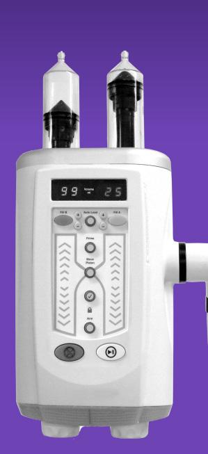
FDA CLEARS ETHOS AUTOMATED ULTRASOUND PROBE CLEANER DISINFECTOR
CS Medical has announced clearance of a new Class II medical device by the U.S. FDA, Ethos Automated Ultrasound Probe Cleaner Disinfector.
“Ethos is the first medical device cleared in North America which provides both cleaning and high-level disinfection of endocavity and surface ultrasound probes,” according to a press release. “Ethos marks a new era in the reprocessing of reusable medical instruments. The health care professional simply places a point-of-care cleaned ultrasound probe into Ethos and follows simple prompts on the color touchscreen display. Ethos guides the health care professional through an FDA validated workflow for effective reprocessing of soiled transvaginal, transrectal or surface ultrasound probes. Electronic record retention is part of Ethos with the ability to store more than 15,000 sessions in system memory. The user interface allows data entry through a scanner or manual keypad entry on the large 7-inch color LCD touchscreen.”
Ethos delivers a new method for cleaning and high-level disinfection of soiled probes with AquaCide cleaner disinfectant, the first of its kind to obtain FDA clearance. AquaCide cleans and disinfects each ultrasound probe in only 3 minutes. The combination of AquaCide cleaner disinfectant and QwikCheck liquid chemical indicator removes the manual process of cleaning, disinfection and user validation of chemistry efficacy. Ethos reduces the time and amount of human labor spent to reprocess an ultrasound probe. By removing the manual process and creating a repeatable and validated process CS Medical and Ethos have taken the guess work out of the reprocessing of endocavity and surface ultrasound probes. An advanced vapor management system is incorporated, as well as a high-quality water
filtration system through an FDA registered Class II device. Ethos rinses each cleaned and high-level disinfected ultrasound probe with a 5 nanometer (.005 micron) water filter that retains bacteria, viruses and endotoxins.
“Today’s news marks a new era in the reprocessing of endocavity and surface ultrasound probes. Health care professionals can now have confidence in the cleaning and high-level disinfection of soiled ultrasound probes. It is now possible within one machine, Ethos, to clean and high-level disinfect. Ethos is the first automated cleaner disinfector for endocavity and surface probes. At CS Medical, we have listened to our TEEClean customer base to design and deliver a product with the same characteristics and ease of use as TEEClean for TEE probes. Ethos automated cleaner disinfector will provide health care professionals with the same ease of use and confidence when reprocessing endocavity and surface ultrasound probes. A new era in the reprocessing of all ultrasound probes is possible through the innovations and commitment of CS Medical and its staff over the past decade. Ethos and TEEClean together can provide any health care facility with confidence and compliance when reprocessing an endocavity or surface ultrasound probe. Ethos is the combination of years of research, development and commitment by our entire staff,” CS Medical President Mark Leath said. “Ethos is another example of our continued commitment to working with health care professionals and other professional organizations in the reduction of HAIs and increase awareness for a better and safer health care system. In addition to our employees, I am thankful and appreciative of the cooperation and assistance given to us by various ultrasound probe manufacturers, without whom Ethos would not be the product it is today.” •
NEWS
ADVANCING THE IMAGING PROFESSIONAL 22 ICEMAGAZINE | AUGUST 2023
NEW TECHNIQUE CAPTURES COVID-19’S IMPACT ON BRAIN
A University of Waterloo engineer’s MRI invention reveals better than many existing imaging technologies how COVID-19 can change the human brain.
The new imaging technique known as correlated diffusion imaging (CDI) was developed by systems design engineering professor Alexander Wong and recently used in a groundbreaking study by scientists at Baycrest’s Rotman Research Institute and Sunnybrook Hospital in Toronto.
“Some may think COVID-19 affects just the lungs,” Wong said. “What was found is that this new MRI technique that we created is very good at identifying changes to the brain due to COVID-19. COVID-19 changes the white matter in the brain.”

Wong, a Canada research chair in artificial intelligence and medical imaging, had previously developed CDI in a successful search for a better imaging measure for detecting cancer. CDI is a new form of MRI that can better highlight the differences in the way water molecules move in tissue by capturing and mixing MRI signals at different gradient pulse strengths and timings.
Researchers at Rotman, a world-renowned center for the study of brain function, saw Wong’s imaging discovery and thought it could likely also be used to identify changes to the brain due to COVID-19. Subsequent tests proved that theory right. The CDI imaging of frontal-lobe white matter revealed a less restricted diffusion of water molecules in COVID-19 patients. At the same time, it showed a more restricted diffusion of water molecules in the cerebellum of patients with
COVID-19.
Wong highlights that the two regions of the brain react differently to COVID-19 and points to two key findings from the research. First, the human cerebellum might be more vulnerable to COVID-19 infections. Second, the study reinforces the idea that COVID-19 infections can lead to changes in the brain.
Not only is the Rotman study one of the few to have shown COVID-19’s effects on the brain, but it is the first to report diffusion abnormalities in the white matter of the cerebellum. Although the study was designed to show changes, rather than specific damage, to the brain from COVID-19, its final report does discuss potential sources of such changes and many link to disease and damage.
In response, Wong suggests future tests could focus on whether COVID-19 actually damages brain tissue. Additional studies could also determine if COVID-19 can change the brain’s grey matter.
“Hopefully, this research can lead to better diagnoses and treatments for COVID-19 patients,” Wong said. “And that could just be the beginning for CDI as it might be used to understand degenerative processes in other diseases such as Alzheimer’s or to detect breast or prostate cancers.”
The study, “Feasibility of diffusion-tensor and correlated diffusion imaging for studying white-matter microstructural abnormalities: Application in COVID-19,” which involves Wong and his student Hayden Gunraj as co-authors, is published in the journal Human Brain Mapping. •
WWW.THEICECOMMUNITY.COM ICEMAGAZINE 23
WEBINAR SHARES BENEFITS OF A 3D LAB
STAFF REPORT
The recent ICE webinar “Developing a 3D Lab to Streamline CT Workflows” was presented by Jessica Chambers, MHA, RT (R)(CT).
She has 22 years of imaging experience with her career starting with the U.S. Air Force as a radiology and CT technologist. After an honorable discharge in 2007, Chambers continued her career with SSM Health as the Director of Clinical Operation Imaging Services, with oversight of three hospitals and four outpatient imaging centers. The webinar was eligible for 1.0 ARRT Category A CE credit by the AHRA (AHRA Reference: LEC12163).
This webinar sparked ideas around why a 3D lab might be beneficial for a CT department, especially for organizations that have multiple CT departments across various campuses. Staffing is a challenge in today’s health care climate, implementing a 3D lab can assist with revenue generation without adding additional hours to the CT schedule.
Chambers walked attendees through the steps her group took to justify this additional department and how it has allowed them to grow volume and revenue while improving technologist and radiologist satis -
faction. At the conclusion of the webinar, attendees logged out with strategies to approach the C-suite about beginning a 3D lab journey within their organization.
Attendees provided feedback via a survey that included the question, “Why do you join ICE webinars?”
“Ease of attendance from my desk at times I can accommodate,” Cabell Huntington Hospital Inc. Director of Radiology Nancy Godby said.
“Learn about best practice and newer technology,” said Krissie Stich, radiology director, East Market, University Hospitals.
“Current knowledge of what is happening in our field,” said Michelle Cousino, radiology manager, University Hospitals.
“CE on topics of interest, this is my first webinar with ICE,” said Greg Laukhuf, RN manager, UHCMC.
The Imaging Community Exchange (ICE) magazine continues to grow and part of the growth is the webinar series where imaging leaders can earn valuable continuing education (CE) credits. •
For information, visit theicecommunity.com/webinars.

NEWS ADVANCING THE IMAGING PROFESSIONAL 24 ICEMAGAZINE | AUGUST 2023
PRODUCTS
Market Report
MEDICAL IMAGING AI MARKET GROWING FAST
ResearchAndMarkets.com recent report on AI in medical imaging is just one predicting continued growth.
“The global AI in medical imaging market size is expected to reach $8.18 billion by 2030. The market is expected to expand at a CAGR of 34.7% from 2022 to 2030,” according to the report.


The report continues, “Moreover, developed countries have been investing in offering people access to inexpensive and advanced health care systems in response to this demographic challenge, ultimately boosting the market growth.”
Additionally, X-ray imaging technology is utilized to identify a variety of disorders, including fractures, infections, various malignancies, and arthritis. As a result, the rise in the incidence rate of these diseases is expected to have a positive impact on the market growth.
COVID-19 had a positive impact on the market as the majority of medical professionals chose imaging as one of the most popular COVID-19 screening modalities as it provided critical diagnostic in a short time. The entire set of imaging data has been used to research the virus and its effect on people of various demographics.
Many hospitals have resorted to medical imaging and image analytics techniques to improve the identification and control of the virus as nearly every healthcare organization is working to manage COVID-19 better and limit its spread. For instance, in June 2020, a hospital in Florida, the Tampa General Hospital employed AI-driven technology to check people for COVID-19 before they interacted with staff members and other patients.
The rising awareness regarding the benefits offered by Artificial Intelligence (AI) technologies and their application in the medical field has led to increased adoption of AI-based solutions, products, and services in the health care market.


Moreover, to develop advanced AI-based applications for the health care industry, several leading health care companies are adopting different strategies such as collaborations, mergers and acquisitions, and partnerships with top AI technology vendors to provide cutting-edge solutions to their clients and strengthen their position in this competitive market environment.
Some highlights of the report include:
• Based on technology, the deep learning segment held the
largest share in 2021 owing to the usage of radiological applications such as object detection, image generation, image transformation, and image segmentation
• The NLP segment is anticipated to expand at the fastest CAGR of 35.7% during the forecast period as it comprehends and presents data in the form of current human language, images, and text
• Based on application, the neurology segment dominated the market with a revenue share of over 35.0% in 2021 owing to the increased use of AI in neurology as it enables higher accuracy, better patient care, and high efficiency

• The breast screening segment is anticipated to expand at the fastest CAGR of 41.5% during the forecast period owing to the rising demand for breast screening procedures due to the growing incidence rate of breast cancer in women
• Based on modality, the CT scanning segment dominated the market with a revenue share of over 35.0% in 2021. A wide variety of AI-based medical imaging solutions are being offered by both major and minor suppliers for use in the CT scan modality
• The X-rays segment is anticipated to grow at the fastest rate during the forecast period. The increased adoption of interventional x-ray equipment, such as C-arms, for image-guided surgeries is the key factor driving the segment
• In 2021, North America dominated the market due to its highly developed healthcare systems and high levels of disposable income. The demand for artificial intelligence in medical imaging has also been driven by the existence of important players and supportive governmental regulations
• Mordor Intelligence’s report states, “The AI Market in medical imaging is expected to register a CAGR of 30.4% over the forecast period from 2022-2027.”
Grand View Research adds, “The global AI in medical imaging market size was valued at $753.9 million in 2022 and is expected to expand at a compound annual growth rate (CAGR) of 34.8% from 2023 to 2030. The rising demand for the influx of large and complex datasets, government initiatives to promote the use of AI-based technologies, increasing attention to lessen radiologists’ workload, Artificial Intelligence (AI) tools in the medical industry, increased funding for AI-based start-ups by private players, and an increase in cross-industry collaborations and partnerships are some of the factors driving this market.” •


STAFF REPORT
ICEMAGAZINE 25 WWW.THEICECOMMUNITY.COM
Product Focus
ARTIFICAL INTELLIGENCE
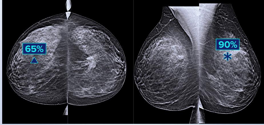
HOLOGIC Genius AI Detection Technology
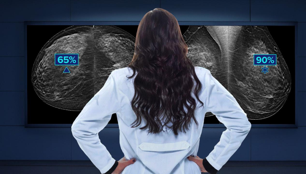
Genius AI Detection technology from Hologic uses deep learning to detect potential cancers in breast tomosynthesis images. 1 Designed to find cancers with improved accuracy as compared to Hologic’s previous generation conventional 2D CAD, the software is fully integrated on the 3Dimensions and Selenia Dimensions systems. Genius AI Detection technology can aid in radiologists’ diagnostic performance with the use of the technology resulting in an improvement of +9% in observed reader sensitivity for detecting cancer cases compared to standalone review. 1 The software can also potentially improve workflow efficiency by displaying results directly at the acquisition workstation to provide point-of-care triaging for Genius AI Detection technology high-priority cases or negative-findings cases.
1
References
1 FDA Clearance K201019. Based on analyses that do not control type 1 error and therefore cannot be generalized to specific comparisons outside this particular study. In this study: The average observed AUC was 0.825 (95% CI: 0.783, 0.867) with CAD and 0.794 (94% CI: 0.748, 0.840) without CAD. The difference in observed AUC was +0.031 (95% CI: 0.012, 0.051). The average observed reader sensitivity for cancer cases was 75.9% with CAD and 66.8% without CAD. The difference in observed sensitivity was +9.0% (99% CI: 6.0%, 12.1%). The average observed recall rate for non-cancer cases was 25.8% with CAD and 23.4% without CAD. The observed difference in negative recall rate was +2.4% (99% CI: 0.7%, 4.2%). The average observed case read-time was 52.0s with CAD and 46.3s without CAD. The observed difference in read-time was 5.7s (95% CI: 4.9s to 6.4s).
*Disclaimer: Products are listed in no particular order.
PRODUCTS
ADVANCING THE IMAGING PROFESSIONAL 26 ICEMAGAZINE | AUGUST 2023
2
GE HEALTHCARE Sonic DL
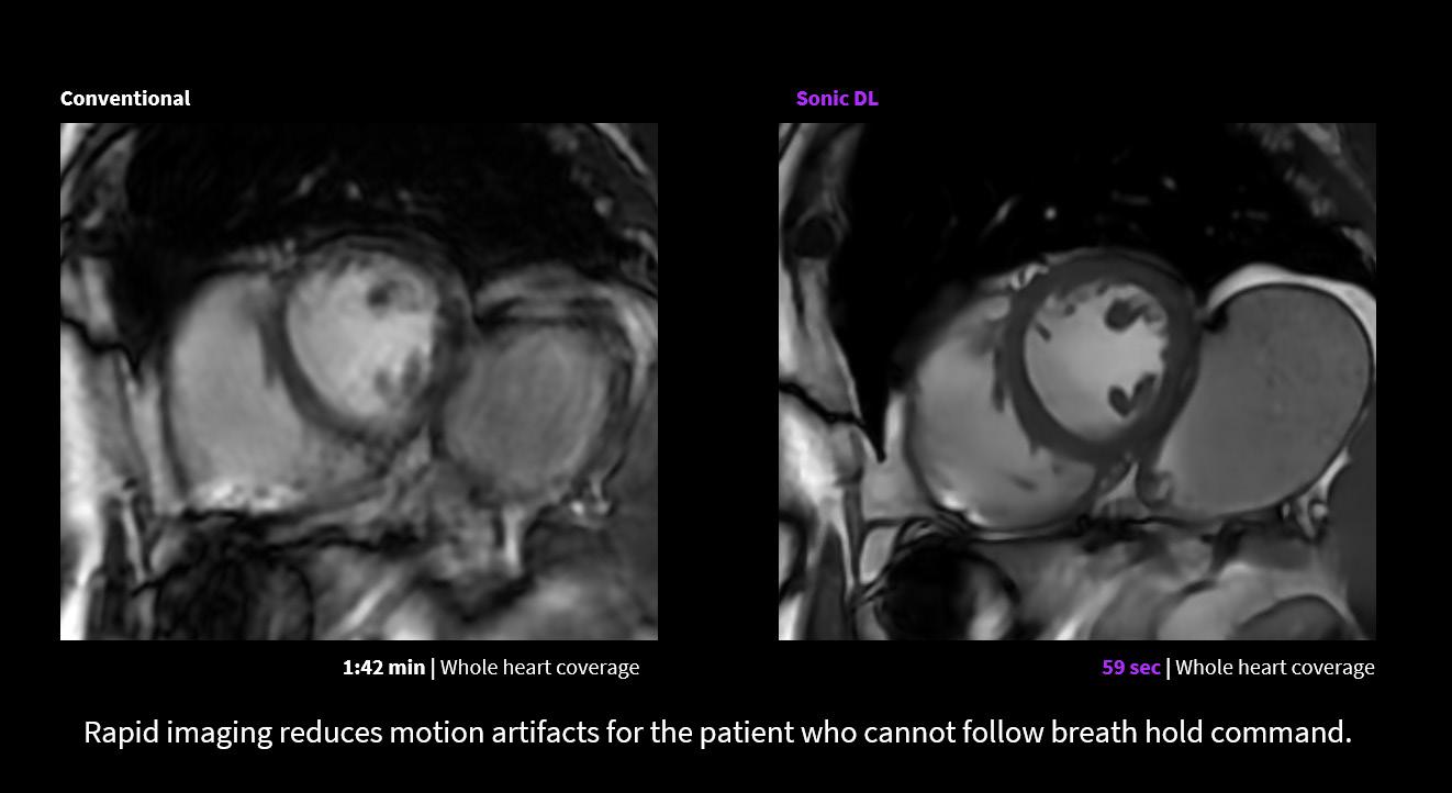
Sonic DL is a deep learning (DL) technology that acquires high-quality magnetic resonance (MR) images up to 12 times faster than conventional methods, enabling cardiac imaging within a single heartbeat. When applied to cardiac MRI, Sonic DL extends access to cardiac patients with arrhythmias and breath-holding challenges, helping to ensure swift, consistent, and more comfortable exams. With up to an 83% reduction in scan time, Sonic DL sets new industry standards for rapid, high-quality MRI.
CARESTREAM AI-Driven Smart DR Room Features
Carestream’s advanced, AI-driven Smart Room features automate tasks and workflow to streamline DR imaging processes. Imaging and Workflow Intelligence solutions help capture anatomy precisely while reducing repeat exams through automated patient positioning and verification of patient positioning; and automatic setting of appropriate acquisition technique – saving time and optimizing dose. Smart Noise Cancellation gives radiographers an important tool to adjust the amount of noise cancellation and exposure to meet the desired imaging quality.
3
ICEMAGAZINE 27 WWW.THEICECOMMUNITY.COM
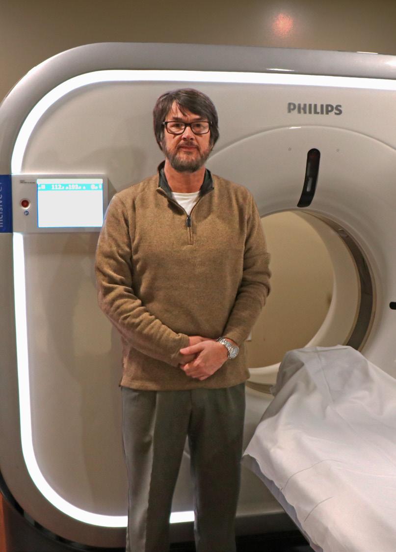
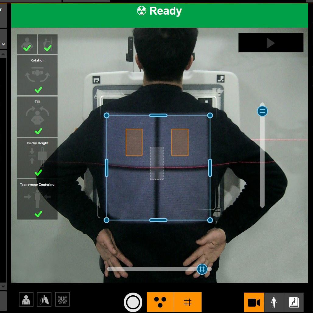
PHILLIPS CT 3500 4
The Philips CT 3500 is a new high-throughput CT system targeting the needs of routine radiology and high-volume screening programs.
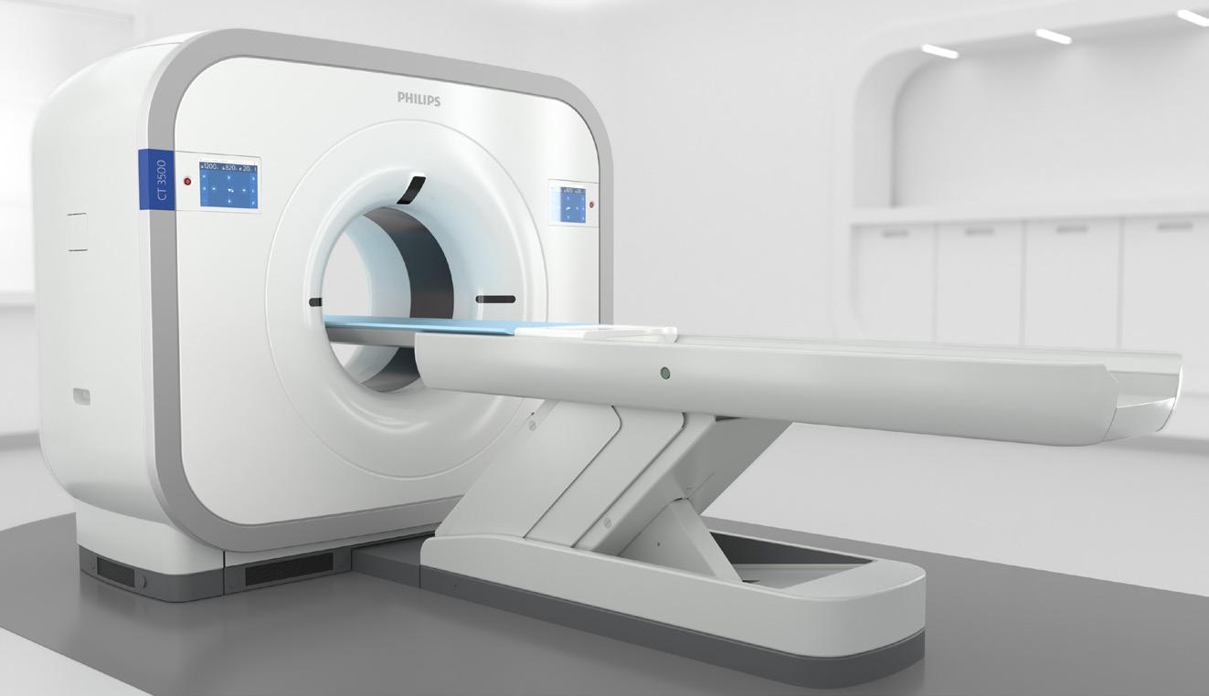
Powered by AI, the Philips CT 3500 includes a range of image-reconstruction and workflow-enhancing features that help to deliver the consistency, speed and first-time-right image quality needed for confident diagnoses by clinicians and increased return on investment – even in the most demanding, high-volume care settings. The CT 3500 features Philips’ latest AI-powered CT Smart Workflow to automate every step in the scanning process. Precise Position uses a camera to automatically determine patient orientation, improving positioning accuracy by 50% while reducing patient positioning time by up to 23%. Precise Planning automatically determines the area to be scanned and the appropriate Exam Card based on the patient’s anatomy. This provides fast exam preparation and can improve inter-operator consistency. Precise Intervention can offer automated setup and treatment guidance for tissue biopsies and other needle-based interventions.
SIEMENS HEALTHINEERS AI-Rad Companion Organs RT
AI-Rad Companion Organs RT, a Siemens Healthineers artificial intelligence-based software assistant in the AI-Rad Companion family, uses deep-learning AI algorithms to automatically contour organs at risk (OARs) on computed tomography (CT) images as part of the radiation therapy planning workflow. These algorithms automate the organ contouring process for various body regions, including the head and neck, thorax, abdomen, and pelvic regions, to accelerate contouring. AI-Rad Companion Organs RT also supports the use of organ template configurations that can be aligned with institutional protocols, which may save time and improve standardization in clinical workflows. AI-Rad Companion Organs RT is available on the company’s teamplay cloud-based digital health platform.
1 teamplay is not yet commercially available in all countries. For regulatory reasons, its future availability cannot be guaranteed.
PRODUCTS *Disclaimer: Products are listed in no particular order.
1
5 ADVANCING THE IMAGING PROFESSIONAL 28 ICEMAGAZINE | AUGUST 2023
FEBRUARY 18-20, 2024 IRVINE, CALIFORNIA ATTENDICE.COM SAVE THE DATE
WHY SHOULD I JOIN THE TALENT NETWORK?
• Completely free to register and apply to any of our listings
• 500+ open positions across the United States (part + full-time, temp, internships and externships)
Personally connect candidates with
Career Center with articles and tips from leading industry professionals

HTMJOBS@MDPUBLISHING.COM EMAIL US & CHECK OUT OURFORWEBSITE MORE INFORMATION!
WHY HIRING MANAGERS SHOULD POST WITH US
• A talent network of 2,500 qualified actively looking HTM and Imaging professionals
• A variety of posting options ranging from singlejob listings to unlimited membership packages
• Social media, print, and eNews promotion
• Personalized recruiting services at a competitive rate
“HTMjobs.com is a remarkable website that can be used by any personnel looking for employment, internships, or just a good read from their Career Center . The site offers job postings from employers in the HTM industry, as well as resources for job seekers to help them prepare for their job search, including resume tips, interview advice, and more.”
Featured
Agiliti, InterMed, TRIMEDX, Renovo Solutions,
Resources, SPBS, Inc., and Universal Medical Resources.
Employers:
Texas Health
THE FUTURE OF CT

COVER STORY
ADVANCING THE IMAGING PROFESSIONAL 32 ICEMAGAZINE | AUGUST 2023
BY MATT SKOUFALOUS
In the 50 years since the first computed tomographic images in the United States were captured by EMI-made scanners at the Mayo Clinic in Rochester, Minnesota, computed tomography (CT) imaging has become an invaluable diagnostic tool. Its persistent evolution spans devices built around xenon gas detectors, to those made with solid-state detectors, to those capable of spiral and multi-slice CT, and eventually, to those capable of spectral imaging: a technology that allows physicians to determine what’s going on in the body at levels that were only theoretical in the previous half-century.
Matthew Fuld, Ph.D., director of photon-counting CT at Siemens Healthineers North America of Malvern, Pennsylvania, described the fundamental value of CT as lying both in its ability to capture exceptionally high-quality images, and at rapid speeds. Optimization of these features has always been tied to the corollary optimization of dose sufficient to achieve these images while limiting exposure of the patient’s body to as little ionizing radiation as possible. Spectral imaging compares data sets collected from different X-ray spectra, to better visualize tissue differences and enhance clinical diagnostic decision-making. In the NAEOTOM Alpha, the first photon-counting CT scanner to achieve FDA clearance, Siemens Healthineers has achieved what Fuld calls “a huge leap” in technology.
The NAEOTOM Alpha photon-counting CT unifies the imaging functions of spatial resolution, speed of image acquisition, and dual-energy, or spectral, capabilities in a single technology platform that also limits the amount of ionizing radiation to which a patient is exposed throughout a study. The clinical value of such functionality in inpatient, outpatient, and research spaces is already being demonstrated across a number of environments, Fuld said, with 175 publications dedicated to the NAEOTOM Alpha in the fewer than two years of its operation. What makes the scanner so distinct from others in the field is its construction, which measures X-rays individually, as electrical signals on the order of 8 nanoseconds, rather than as light, he said.
“The idea is one that the physics community has worked on for many years,” Fuld said. “The challenge with clinical imaging is that we use a large number of X-rays to generate an image. The proof of concept of measuring an individual X-ray was always done with a very low amount of X-rays that would never be sufficient or fast enough to capture a clinical image. With the NAEOTOM Alpha, we’ve been able to get just shy of 45 line-pairs per centimeter, which offers tremendous detail in bone structure, coronary arteries, the tiny vasculature in the
brain and the liver; anywhere in the body, you can see detail you never thought possible. Because we’ve eliminated light, we are measuring each X-ray on its own.”
By differentiating the energy of an individual X-ray from background noise, a photon-counting CT scanner can create better quality images; by comparing the electrons to one or more energy thresholds, it can present dual-energy information, Fuld said.
“Whether that’s separating iodine and calcium to reduce the blooming artifact in a coronary artery, or to calculate iodine uptake in a pancreatic tumor, the inherent spectral sensitivity of the detector allows us to do spectral imaging on every scan, no matter how it’s operated,” he said. “It has the speed of dual-source, it’s lower radiation dose, and it’s giving spectral information at the same time.”
The power of photon counting technology has already been demonstrated in the cases of patients with spontaneous intercranial hypertension, in which leaking cerebrospinal fluid causes debilitating painful headaches. Traditional methodologies for finding the sources of those leaks in Type II CSF don’t work anywhere near as well as those based around photon counting do, Fuld said.
“It’s changing care for patients,” he said.
The bottom line is that photon-counting CT simply “makes everything better,” he said, by offering a clearer look at the inner workings of the body through precise, structural measurements taken at full speed. It’s particularly useful in pediatric applications, cardiac cases, lung diseases, and differentiating patients who need interventional procedures from those who need diagnostics.
“It gives us considerably more insight into things we never would have thought before,” Fuld said. “In the past, what we believed to be certain clinical presentations, when we scan them with photon counting, what was once blotchy or blurry is now crisp, with strong details, and it allows us to change the characterization of these patients. My personal opinion is that this will lead to better treatment in the future.”
As the technological components of next-generation CT scanners continue to improve alongside new data transmission and computing capacities, Fuld believes that patients and imaging technicians alike will continue to benefit from the automation of technology that can compensate for the variability of one study to the next, and save a lot of time along the way. Manufacturers like Siemens Healthineers will continue to focus on providing greater clinical value with less operational expense, the better to optimize the process.
“If I’m talking to a clinician who would operate this equipment, they don’t want complexity, they want simplicity,” he said; “make the user experience high-level. The technologist

COVER STORY
ICEMAGAZINE 33 WWW.THEICECOMMUNITY.COM
doesn’t have to make manual choices; it will make them automatically in the background. They’re helping that patient off the table, making sure their experience is good, and on to the next one, with the throughput that hospitals need.”
“We have a wide variety of clinical questions, and the more capable the technology becomes, the more different questions we can ask, and the context of where you ask them expands,” Fuld said. “If you have the right level of clinical capabilities, we can achieve high clinical value. Technology is giving us amazing clinical insight. How well we can take this information and use it collectively to take care of patients?”
Chief among the critical functions of advanced CT technology is the potential expansion of clinical screening, particularly for lung cancers and heart disease. Olivia Egan, director of CT product marketing at Siemens Healthineers North America described how dramatically lung cancer survivability rates climb with early detection, and how expanded eligibility in concert with more sensitive imaging equipment can save higher orders of lives.
“If you catch lung cancer early, it’s 70 to 90 percent survivable,” Egan noted; “it’s less than 20 percent if you don’t. In the U.S., although there’s been lung cancer screening established since 2013, only six percent of all those eligible are actually being screened.”

In November 2021, the Centers for Medicare and Medicaid Services (CMS) released a final ruling on the Medicare Outpatient Prospective Payment System (OPPS), expanding nationwide eligibility criteria for lung cancer screening to cover 14.5 million Americans (up from 8 million in 2013), and increasing reimbursement rates for hospital outpatient lung cancer screening by around 37 percent (up to around $111 from $80).
Despite this expanded eligibility and higher reimbursement rate, Egan said additional efforts are required to reach the atrisk patients who would benefit most from early detection of lung cancer.
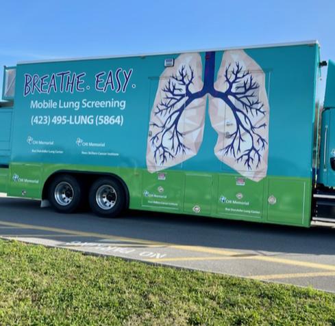
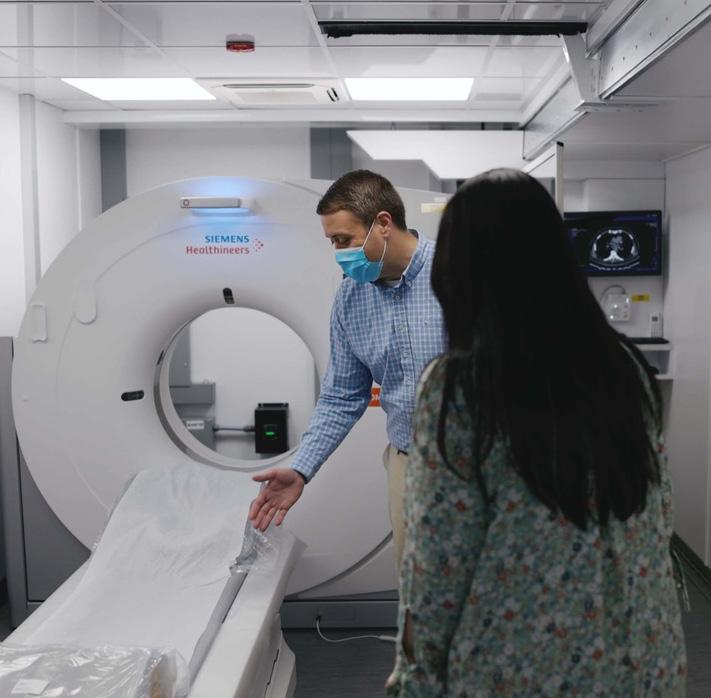
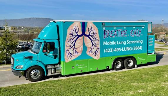
In North America, Siemens Healthineers expanded its CT scanning portfolio to include a dedicated mobile CT solution for lung cancer screening. This mobile CT configuration incorporates the SOMATOM go. Up CT scanner, which uses tin filter technology that is unique to Siemens Healthineers. The tin filter cuts out lower photon energies to reduce dose and optimize image quality, and has direct benefits in lung imaging, resulting in ultra-low-dose CT lung cancer screening examinations. This mobile, self-contained vehicle can support community outreach programs dedicated to connecting with people in rural and farther-flung areas of the country by taking lung cancer screening directly to them.
In Chattanooga, Tennessee, Rob Headrick, chief of thoracic surgery at CHI Memorial Rees Skillern Cancer Institute, worked to develop a rural outreach lung cancer low-dose screening project, outfitting a Winnebago shell with a 16-slice refurbished CT scanner as his prototype. The data Headrick was able to gather from that outreach in Spring City, Tennessee, a rural hamlet with a population of 2,400 people, literally changed the fabric of the community.
“One death in a community like that doesn’t go unnoticed,” Headrick said. “The pessimism of lung cancer hangs over you. After five years, we have screened half the eligible people in that community with a dozen lung cancer patients alive and walking around. Now the people in that community sign themselves up.”
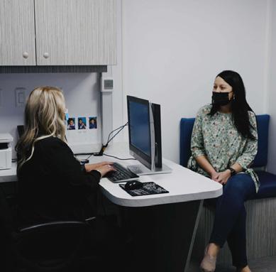
For Headrick’s team, the program was successful not necessarily because it was revolutionary, but more so because it paired advanced health care technologies like AI-powered computing, wireless data transmission, and HIPAA-compliant text messaging to rapidly acquire images, communicate results of studies, and share same-day results with practitioners who can develop care plans and treatments for those who need them. The benefit of that early-stage detection is quite literally the difference between life and death, Headrick said.
“The people who need it most are in rural communities,” he said. “They’ll still come to Nashville, Knoxville, or Chattanooga for health care, but when it’s too late. The key to lung screening is high volume, and how quickly you can manage the process. What started as a local solution for our geographical barriers to health care is now becoming a national solution.”
Headrick is now working on what he calls “a traveling show,” aiming to develop a network of bus-mounted CT scanners that
COVER STORY ADVANCING THE IMAGING PROFESSIONAL 34 ICEMAGAZINE | AUGUST 2023
could rotate among Veterans Affairs clinics, giving rural community hospitals a modern solution to reach the population they serve. He said that hospitals from Texarkana to southern Georgia to upstate New York are also jumping on board, aiming to improve the poor survivability rates currently associated with lung cancer. More than 20 different hospitals are fundraising towards the goal of a program like the one that debuted in Spring City, Tennessee.
“You’re taking health care to the people who don’t have options, but they’re still lives that matter,” Headrick said. “We can spend an hour driving, scan 45 people in a seven-hour window, drive the bus back, and put it up. This is a more realistic solution than constructing imaging centers in these rural communities.”
“In the state of Tennessee, almost half our population is going to die from a heart attack or lung cancer,” he said. “By scanning somebody who’s high-risk for lung cancer, I can reduce their risk of mortality by 20 to 30 percent. I can do the same for heart disease by adding a calcium score to that scan.”
“What would it do for the state of Tennessee if I cut the mortality rate for both these diseases by 30 percent?” Headrick asked. “That changes our workforce stability; it changes the economics of the state. It saves many lives. You now have the affordability, processing power, and truly can find disease before there’s a problem. I think it will be the future of how we deliver health care in a much more affordable manner.”
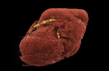
Headrick also believes that the future of CT lies in making use of the additional data from studies of the torso. While visualizing the lungs in a three-minute screening, for example, images captured can display information about coronary calcification, heart chambers, visceral fat content, bone density, and core muscle mass; organs like the liver and gall bladder, and more. Without reducing the length of time of that screening, physicians could leverage the low-dose, high-quality images captured “and bring value back to health care,” he said.
“We may have the first screening test that not only reduces mortality from a specific disease, but also offers information on how to improve someone’s quality of life and may be the first screening test that could improve overall longevity,” Headrick said. “That scan is now at a price point, and given the ability to acquire and process the images so quickly, it’s
now something you can easily scale up.
“The previous bottleneck was handling all the data present on a CT scan – how much time does it take for the radiologist to measure the aorta, or calculate bone density,” he said. “AI has solved that problem. The national lung cancer screening trial database has also allowed so many people to develop software and test it on this high-risk population dataset. We now have an opportunity to move the hospital closer to the patients; to move the imaging closer to the patients, and bring meaningful health care to those who need it the most.”

Similarly, Dr. Omar Khalique, director of the division of cardiovascular imaging at Saint Francis Hospital and Heart Center Catholic Health in Roslyn, New York, believes that clinicians will only come to gain more and better information faster from advances in CT technology. The modality offers the highest growth potential for cardiac imaging, not only for clarity of images captured, but for the fullness of seeing three-dimensional anatomy for a variety of observers, from imaging experts to cardiologists and researchers.
“With coronary CT, we probably started too early from the technology standpoint,” Khalique said. “We knew what we wanted to look for, but because of the technological limitations, we couldn’t see it then. Imaging of percutaneous valves was first approved in 2011, and now has grown exponentially. Photon-counting CT has superior detectors for spatial resolution. We’re much closer to seeing what we wanted to see 10 years ago.”
Previous cardiac imaging technologies suffered from “blooming,” which makes calcium deposits look larger, and obscures plaques, Khalique said. But because contemporary CT imaging technologies, like the photon-counting NAEOTOM Alpha, allow cardiologists to see more inside of coronary arteries, the requirements for any calcium score cutoffs can be eliminated. Stent imaging is now possible. Patients have the opportunity of non-invasive testing instead of more invasive angiography when they need diagnostic information alone.
“I think the precision of the analysis is just going to keep increasing,” Khalique said. “Photon counting has been the Holy Grail; now that they’ve pushed the envelope, I’m hoping that detectors not only get wider, but offer better resolution. I’m sure others will follow.” •
COVER STORY ICEMAGAZINE 35 WWW.THEICECOMMUNITY.COM
BY BETH ALLEN DIRECTOR’S CUT
END OF AN ERA
We have reached an end of an era. Dr. Threasa Frouge, MD, has served as Banner Imaging’s chief medical officer since our creation in April of 2019 and will retire this month. She was instrumental in our success long before that.

I have been privileged to work with her for most of my career and wanted to give her the opportunity to share her thoughts about where imaging has been and where we are going, in her words.
“I arrived in Phoenix in July 1988, after completing medical school at Duke University and Residency and Fellowship training at the Mallinckrodt Institute at Washington University.
Dr. Denise Smith and I arrived and joined Valley Radiologists the same month. We were the first women to join our group.
All of my training was immediately put to good use in covering a very busy radiology practice that served several hospitals, as well as a group-owned outpatient imaging practice.
When I joined in 1988, we were all fellowship trained; however, private practice and radiologist staffing, with no PACS or remote access to images, required that everyone be proficient in all aspects of radiology. After two years of fellowship training in abdominal imaging/interventional and nuclear medicine, I found the need to quickly study and brush up on other areas of radiology. I soon discovered that my career would be a lifetime of learning.
Radiology continues to progress and expand tremendously; this requires continuous learning in order to diagnose and treat our patients and support our clinical colleagues.
After becoming a partner in Valley Radiologists, I served as department chairman and med-
ical director of various hospitals, most recently Banner Estrella. Working in a hospital environment taught me the value of service, teamwork, organization, efficiency and commitment to quality and safety. I felt very fortunate to serve at Banner Estrella, where I witnessed everyone working together to achieve these goals.
As I was doing this, I was also serving my group in a leadership role, that included oversight and development of our outpatient imaging practice that would eventually become a part of Banner Imaging.
It was exciting to learn the business aspect of successful, high-quality ambulatory imaging. As a physician-owned practice, we were responsible for maintaining a collegial and supportive culture for our imaging staff and radiologists, promoting quality, safety, and service, while being efficient and financially responsible. Financial success meant the ability to open new sites and purchase new equipment to better serve our growing community.
As Banner Health considered expansion into ambulatory imaging, the outpatient imaging practice that we (Valley Radiologists) had built and nurtured since the 1960s paired with the equivalent practice of our east side sister radiology group (EVDI/ARL) seemed a natural launching point for Banner.
Becoming Banner Imaging seemed a natural transition given our long history with Banner Health and the increasing complexity of managing and growing successfully into the future, where large well integrated structures will be needed to provide comprehensive care in multiple settings in all phases of life.
As Banner Imaging was getting off to a great start, the COVID-19 pandemic hit. The value of being part of a large, proactive health system became immediately apparent. Banner Imaging
INSIGHTS
ADVANCING THE IMAGING PROFESSIONAL 36 ICEMAGAZINE | AUGUST 2023
received immense support to continue safely operating and serving the community throughout the pandemic. None of our offices closed. Banner Imaging staff continued to work in our offices or fulfill needs at other Banner facilities.
After having successfully navigated the pandemic and learned lessons of fortitude and resilience, Banner Imaging is poised to meet the challenges of future growth, both geographically and scope of service.
The best aspect of practicing radiology in both the acute care and ambulatory setting is working with the imaging team to provide direct patient care.
Radiology is a team sport. Everything we offer our patients and referring clinicians involves the entire team, from scheduling to front desk staff to technologist, medical assistant, imaging leadership and radiologist. We all have an important role to play in providing excellent and correct imaging, compassionate care and an accurate diagnosis and/or procedure.
Our patient is the focal spot of everything we do. We must always remember that any day at work may seem routine for us, but may be the day that one of our patients receives a cancer diagnosis or some other life changing news.
Many patients come to us under duress from illness and/or worry. I am always amazed at how our imaging staff is able to care for patients and defuse stressful situations. We do this by working together and supporting each other as we do the work of caring for others.
It has been rewarding to watch team members learn, problem solve, cross train, meet new challenges and advance to new positions in our organization. There are team leaders that I remember as RT students many years ago.
One of the greatest complements I received was a team member describing a problem that she was trying to solve … while saying to herself, ‘What would Dr. Frouge do?’
Solving problems, creating new and better processes and workflows, building new offices and service lines … these are all amazing activities that I have shared with our team over the years.
Looking back, there is not so much that I would have done differently, but certainly plenty of opportunity to learn and improve moving forward. Every shortcoming or misstep is really a chance to learn and try something different in the future.
For example, we realized that we always built offices “too small” at Valley. After 3 to 5 years, we were faced with investing more to expand assuming that we were not “land locked” by neighboring tenants. In future offices, we included “soft space” for future expansion. We started “building the future” into our imaging center floor plans.
Our challenges in the future may include doing more with fewer resources. The cost of caring for an aging population who will need more imaging may stress current resources.
We may need to recruit and train more imaging staff and expand days/hours of operation with our existing offices and equipment.
Service focused imaging leads to success. By serving patients, clinicians, and each other, we make the right choices. We place quality and safety first. We understand the clinical question, do the correct exam, provide accurate results in a timely manner, and contribute to the treatment and overall health and wellbeing of Sophia. We support each other as we do this work, creating a welcoming and collegial environment that will provide a positive experience for a patient. We train new workers to join our team. We interact positively with our health system and community in our endeavor to be ‘the easy button’ … ‘making health care easier, so life can be better!’ ”
We are proud to share this as evidence of success with a supportive leader. Frouge will move on to a well-earned retirement, but her Southern hospitality will continue to be evident in the way we welcome our patients. Our team is so fortunate to have had such an inspirational example.
Thanks for all you do! •
ICEMAGAZINE 37 WWW.THEICECOMMUNITY.COM
THE TIME HAS COME FOR A NEW AI AMBASSADOR
PACS/IT
BY MARK WATTS
Biomedical professionals may be on the verge of a new key skill tree development. It would be an extension of their known skill set and a welcome ambassador in the imaging ecosystem. The recent moves in the market by Bayer and Guerbet in acquiring AI workflow orchestration or hosting platforms has caused this question to be asked by many in the community. This would mean the contrast injector may become the chosen route for AI deployment and support.
Bayer, Bracco and Guerbet develop, sell and maintain powered contrast injection delivery systems and complimentary or adjacent analytical tools and information. GE HealthCare has more fully entered the injection system market via a very close relationship with Ulrich.
This omnipresence of their device and software solutions gives them access and visibility into the diagnostic imaging market and buying centers in health systems. Nearly every MRI, Nuc Med/PET-CT and CT scanner has a contrast injector associated with it (approximately 65% of all CT exams involve IV contrast) and every angio/cath lab will have an injection system in it. In the form factor of an injector platform, there are complex, distributed, electromechanical systems that also have matured to include a full suite of capabilities that entail the collection, computation, and transmission of data to and from the scanner, PACS, RIS and other departmental or enterprise systems.
A friend of mine spent years developing the informatics architecture and software tooling for Medrad and Bayer including the incorporation of operating system software stacks, interoperability via DICOM, HL7 and web-services. There is a specific DICOM SR SOP class for injection data.

Quietly we have seen a software and analytics evolution pattern across each of the major contrast delivery and contrast agent manufacturers – Bayer, Bracco, GE and Guerbet. Each has their own brand of contrast injection/delivery systems, remote service capabilities, their own suite of connected analytic solutions for contrast and radiation analytics or/and optimization, and scanner and imaging IT interoperability and connectivity solutions. Some also distribute or have distributed software applications. They have national scale sales, service and clinical application teams touching nearly every hospital as
well. Those service and apps teams are critical for getting any medical innovation into use. They also have strong relationships with departmental admins, radiologists and, because of the smart capabilities of the systems, connections, and relationships with service-line leaders in radiology, cardiology, and nuclear medicine, and often enterprise IT. These are very different capabilities and relationships from those developed and needed when representing contrast agents.
The number of sales reps for contrast agents has decidedly decreased in the last 10-plus years (and contrast doesn’t need a service team). However, those reps have strong relationships with health care system purchasing, GPOs and supply chain. They can and will help influence or at least open the right doors at a health care delivery organization for other teams in their commercial organization. A commercial team that has relationships with purchasing, GPOs, IT, and service-line teams is valuable and something that pure-play, health care IT or software vendors will find difficult to replicate. I am recognizing biomed professionals as key players and owners of crucial relationships for advancing innovation into healthcare operations Software as a Service IT-focused startups do not realize this until way too late in their lifecycle.
Scanner sales teams don’t like to get stuck in long sales cycles/evaluation for software applications that might delay or put at risk a million-dollar scanner sale. Compare that dynamic to a $25,000-$45,000 contrast injector and the add-on value of AI applications can be more easily deployed.
Who can help health care organizations and private practices identify the most appropriate and useful clinical analytical tools and facilitate measurement of utilization and demonstrate ROI. I think biomedical professionals will be given an opportunity to help AI vendors with product processes, management and life-cycle planning. They will help define and refine the market needs and scope, customer fit and value proposition.
I have developed learning modules for a national continuing education company in Milwaukee, Wisconsin. We can make this happen together. •
INSIGHTS ADVANCING THE IMAGING PROFESSIONAL 38 ICEMAGAZINE | AUGUST 2023
Mark Watts is an experienced imaging professional who founded an AI company called Zenlike.ai.


EXCLUSIVE CONTENT, INCLUDING: • BREAKING NEWS • DIGITAL ISSUES • WEBINARS • PRODUCT SPOTLIGHTS MAGAZINE FOLLOW US ON SOCIAL MEDIA! GE • TOSHIBA SIEMENS • PHILIPS Give us a call at 213-276-8209 (844-PMIM-MRI) or visit www.pmimagingmgmt.com LOOKING TO PURCHASE OR SELL AN MRI, PET/CT, CT, OR X-RAY? LOO K N O FURTHER . PROOF LOOKING TO PURCHASE OR SELL? LOOK NO FURTHER. 213-276-8209 (844-PMIM-MRI) PMIMAGINGMGMT.COM GE • TOSHIBA • SIEMENS • PHILIPS JOIN AT THEICECOMMUNITY.COM/SUBSCRIBE Magazine and stay up to date on industry trends, news, webinars and conferences! LOVE FREEBIES? HOW ABOUT A FREE SUBSCRIPTION Use the QR code to sign up today! ICEMAGAZINE 39 WWW.THEICECOMMUNITY.COM
CODING & BILLING
BY MELODY W. MULAIK,
ARE YOU APPROPRIATELY CAPTURING ALL YOUR VISITS?
Evaluation and management (E/M) guidelines have changed significantly the past couple of years, and this is appropriately challenging many radiology practices to re-evaluate their coding and billing practices.

Historically ensuring the performance and necessary time to document the required components for these services has been challenging for many practices so the return on investment was not always present to justify coding and billing, especially for inpatient services. In some practices, interventional radiologists not only provide requested treatments, but they also directly manage the care of certain patients. In these circumstances, it is often appropriate to bill for an E/M service.
The specific changes for 2023 were focused on services provided in the hospital, patients that are inpatient or observation status. At a high level, the major changes made by the American Medical Association (AMA) most directly impacting interventional radiologists include the following:
• Inpatient and observation services were combined into one code set;
• Only the choice to use time or medical decision making (MDM) in determination of the code level;
• Revised time values assigned to the code levels;
• Inclusion of all the time spent on the date of the visit; and
• Eliminating the ability to use the history and exam or time in combination with the MDM to select the final code level.
While it is easier to meet the requirements of billing for an E/M visit for an inpatient, procedure global periods cannot be overlooked. Medicare’s global surgery policies are found in the Medicare Claims Processing Manual, Chapter 12, Section 40 (“Surgeons and Global Surgery”) and in the National Correct Coding Initiative Policy Manual for Medicare Services (NCCI Manual), Chapter I. Under these policies, the physician’s payment for a procedure includes payment for related E/M services during a specific period of time, known as the global period.
The table below shows Medicare’s global period categories. You can determine which category a procedure belongs to by looking up the CPT®/HCPCS code using the Physician Fee Schedule Look-Up tool on the CMS website.
Most interventional procedures are listed with an indicator of “000”, “010” or “XXX.”
A minor procedure is one that has a zero-day or a 10-day global period. Regarding E/M services on the same day
INSIGHTS
ADVANCING THE IMAGING PROFESSIONAL 40 ICEMAGAZINE | AUGUST 2023
MSHS, CRA, RCC, RCCIR, CPC, COC, FAHRA
as a minor procedure, the NCCI Manual states:
“In general E&M services on the same date of service as the minor surgical procedure are included in the payment for the procedure. The decision to perform a minor surgical procedure is included in the payment for the minor surgical procedure and shall not be reported separately as an E&M service. However, a significant and separately identifiable E&M service unrelated to the decision to perform the minor surgical procedure is separately reportable with modifier 25. The E&M service and minor surgical procedure do not require different diagnoses. If a minor surgical procedure is performed on a new patient, the same rules for reporting E&M services apply. The fact that the patient is ‘new’ to the provider is not sufficient alone to justify reporting an E&M service on the same date of service as a minor surgical procedure.”
The NCCI Manual also provides the example of a patient who is being seen for a head laceration. The physician who repairs the laceration should also not report an E/M code if the interaction with the patient involves simply checking allergy and immunization status and obtaining informed consent. However, the physician can report an E/M code with modifier 25 if he or she performs a “medically reasonable and necessary full neurological examination” in addition to the wound repair.
If a true interventional consultation is ordered, and
not just a request for a specific procedure, the encounter at which the physician makes the decision to perform the procedure is separately billable if there is supporting documentation and medical necessity. For example, if the interventionalist sees the patient in consultation and recommends that a procedure be performed, the initial encounter should be separately paid even if it occurs on the day prior to the procedure or the day of the procedure. Appropriate modifiers will need to be applied.
It is important that every radiology group carefully review orders, documentation templates, physician workflows and billing processes to ensure the systems are in place to bill confidently and compliantly. The recent coding updates should encourage every practice to re-evaluate their opportunities to bill for these services and potentially increase revenue opportunities. •
Indicator Definition Global Period 000 Endoscopic or minor procedure The day of the procedure 010 Minor procedure The day of the procedure and 10 days following the procedure 090 Major surgical procedure The day before the procedure, the day of the procedure, and 90 days following the procedure XXX Non-surgical procedure Global period does not apply YYY Contractor-determined Global period is set by the local Medicare contractor ZZZ Add-on code Included in the global period of the base code
ICEMAGAZINE 41 WWW.THEICECOMMUNITY.COM
ESSENTIAL LEADERSHIP RESPONSIBILITIES
EMOTIONAL INTELLIGENCE
BY DANIEL BOBINSKI
If an organization is to succeed it needs good leadership. But what is that? Before I define leadership responsibilities, let me first state what leaders do not have to do.
Leaders are not heroes who ride in to save the day. That myth comes from movies and novels. People love the idea of someone being fearless and infallible, either because that’s what they’d like to see in themselves or because they want to feel safe knowing someone like that is in charge.
Besides, a good leader should not have to ride in to save the day. A better approach is to ensure everyone is trained to the best of their abilities. That way it’s extremely rare that a day would need to get saved.
Also, leaders do not have to be gregarious extraverts who go around making others feel encouraged and empowered. Not that this is a bad thing, but some of the best leaders are reserved individuals who do more listening than talking, carefully weighing their choices after gathering much information and considering the potential ripple effects of each option.
In the same way, leaders do not need to have dominating personalities. Those with dominant styles tend to make fast decisions and can jump on problems as soon as they arise. That can be a strength, but sometimes that approach causes more problems than it solves. Different problems can require different solutions. A good leader recognizes that.
At a fundamental level, a good leader recognizes the fundamental areas of responsibility that should be overseen, and what must be done in each of those areas to achieve the most effective outcome.

AREAS OF RESPONSIBILITY
From my perspective, three fundamental areas of responsibility for leaders exist, and they are ideas, the horizon, and the organization’s capabilities. Certainly, more areas of responsibility must be attended to, but I believe that these three fundamental areas are too often overlooked.
First on the list is ideas. More specifically, it’s a leader’s responsibility to collect and consider ideas. With that, if leaders want to excel, they should seek input from every level in the organization, because everyone has ideas, and many times some great ideas come from front-line workers who have never stepped foot in the executive conference room.
But let me also emphasize that ideas must be evaluated. Some ideas are great, others aren’t. But even some of the bad ideas end up leading to good ideas.
Perhaps you’ve seen the TV show “Undercover Boss.” In production now for more than a decade, the show features high-level executives or business owners going undercover as entry-level employees in their own companies. What they learn is often eye-opening, and the end result is leaders collecting great ideas for how to make their company better. However, leaders need not go to this length to gather useful ideas. Genuine curiosity when asking questions goes a long way.
The second area of responsibility for leaders is the horizon. The word “horizon” means our range of perceptions or the extent of what we can see. Leaders should have multiple horizons on their radar, such as economic horizons, geo-political horizons, local market horizons, technology horizons … the list goes on. Good leaders take time to read and learn about these various horizons and also confer with their colleagues and support teams, discussing the implications of what’s happening “out there.”
That said, let me elaborate on how this area of responsibility correlates to the first area, collecting ideas from all levels within an organization. Picture a leader as the pilot of an airplane flying at 40,000 feet. At that height, the leader has a good view of the horizon, but the viewpoint is primarily forests, not trees. If leaders confer only with others who fly at 40,000 feet, they may make decisions that seem right at the time but will be expensive and time-consuming due to not having enough information.
For example, what may look like prime woodland at
INSIGHTS
ADVANCING THE IMAGING PROFESSIONAL 42 ICEMAGAZINE | AUGUST 2023
40,000 feet might really be a swampy muck underneath. People on the front line with experience in that forest can provide valuable advice on what can be done to achieve the best return on investment.
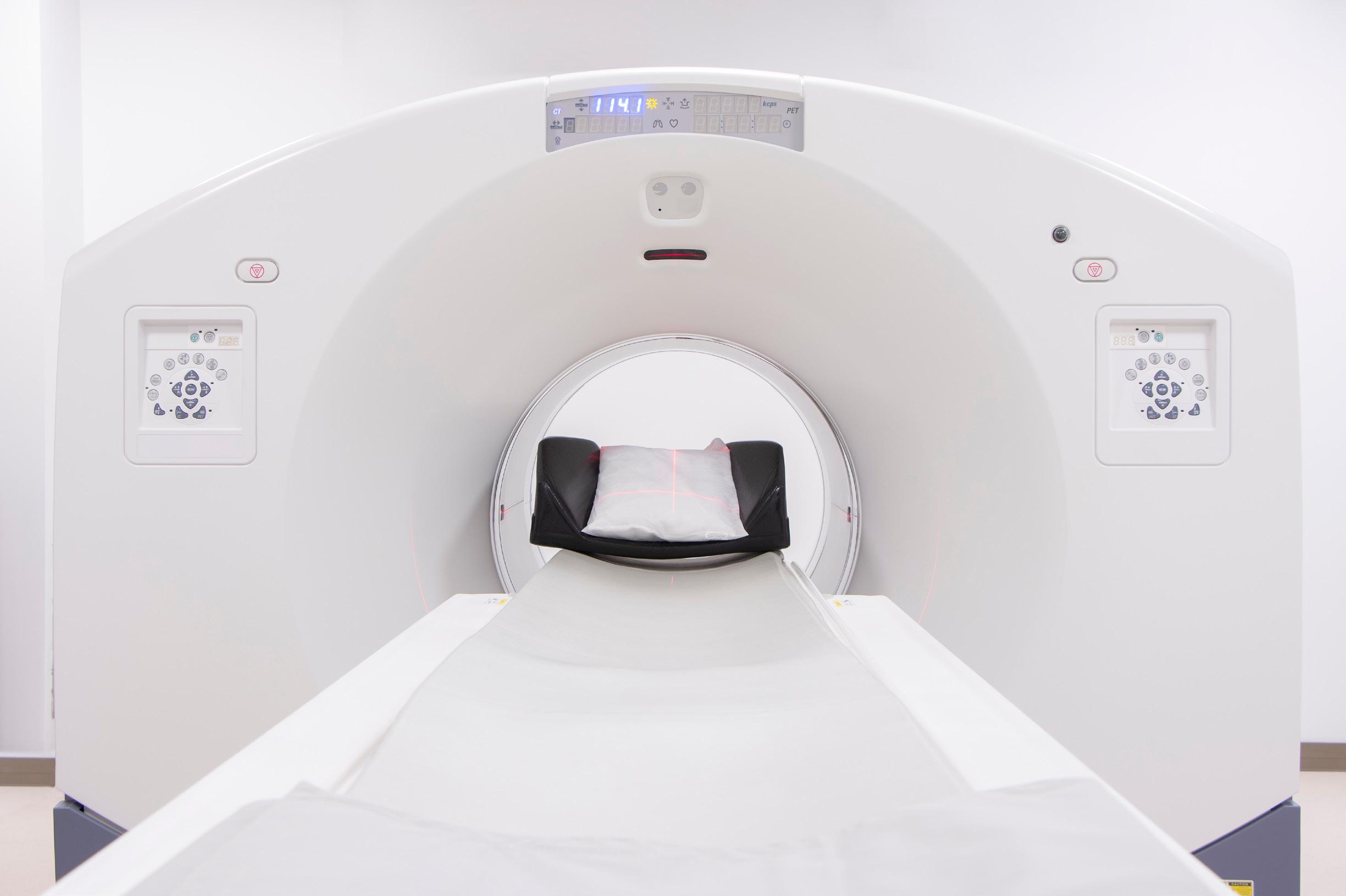
One company president I know says, “Nobody is omni scient, so don’t pretend to know all the answers.” And, since that president believes his core responsibility is making decisions to move the company in an effective direction, he purposefully seeks input from people who are well-versed in the various horizons so he can make good decisions.

The third area of responsibility is the organization’s capabilities. In other words, leaders should be aware of the capabilities and limitations of the organization’s equipment, personnel, policies and procedures. This is not advocating for micro-management. Leaders just need to know what the organization can and cannot do. From there, leaders must be proactive in providing funding for improvements if a cost-benefit analysis shows the investment would be worth it.


THE BIGGER LEADERSHIP PICTURE
Naturally, leaders are responsible for much more than what I’ve listed above. Other responsibilities include setting and communicating the vision and mission as well as budgeting and financial management, just to name a few. But I’m a firm believer that way too often, ideas, the horizon and the organization’s capabilities are three areas of responsibility that get shoved too far down the priority list. That’s because none of these three are necessarily urgent. All three are vitally important, but rarely are any of these things urgent. And so they get shoved aside way too easily.
The fix for this is to become disciplined. Firefighting on the latest major problems can easily consume one’s day, so finding ways that help with regimentation on these core fundamentals is essential. Perhaps it’s establishing an accountability partner, which might be as easy as creating standing meetings with administrative assistants where standard questions are asked and priorities are set. If not someone from within the organization, perhaps someone from without, such as a peer or an executive coach.
Whatever way works best, leaders must devote sufficient time to each of these areas so these vital fundamentals don’t fall through the cracks. •
Daniel Bobinski, who has a doctorate in theology, is a best-selling author and a popular speaker at conferences and retreats. For more than 30 years he’s been working with teams and individuals (1:1 coaching) to help them achieve excellence. He was also teaching Emotional Intelligence since before it was a thing. Reach him by email at DanielBobinski@ protonmail.com or 208-375-7606.
WHERE KNOWLEDGE, EXPERTISE, AND INTEGRITY MEAN NO WORRIES 512 477 1500 KEIMEDICALIMAGING COM 800-541-0632 brandywineimaging@comcast.net Offering the best reliability and support in the medical industry, just for you.
ICEMAGAZINE 43 WWW.THEICECOMMUNITY.COM
Digital Imaging, X-Ray Sales and Service Company
Established
MRI CT SERVICE, SALES & PARTS

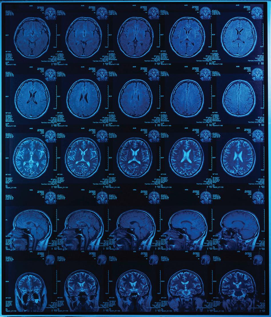


Imaging Service Solutions global provider of OEM quality pre-owned and refurbished medical imaging parts. ISS specializes in supplying parts for Philips and Siemens MRI and CT scanners.
in 2009 in Baltimore, MD, we proudly work closely with hospitals, service providers, and manufacturers globally.
SELL US YOUR MRI AND CT SYSTEMS! We purchase installed systems at fair market value and provide quality replacement parts for your equipment at a great value. We can remove purchased equipment at no cost as well. (410) 530-5548 | info@imagingss.com | 4001 Washington Blvd | Baltimore, MD 21227 Find us on Dotmed at dotmed.com/webstore/118100 imagingss.com SERVICES • OEM trained service engineers • Repair and maintenance • Professional installation and deinstallation • Rigging • Crating • Worldwide shipping • Trade-in equipment ADVANCING THE IMAGING PROFESSIONAL 44 ICEMAGAZINE | AUGUST 2023
EXPERT BODY LANGUAGE
THE ROMAN REVIEW
Iwas watching an interview regarding extraterrestrials. Yep, ET. The man being interviewed is an “expert” on the many ETs that have seeded and influenced the progress of mankind on earth. Obviously, my mind is open to anything, especially when I am bored.
I was surprised that the man had an amazing control of his verbal and nonverbal presentation. By this I mean that he appeared sincere and truthful over the seven or so one-hour episodes. Even trained politicians demonstrate signs of discomfort when conducting presentations or being interviewed. This man only showed discomfort when the interviewer said something that he had to clarify or correct. His discomfort appeared only during these times. It seemed to me that he was concerned for the interviewer’s feelings, not with what he was saying.
Either the man really does engage in ongoing contact with ETs, he actually believes it or he has extraordinary nonverbal control. Much has been said regarding communication including that the words themselves carry only about 7% of the message. For example: How do you interpret “Let’s eat, gramma” vs “Let’s eat gramma.” The words are the same, however how you say them, the tone caused by the comma, changes the meaning. Tone is said to carry 38% of the message. The remaining 55% of the message is conveyed by the nonverbal cues called the body language.
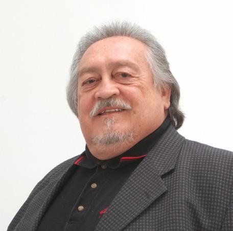
Some say that within five minutes you can evaluate people with 70% accuracy, which means that the other 30% can get you in much trouble. I subscribe to Barking Up The Wrong Tree by Eric Barker which is an the excellent newsletter. In “4 secrets to reading body language like an expert” he outlines ways that we get body language wrong.
• We ignore context : The defensive posture of crossed arms may be invalid if the room is cold. I have a friend that always crosses his arms when he
is conducting standing communication, even when he is the presenter.
• We don’t look for clusters : A single gesture does not make a body language sentence. Scratching the side of the nose, or the corner of the eye may demonstrate discomfort with what they are saying, however look for additional confirming gestures. In my opinion, there must be at least three body language cues indicating consistency to have true meaning.
• Ignoring biases : Our personal biases may cloud our judgement. Do you already have a positive or negative feeling about the individual or similar circumstances, etc.?
Mr. Barker suggests that we focus on “unconscious behaviors that are not easily controlled and convey a clear message.”
• Mirroring actions : Mirroring is when individuals speak in a similar language, speed and tone as well as assuming continuously similar body positions. This is easy to observe and very difficult to fake.
• Activity levels : The level of activity indicates interest and excitement. Higher activity equals higher interest.
• Consistency of emphasis and timing : This signifies focus, control and confidence.
It is beneficial to establish a baseline of an individual’s actions and then note any unconscious deviations, in clusters of course. Communication is a relatively difficult process involving multiple factors which can easily be misinterpreted. Becoming more proficient at interpreting body language will enhance your ability to determine the real meaning. Controlling your own will also help. I only touch my nose when I am alone speaking inside my head. •
Manny Roman, CRES, is association business operations manager at Association of Medical Service Providers.
MANNY ROMAN
INSIGHTS ICEMAGAZINE 45 WWW.THEICECOMMUNITY.COM

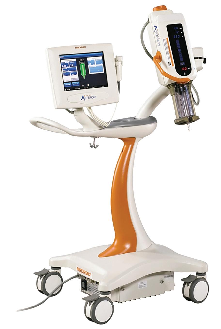


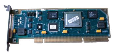
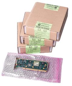


Contrast Injector Training • 100s of Error Codes not found in OEM Lit • Online and Onsite Training Available • Training BMETs since 2008 • Up to $5,500 in FREE Parts, PM Kits and Service • Massive Troubleshooting Library WWW.MAULLBIOMEDICAL.COM | 440-724-7511 | STEVE@MAULLBIOMEDICAL.COM ON-SITE AVAILABLE X-R AY T UBES & P ARTS FOR M EDICAL I MAGING E QUIPMENT Email: sales@w7global.com Toll Free: 855-W7GLOBA (974-5622) Online: www.w7global.com X-Ray Tubes for CT, Cath/Angio, and X-Ray • Tubes for All Major Equipment Brands • New, Used, and Refurbished • OEM Replacement Parts FOR 2022 Parts Vendors Certified ADVANCING THE IMAGING PROFESSIONAL 46 ICEMAGAZINE | AUGUST 2023
IT ALL STARTS ONLINE ICEWEBINARS .LIVE
1 2 3
REGISTER
Register to view live webinars each month.
WATCH Watch recorded webinars on-demand.


EARN
ARRT Category A CE credit is pending approval by the AHRA.

866-507-4793 allpartsmedical.com apmsales@philips.com AllParts Medical, a Philips company Portable X-Ray R&F and Fixed X-Ray C-Arms CT Confidence and reliability when you need it most. Cath lab MRI Biomed Ultrasound ADVANCING THE IMAGING PROFESSIONAL 48 ICEMAGAZINE | AUGUST 2023
1 Have title to
2 Troubles often caused by spinal problems


3 Venue
4 Board member, abbr.
5 It receives mucus from the sinuses
Visit theicecommunity.com for the solution.
Quote of the Month: 1 2 3 4 5 6 7 8 9 10 11 12 13 14 15 16 17 18 19 20 21 22 23 24 25 26 27 28 29 30 31 Across 1 Vital step for new employees 6 Top grades 9 Type of medicine that is a specialized area of radiology 10 Images providing diagnostic results 11 Key area of the brain 13 Sharp part of a blade 15 Part of the breathing cycle 17 ____ mater (outermost layer of three meninges) 20 Diagnostic test usually performed in the lumbar spine 21 Noninvasive med. procedure, abbr. 23 Capture 24 Types of billing banned by the Consolidated Appropriations Act 29 Liver secretion 30 Stomach area 31 Imaging tool that looks at moving bodily structures Down 1 Have title to 2 Troubles often caused by spinal problems 3 Venue 4 Board member, abbr.
It receives mucus from the sinuses
Software's operation
The gold standard for assessing the structure and function of the heart, 2 words 11 Direction of a market 12 Raise 14 Pacific territory of the United States 16 Zodiac sign 18 Emergency situation 19 Cleaning cloth 22 Lung rattle 23 Dosage amount, abbr. 25 Website symbol 26 Expert 27 Company going public 28 And so forth 1 2 3 4 5 6 7 8 9 10 11 12 13 14 15 16 17 18 19 20 21 22 23 24 25 26 27 28 29 30 31 Across 1 Vital step for new employees 6 Top grades 9 Type of medicine that is a specialized area of radiology
Images providing diagnostic results
Key area of the brain
Sharp part of a blade
Part of the breathing cycle
____ mater (outermost layer of three meninges)
Diagnostic test usually performed in the lumbar spine 21 Noninvasive med. procedure, abbr. 23 Capture 24 Types of billing banned by the Consolidated Appropriations Act 29 Liver secretion 30 Stomach area 31 Imaging tool that looks at moving bodily structures Down
ICE Break
IMAGING CROSSWORD
5
7
8
10
11
13
15
17
20
1 Have title to
2 Troubles often caused by spinal problems 3 Venue
4 Board member, abbr.
5 It receives mucus from the sinuses
7 Software's operation
structure and
of the heart, 2 words
Direction of a market
Raise
Pacific territory of the United States
Zodiac sign
Emergency situation
Cleaning cloth
Lung rattle
Dosage amount, abbr. 25 Website symbol 26 Expert
Company going public 28 And so forth 1 2 3 4 5 6 7 8 9 10 11 12 13 14 15 16 17 18 19 20 21 22 23 24 25 26 27 28 29 30 31 Across
Vital step for new employees 6 Top grades 9 Type of medicine that is a specialized area of radiology
Images providing diagnostic results
Key area of the brain
Sharp part of a blade
Part of the breathing cycle
____ mater (outermost layer of three meninges)
Diagnostic test usually performed in the lumbar spine
Noninvasive med. procedure, abbr.
Capture 24 Types of billing banned by the Consolidated Appropriations Act 29 Liver secretion 30 Stomach area
Imaging tool that looks at moving bodily structures Down
8 The gold standard for assessing the
function
11
12
14
16
18
19
22
23
27
1
10
11
13
15
17
20
21
23
31
7 Software's operation
2
Direction of a market
Raise
Pacific territory of the United States
Zodiac sign
Emergency situation
Cleaning cloth
Lung rattle
Dosage amount, abbr. 25 Website symbol 26 Expert 27 Company going public 28 And so forth ICEMAGAZINE 49 WWW.THEICECOMMUNITY.COM
8 The gold standard for assessing the structure and function of the heart,
words 11
12
14
16
18
19
22
23
“The purpose of our lives is to be happy.”
— Dalai Lama
ADVERTISER INDEX
626 Holdings p. 9
Advanced Health Education Center p. 44
HTM Jobs p. 30
ICE Webinars p. 47


PM Imaging Management p. 39
Radon Medical LLC p. 21
AllParts Medical p. 48

Imaging Academy p. 2
Ray-Pac® Ray-Pac p. 52
Banner Imaging p. 19

Brandywine Imaging p. 43
Imaging Service Solutions p. 44

Imaging Service Solutions global provider of OEM quality pre-owned and refurbished medical imaging parts. ISS specializes in supplying parts for Philips and Siemens MRI and CT scanners.


MRI CT SERVICE, SALES & PARTS


SELL US YOUR MRI AND CT SYSTEMS!
RIchardson Healthcare p. 5
SERVICES
CM Parts Plus p. 37
Engineering Services p. 3

KEI Medical Imaging p. 43
Established in 2009 in Baltimore, MD, we proudly work closely with hospitals, service providers, and manufacturers globally.

We purchase installed systems at fair market value and provide quality replacement parts for your equipment at a great value. We can remove purchased equipment at no cost as well.


imagingss.com
• OEM trained service engineers
• Repair and maintenance
• Professional installation
• Crating
• Worldwide shipping
RSTI p. 13
• Trade-in equipment
(410) 530-5548 | info@imagingss.com | 4001 Washington Blvd | Baltimore, MD 21227 Find us on Dotmed at dotmed.com/webstore/118100
Maull Biomedical p. 46

Metropolis International p. 48
Technical Prospects p. 4
SOLUTIONS
TriImaging Solutions p. 51 W7 Global, LLC. p. 46
INDEX ADVANCING THE IMAGING PROFESSIONAL 50 ICEMAGAZINE | AUGUST 2023
The Total Package
Hands-on technical training for all imaging modalities in Nashville, TN. Now offering an Apprentice program!
Service support for all major imaging manufacturers including GE, Philips, Siemens, Hologic and Toshiba/Canon.
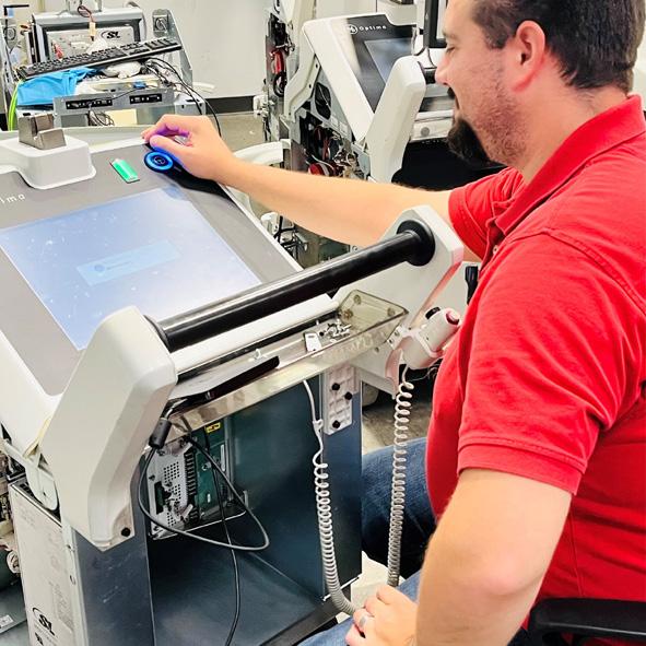

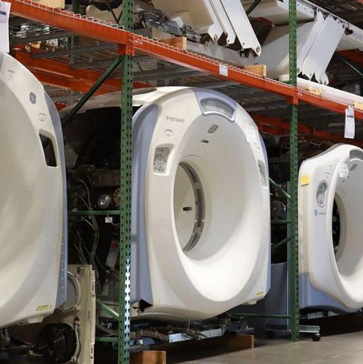
FOR EMPOWERING THE IMAGING PROFESSIONAL 1 REPLACEMENT PARTS
for CT, Mammography,
Cath labs,
X-Ray 2
TRAINING
Quality Imaging parts
MRI,
and general
TECHNICAL
4 PRE-OWNED EQUIPMENT
tested, and
our
Nashville, TN. 3 TECH SUPPORT & SERVICE
Inspected,
certified by
team in our 60k square foot facility in
SOLUTIONS triimaging.com | 855.401.4888 Located in Nashville, TN • ISO 13485:2016 certified
E7254 with RAD-21 and RAD-60 options now available. E7252 with RAD-14 options.
Plus Replacements for: E7239, E7242, E7252, DRX3724