

JULY 2023 | VOLUME 7 | ISSUE 7 MAGAZINE ADVANCING IMAGING PROFESSIONALS THEICECOMMUNITY.COM LOWERING SHIELDS THE PAGE 40 PRODUCT FOCUS: X-RAY PAGE 35 COMPANY SHOWCASE PAGE 12 COMPANY SHOWCASE PAGE 22


To learn more visit theimagingacademy.com or call 210.570.1452. WE ARE BUILDING THE NEXT GENERATION. As a student, you deserve more. THE WORLD MOVES FAST. WE MOVE FASTER.™ + powered by For the class schedule visit us at theimagingacademy.com. New Classes Begin February 13th Enroll Today! More versatile More valuable More job satisfied More upwardly mobile At Imaging Academy, we give you more.
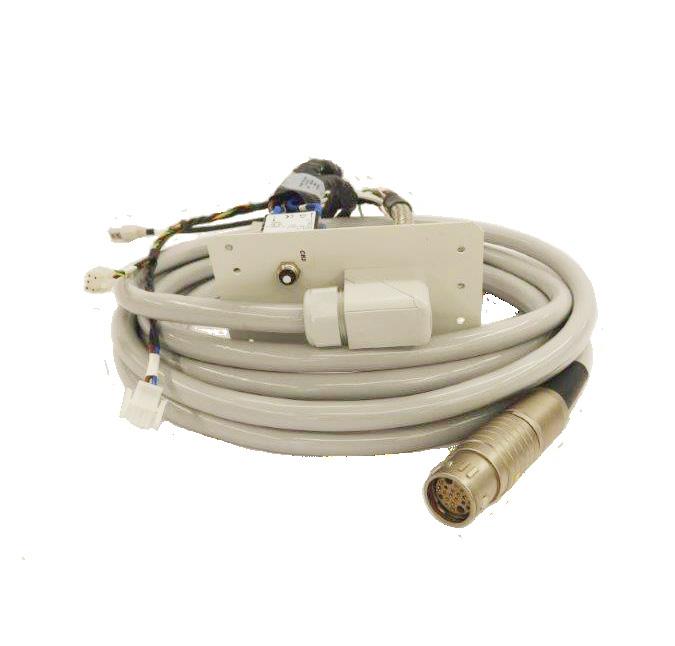
GE & OEC Cables SUPPLYING YOUR AT A PRICE YOU CAN AFFORD
Kenneth Saltrick, President of Engineering Services in Twinsburg, Ohio, knows from his long experience that C-arm machines themselves are absolute workhorses.
For customers looking to blend the gap between expensive OEM and unreliable used assemblies, WE have your solution.
Our complete repair contains a new cable assembly, utilizing all OEM cable and components with a harvested plate and connector housing as they are proprietary items. These completely repaired products will have a significant cost savings with build quality above new OEM products and carry a warranty of 180 days, which is untouchable in the market.

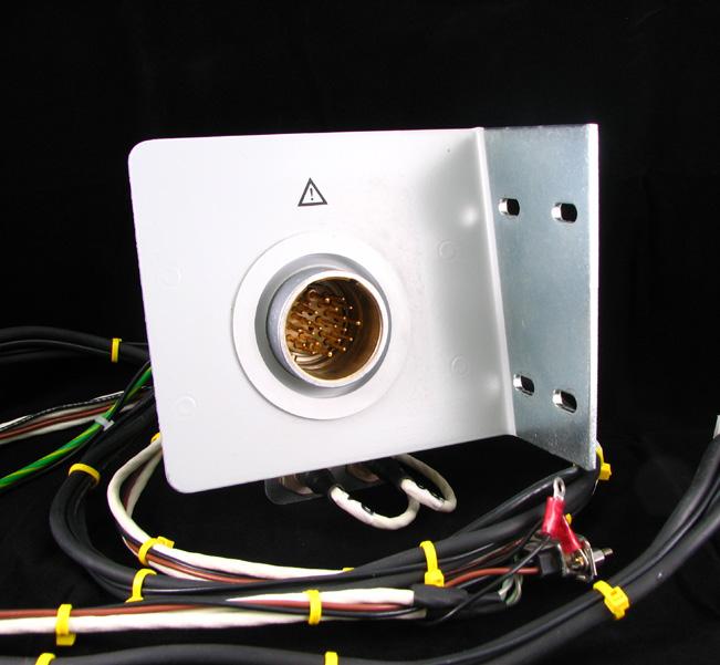
CONTACT US TODAY! 330.425.9279 X.11 | WWW.ENG-SERVICES.COM INFO@ENG-SERVICES.COM 9900, 9800 RECEPTACLE CABLES 9900, 9800 INTERCONNECT CABLES 5435446-01 AND 5840677-01 ELITE INTERCONNECT CABLE
Our technical support experts are ALWAYS AVAILABLE to assist.
Experts in Siemens Medical Imaging
DISASTER STRIKES! GOT BACKUPS?
System uptimes are more important than ever. Technical Prospects would like to remind everyone to practice responsible backup management.
Your system and calibration files as well as your protocols need to be backed up. Disaster recovery of mission critical equipment is a team effort and crucial to business continuity.

WHO SHOULD BE RESPONSIBLE FOR YOUR BACKUPS?
• Service Engineers
» Typically, during a PM or after a major repair.
• Department Managers or Lead Technologists
» Specifically, after a protocol or exam set changes.
Even if your system has an internal service hard drive for backups, we strongly recommend maintaining external copies.
As part of a responsible Disaster Recovery Plan, external backups are crucial.
877.604.6583 / 920.757.6583 | sales@technicalprospects.com | technicalprospects.com
TECHNICAL PROSPECTS
OEM NE W


FEATURES
DIVERSITY
How can I help to improve health equity?
COVER STORY
When colleagues say that lead shielding “doesn’t really provide any safety for the patients, but it’s the way it’s always been done,” it creates a moment for conversations.
RISING STAR
Amy Jeffreys “brings a lot of value to the Ft. Jesse imaging team.”

10 50 40 ADVANCING THE IMAGING PROFESSIONAL 6 ICEMAGAZINE | JULY 2023
IMAGING NEWS
Catch up on the latest news from around the diagnostic imaging world.
PRODUCT FOCUS
A look at X-ray devices and systems with features for today’s health care environment.
EMOTIONAL INTELLIGENCE
When disputes arise in healthy relationships, the issues in question are put on the table and discussed with objective language.

JULY 2023
26
54
35 ICEMAGAZINE 7 WWW.THEICECOMMUNITY.COM
CONTENTS SPOTLIGHT 10 Rising Star Amy Jeffreys 12 Company Showcase KMG 14 In Focus Pamela K. Woodard, MD 16 Off the Clock Zach Johnson, National Sales, Kings Medical Group of Richfield Ohio 20 Rad Idea Innovative AI Research 22 Company Showcase The InterMed Group NEWS 26 Imaging News A Look at What’s Changing in the Imaging Industry 33 Webinars Webinar Shares Effective Communication Tips PRODUCTS 34 Market Report 35 Product Focus X-ray INSIGHTS 46 Director’s Cut Unlocking the Potential of Your Team: Strategies for Effective and Engaging Training 48 SPONSORED: Avante Health Solutions Dose Regulation in a Vascular Lab Environment 50 Diversity My 4-Step Plan 52 PACS/IT The Mental Stress for Radiologists and Medical Imaging AI 54 Emotional Intelligence Resolve to Resolve Conflict 56 Roman Review Give Their Time Back 65 ICE Break: Imaging Crossword 66 Index ICE Magazine (Vol. 7, Issue #7) July 2023 is published by MD Publishing, 1015 Tyrone Rd., Ste. 120, Tyrone, GA 30290. For subscription information visit www. theicecommunity.com. The information and opinions expressed in the articles and advertisements herein are those of the writer and/or advertiser, and not necessarily those of the publisher. Reproduction in whole or in part without written permission is prohibited. © 2023 MD Publishing 1015 Tyrone Rd. Ste. 120 Tyrone, GA 30290 Phone: 800-906-3373 President John M. Krieg john@mdpublishing.com Vice President Kristin Leavoy kristin@mdpublishing.com Vice President of Sales Jayme McKelvey jayme@mdpublishing.com Group Publisher Megan Cabot megan@mdpublishing.com Editorial John Wallace Editorial Board Jason C. Theadore Nicole Dhanraj Melody W Mulaik Verlon E. Salley Rachel Thiesse-Yount Traci Foster Sales Emily Hise Art Department Karlee Gower Taylor Hayes Kameryn Johnson Events Kristin Leavoy Webinars Linda Hasluem Digital Department Cindy Galindo Kennedy Krieg Haley Wells Accounting Diane Costea 8 ICEMAGAZINE | JULY 2023 ADVANCING THE IMAGING PROFESSIONAL


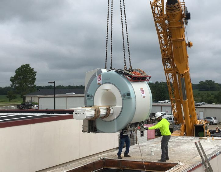
626 acquisitions to expand our services to our customers ICE thrives on providing specialized maintenance and repair of cryogenic refrigeration systems including cold heads, cryopumps, helium compressors, activated carbon oil adsorbers and other ancillary devices that cool superconducting magnets in MRIs. Call us and we will show you how we improve your service while we manage your cost! (800) 516-0990 | weare626.com You asked for a multi OEM, MRI Magnet Solution provider. WE ARE ISO 13485:2016 CERTIFIED
RISING STAR
AMY JEFFREYS
Amy Jeffreys holds the title of practice liaison at Ft. Jesse Imaging & Gale Keeran Center for Women. She holds a Bachelor of Science-Communication from Illinois State University and an Associate Degree in Radiography from Heartland Community College.

Jeffreys is also an imaging leader in the making, according to atleast one colleague.
“Amy goes above and beyond for both the Ft. Jesse Imaging & Gale Keeran Center for Women team and her community. She was part of a team that recently brought a new MRI magnet to their center along with some amazing patient experience lighting with a beautiful new PDC CaringMRSuite that includes LED Illuminated Image Ceiling and Patient-personal RGB LED Lighting,” Megan Shoaf shared in her nomination. “She cares about the imaging team and patients and has moved from a tech role to more of a marketing role to help the team continue to be successful. From the vendor site, we have really enjoyed working with Amy and feel she brings a lot of value to the Ft. Jesse imaging team in her marketing role.”
ICE found out more about this Rising Star in a recent question-and-answer session.
Q: WHERE DID YOU GROW UP?
A: Born and raised in Bloomington, IL, still here.
SPOTLIGHT
ADVANCING THE IMAGING PROFESSIONAL 10 ICEMAGAZINE | JULY 2023
Amy Jeffreys wants to make sure patients have the best experience possible.
Q: WHERE DID YOU RECEIVE YOUR IMAGING TRAINING/EDUCATION? WHAT DEGREES/CERTIFICATIONS DO YOU HAVE?
A: Associates in Radiography from Heartland Community College. I let go of my ARRT certification when I realized my path was in marketing and sales. Prior to that, I attended Illinois State University and received my bachelor’s in communication with a focus in public relations.
Q: HOW DID YOU FIRST DECIDE TO START WORKING IN IMAGING?
A: After graduating with a four-year, I realized my passion was in health care. My local community college has a reputable radiography program, which I applied for and was accepted. It was a tough, but rewarding two years … I learned a ton and met some wonderful people in the field, many who I still get the privilege to work with on various projects. I am so grateful for my educational path that got me where I am today!
Q: WHY DID YOU CHOOSE TO GET INTO THIS FIELD?
A: Easy, getting the opportunity to help and care for people.
Q: WHAT DO YOU LIKE MOST ABOUT YOUR POSITION?
A: Everyday is different … you can find me in a doctor’s office, at a community event, on the phone helping solve a problem or get a patient in for an exam faster, networking with our wonderful community members over a meal or a drink, working with a facility on how we can best serve their patients, or working from my car! I get the best of both worlds, working with our medical and consumer community, but also helping patients from behind the scenes.
Q: WHAT INTERESTS YOU THE MOST ABOUT THE IMAGING FIELD?
A: It truly is the key to many diagnoses. Imaging gives physicians and patients answers to what their next move may, or may not, be. The technology is fascinating to me, and I love how much there is to learn!
Q: WHAT HAS BEEN YOUR GREATEST ACCOMPLISHMENT IN YOUR FIELD THUS FAR?
A: Simply being with my imaging center for 10 years and seeing the enormous growth, not only with volume, but our wonderful team of technologists and administrative folks. There are so many projects we as a team have rolled out and introduced to the community, from 3D mammography, ABUS, endless new technologies/equipment/AI and exams, to the most recent roll out of our 3T Wide Bore MRI CaringSuite!
Q: WHAT GOALS DO YOU HAVE FOR YOURSELF IN THE NEXT 5 YEARS?
A: Well, as someone who takes life a day at a time, this is a hard one to answer. There is so much change happening in our local medical community, and so much opportunity for our center. Without a doubt, I only see growth for our center. We have so much to offer being an independent outpatient imaging center, and currently the only one in our market. Personally, my goal is to continue to pour my heart into this center and make sure patients have the best experience possible. I also want to explore new opportunities for professional development in the field. •
FUN FACTS
FAVORITE HOBBY:
Golf (trying at least), working in the yard, and spending time with my sweet family and a great circle of friends.
FAVORITE SHOW: My guilty pleasure … anything on Bravo.
FAVORITE FOOD: Lobster and steak
FAVORITE VACATION SPOT: Marco Island, Florida
1 THING ON YOUR BUCKET LIST:
To visit Italy and the Mediterranean Coast
ICEMAGAZINE 11 WWW.THEICECOMMUNITY.COM
SOMETHING YOUR CO-WORKERS DON’T KNOW ABOUT YOU: I am a pretty open book!
COMPANY SHOWCASE
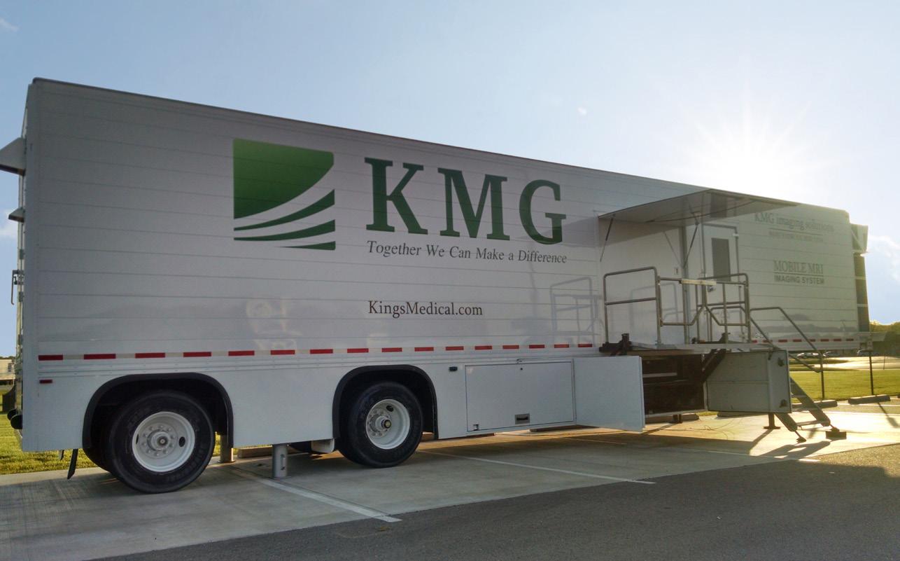
In the fast-paced world of health care, hospitals and imaging centers are constantly grappling with numerous challenges that hinder their efficiency and profitability. Patient backlog, lack of continuity of care and missed returns on investments (ROI) are just a few of the recurring obstacles they face daily. However, with the expert guidance and innovative imaging solutions provided by KMG, these challenges can be overcome, leading to improved operations, enhanced patient experiences and optimized financial outcomes.
1. PATIENT BACKLOG: UNLOCKING THE DOORS TO DELIVERING EFFICIENT CARE
One of the most pressing challenges faced by hospitals and imaging centers is the ever-growing patient backlog. As demand for medical imaging scans continue to rise, organizations struggle to manage and streamline their patient flow effectively. This not only leads to prolonged wait times and frustrated patients but also puts a strain on resources, hindering the overall efficiency of the facility.
At KMG, we recognize the urgency of addressing this issue head-on. By partnering with us, you can provide your patients with mobile MRI, CT and PET/CT imaging services for any desired amount of time. Your KMG sales representative will be able to review short or long-term leasing options to help you decide what makes sense for your facility.

Your mobile unit (trailer) will come fully equipped with everything you need such as:
• Coils (head, neck, spine, body, small flex, large flex, shoulder, wrist, knee)
• Injector
• Changing curtain
• IT/Connections
• Chairs and Desk
• Stereo System
• Fire Extinguishers
• Mobile Safe Gurneys
Staffing struggles can also impact efficient patient care, creating longer wait times, delayed exam results and can impact overall clinical operations. At KMG, we help alleviate these staffing issues by only offering ARRT certified clinical staff. All of our patient-facing medical technologists and support staff are handpicked to ensure that they are a seamless fit for you and your facility. What’s more, we require rigorous drug and background screenings and hold professional medical liability insurance on all our employees that are providing medical services to patients.
2. LACK OF CONTINUITY OF CARE: CONNECTING THE DOTS FOR PATIENTS AND HEALTHY OUTCOMES
ADVANCING THE IMAGING PROFESSIONAL 12 ICEMAGAZINE | JULY 2023
INTRODUCING KMG: TRANSFORMING MEDICAL IMAGING SOLUTIONS FOR HOSPITALS AND IMAGING CENTERS SPONSORED CONTENT SPOTLIGHT
Another critical challenge faced by health care providers is the lack of continuity of care. When hospitals or imaging centers do not have the proper imaging equipment or setup, it is impossible to keep up with the imaging demand. The inability to provide these services leads to potential patient and revenue loss.
KMG specializes in developing customized solutions that foster collaboration and enhance care coordination across the health care ecosystem. Proper medical imaging solutions allow your facility to maintain the highest standards of care for your patients. With a mobile or interim mobile imaging unit from KMG, you will no longer need to send your patients out for scans – potentially losing them to another provider or facility. This continuity of care promotes patient-centered care, establishing a trusting relationship between the patient and the provider. When the patients have ongoing relationships with their health care team, they feel more comfortable discussing their health concerns, sharing personal information and participating in shared decision-making. This leads to better health care outcomes and patient satisfaction.
3. MISSED ROI: MAXIMIZING YOUR HEALTH CARE AND IMAGING INVESTMENTS
In an era of tightening budgets and increasing financial pressures, hospitals and imaging centers face the constant challenge of achieving a return on their investments. A loss of ROI can have a cascading effect on equipment, staffing, patient care, research, facility maintenance and financial stability. It may also hinder the department’s ability to provide high-quality services, impede technological advancements, and compromise patient outcomes. Therefore, it is essential for hospitals and imaging centers to prioritize their investments and financial sustainability to ensure continued success.
KMG understands the significance of optimizing ROI for health care and imaging investments. With our mobile or interim imaging solutions, our many leasing and financing options – you can continue to grow and sustain your patient populations and advance your technology all while maintaining a low monthly cost. Our site planning guide
will help address every possible need and contingency to ensure that you have the right mobile imaging equipment and support along the way.
PARTNERING WITH KMG FOR A FUTURE-PROOF HEALTH CARE LANDSCAPE
Being an employee-owned company with over 40 successful years in the imaging industry, KMG knows what it takes to provide seamless turnkey imaging solutions. Through our tailored approach, we empower hospitals and imaging centers to overcome patient backlog, enhance continuity of care and unlock missed ROI opportunities. By leveraging our deep industry expertise, cutting-edge technologies and unwavering dedication to client success, health care organizations can thrive in an increasingly competitive environment while providing the highest standard of care to their patients.
In addition to Interim mobile imaging solutions, KMG also offers:
• Modular Units
• Fixed-Base/In-House Solutions
• Short-Term and Long-Term Solutions
• Leasing and Financing
• Equipment Service
• Staffing
• Site Survey
• Project Planning and Management
Download our Site Planning Guide here: resources.kingsmedical.com/planning-basics-guide
KMG Website: kingsmedical.com
Email: contact@kingsmedical.com
Phone: (800)-854-9061
KMG installing a fixed-base MRI Unit into a hospital. KMG has the experience to give you the fixedbase imaging suite you and your patients deserve.
ICEMAGAZINE 13 WWW.THEICECOMMUNITY.COM
Where flexibility meets function. One of KMG’s Mobile MRI Units, set-up and ready to scan in an imaging center parking lot.
FOCUS IN
PAMELA K. WOODARD
BY TAMERA BHANDARI
Pamela K. Woodard, MD, a national leader in cardiothoracic imaging, has been named head of the Department of Radiology, Director of Mallinckrodt Institute of Radiology (MIR) and the Elizabeth E. Mallinckrodt Professor of Radiology at Washington University School of Medicine in St. Louis. She will begin her new role July 1.

In this new role, she will leverage her extensive research, clinical and leadership experience in the field of radiology. She is currently the Hugh Monroe Wilson Professor of Radiology, senior vice chair and division director of MIR’s Radiology Research Facilities, director of the Center for Clinical Imaging Research, head of Advanced Cardiac Imaging CT/MRI, director of the Radiology Research Residency Program, and director of TOP-TIER, a clinician-scientist training program for residents and fellows.
“Dr. Woodard was unanimously selected by our leadership team from a deep and impressive group of
candidates,” said David H. Perlmutter, MD, executive vice chancellor for medical affairs, the George and Carol Bauer Dean of the School of Medicine, and the Spencer T. and Ann W. Olin Distinguished Professor. “We believe she can lead us in further defining the career of radiologists and imaging scientists, the role of imaging in personalized medicine, and new strategies for diagnosis and treatment through interventional, minimally invasive and even noninterventional approaches that will advance human health. Her experience in collaborative work with other clinical and preclinical departments is an essential ingredient of the virtuous cycle of academic medicine that exemplifies the partnership of WashU Medicine and BJC HealthCare.”
Also a professor of medicine, of pediatrics and of biomedical engineering, Woodard conducted seminal research that led to the translation of cardiac magnetic resonance imaging (MRI) into clinical practice, including methods to improve imaging quality by suppressing respiratory motion. Such methods are in use in pediatric cardiac and congenital heart imaging.
She also led a team that devel-
SPOTLIGHT
Pamela K. Woodard is head of the Department of Radiology, Director of Mallinckrodt Institute of Radiology (MIR) and the Elizabeth E. Mallinckrodt Professor of Radiology at Washington University School of Medicine in St. Louis
ADVANCING THE IMAGING PROFESSIONAL 14 ICEMAGAZINE | JULY 2023
oped a nanoparticle-based imaging agent for atherosclerotic plaques in blood vessels. The imaging agent detects a protein associated with unstable plaques that are prone to causing sudden major problems such as a heart attack or stroke.
Woodard is involved in the translation of novel positron emission tomography (PET) agents to assess blood flow through heart muscle. Poor blood flow is a sign of cardiovascular disease that could cause serious problems such as heart attacks. Her work led to the development of an imaging approach now widely used to assess blood flow through heart muscle, crucial information that doctors use to determine optimal treatment for each patient.
Woodard’s early work involved novel approaches to imaging blood clots in the lungs. In 1995, as a resident at Duke University, she published one of the early papers showing that such clots could be detected by multidetector spiral CT scan, then a developing technology. This type of CT scan uses an array of detectors to acquire multiple images simultaneously. Later, as an assistant professor at Washington University, she was a principal investigator on a clinical trial funded by the National Institutes of Health (NIH) that resulted in a landmark paper in The New England Journal of Medicine and established multidetector CT as the standard of care for diagnosing blood clots in the lungs.
The Department of Radiology is a world leader in radiological innovations that advance the science of imaging to improve patient care and further biomedical research. It has a long history of national leadership in the practice and science of radiology, and in educating the next generation of radiologists and radiology researchers.
“I am honored to serve and lead Mallinckrodt Institute of Radiology at Washington University School of Medicine in this important role,” Woodard said. “The Department of Radiology has a long tradition of excellence and innovation in clinical radiology, radiology education, and imaging research. I am delighted to lead our world-class faculty and trainees in radiology into the next decade in collaboration with our partners at BJC HealthCare and across the Medical Campus.”
Woodard earned her bachelor’s and medical degrees at Duke. She completed her internship in internal medicine at the University of North Carolina at Chapel Hill and her residency in radiology at Duke before coming to Washington University for a clinical fellowship in cardiothoracic radiology. She joined the School of Medicine faculty in 1997.
She is a fellow of the American Association for the Advancement of Science, the American Institute for Medical and Biological Engineering, the American College of Radiology and the American Heart Association. She serves on the Board of Chancellors for the American College of Radiology, the Executive Committee of the Board of the Academy for Radiology and Biomedical Imaging Research, and the Board of the Society for Cardiovascular Computed Tomography.
Woodard will succeed Richard L. Wahl, MD, who has led the department for nine years. Wahl will continue to lead a research laboratory as a professor in the department.
“Dr. Richard Wahl’s leadership has continued the strong tradition and legacy of Mallinckrodt Institute of Radiology and positioned the department for even greater potential in the future,” Perlmutter said. “Dr. Pamela Woodard is the ideal person to take on the mantle to take the department into the next era of foundational accomplishments.” •
FEBRUARY 18-20, 2024 IRVINE, CALIFORNIA CALL FOR PRESENTERS ATTENDICE.COM ICEMAGAZINE 15 WWW.THEICECOMMUNITY.COM
Clock Off THE
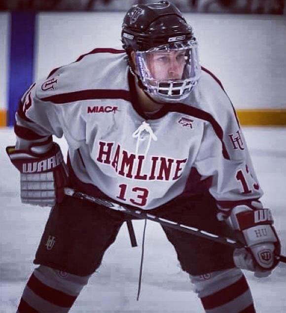
ZACH JOHNSON, NATIONAL SALES, KINGS MEDICAL GROUP OF RICHFIELD OHIO
BY MATT SKOUFALOS
Like many kids growing up in Minnesota, Zach Johnson was out on the ice, learning to skate, not long after he could walk. He grew up on frozen ponds and outdoor rinks, shooting pucks around the unfinished basement of his parents’ home in Rogers, and playing one-on-one with his dad. At three, he started playing Mini Mites hockey in Elk River; then moved up to Squirts and Pee Wees after the family moved to Saint Michael. By the time Johnson was approaching his 13th birthday, he was an eighth-grader, about 5-feet-eightinches tall and 165 pounds. Saint Michael added him to its varsity hockey team, where Johnson lettered through 10th grade.
Although every young hockey player dreams of someday playing in the National Hockey League, in Minnesota, their first dream is to compete in the state high school hockey championship tournament. When Johnson moved from Saint Michael to Mound, it felt closer than ever.
“It’s always the dream; the thing you idolize, you go do,” Johnson said. “You take a week off of school. You’re downtown at the Xcel Energy Center, watching the state tournament, eating junk food, and going to Tom Reid’s [Hockey City Pub]. You’re running around Eagle Street; it’s something you look forward to, whether you’re in it or not.”
Johnson’s team at Saint Michael was “OK,” he said, but too small to be competitive. At Mound Westonka High School, the team lost in the semifinal qualifiers of his junior year, and then again to a team from Breck School during his senior year at a time when private academies were creating super-teams comprised of young transfer players. Those schools boasted deeper benches and the ability to roll three or four lines; at Mound, Johnson and his linemates were on the ice for most of the game, pivoting between forward and defense positions.
“You maybe take a minute breather, and you’re back out,” Johnson recalled. “You had to be in tip-top shape. But if you get me on the ice, I don’t want to come off if I don’t have to. I pretty much played year-round.”
In the summer, Johnson had begun playing with a Triple-A youth development team called the Minnesota Thunder; from there, he went to another, even more influential developmental club called the Minnesota Lightning. When he wasn’t skating with his school teams, Johnson was on the ice two to three nights a week, practicing, and traveling every weekend for tournaments.

“My coaches taught us to battle and compete,” he said. “I don’t think we lost a game that summer. A group of guys from all over Minnesota; one of them’s still playing in the show: Nick Jensen, a pretty stellar defenseman for the Washington Capitals. Jake Gardiner, Nic Dowd; those summer-league teams were always going.”
“I thank my parents every day, because you look at how much that game costs, and the tournaments we were always traveling to and playing in,” Johnson said. “I really learned the game at that time in my career. That team is one I truly, truly enjoyed, and look back at some of my favorite memories from there, growing up.”
After graduation, Johnson had an opportunity to consider extending his athletic career in a professional hockey league. At 18, he was drafted by the Junior A Texas Tornado, but the club folded. Johnson was invited to play in the Ted Brill Great Eight, a weekend tournament composed of the top 20 players from each high-school conference in Minnesota, created largely as a scouting showcase. After that event, Johnson was invited to play with the Wenatchee Wild, a new Junior A club that was rolling out in the North American Hockey League (NAHL).
“I got a call from Paul Baxter, Ryan McKelvie, and Chris Clark, who were starting a new team called the Wenatchee Wild. Clark and McKelvie were both Mankato Mavericks [from Minnesota State University],” Johnson said. “Paul Baxter had
SPOTLIGHT ADVANCING THE IMAGING PROFESSIONAL 16 ICEMAGAZINE | JULY 2023
fists like a boxing mitt. He’d fought guys like [Bob] Probert and all the big dogs back in the show; but he was not much bigger than me.”
“I was 5’ 8” and maybe 195,” Johnson recalled. “It was still a rough-and-tough junior league. There wasn’t any limit to fights you could have in a season. Opening day, we were playing the Topeka RoadRunners, and ended the weekend with a bench brawl.”
For an 18-year-old still looking to find his game, the BCHL offered an opportunity for Johnson to play professional hockey until he aged out of the league at 21. Baxter, Clark and McKelvie were bringing the game to a city of 30,000 people who had had no idea what it was, and youngsters like Johnson were instant celebrities.

“You had a $58-million arena, 5,000 fans, and it was packed every night,” Johnson said. “We never paid for meals. You’re signing autographs for kids; people are buying your jersey. It was wild.”
Johnson had been a prolific goal-scorer throughout his youth hockey career, but at Wenatchee, he learned his role would need to evolve. The coach, Baxter, had been an enforcer at the pro level: a role player who does the work of fighting to limit the opposition taking liberties with the star skilled players. He pulled Johnson aside before the season started,
and told him about his expectations.
“He said, ‘You got voted captain by your teammates. Here’s what I want you to do. You don’t complain about anything in practice. You go your hardest. And if I tap you on the shoulder in the middle of the game, I want you to find the biggest guy on the other team and beat the hell out of him.’ ”
“You learn to fight for your teammates,” Johnson said. “You bleed for them; whatever it takes. You find a solution, you back your team up. It’s the one thing that’s translated from my life outside of hockey. You’re part of a team? You do whatever you can for that team until you can’t anymore.”
Johnson did as he was asked. He killed penalties, grinded out shifts on the third line, checking against the other team’s top forwards, and, when it came down to it, threw hands with the guys on the other side. Nagging injuries from his high-school career continued to impede his development, however. So when his eligibility for the Junior A league ended, Johnson joined the team at Hamline University, back in Saint Paul, Minnesota, as a 21-year-old freshman. He graduated with a degree in biology, and a roster full of friends he’s still in touch with to this day.

“We were one, no matter what,” Johnson said. “It was one of the best teams to be a part of. We had the best record
ever as a Hamline hockey team, won the regular season championship, won the playoffs, and ended up getting beat by the national tournament champions, St. Norbert, in their home barn.”

Wherever he played, Johnson always dove into the organization to make himself more valuable to it. On off-weekends, he’d sign autographs, do interviews, and pay his respects to the people who put me there. After college, Johnson coached women’s hockey, and discovered he might have even been a better coach than he was a player.
The competitive mindset that served him well as an athlete and teammate made him a valued asset to Kings Medical Group of Richfield, Ohio, where he works in national sales. His hockey play is confined mostly to a men’s chapel league on Wednesday nights, or the occasional private lesson.
But to this day, when Lake Minnetonka freezes over, you can find Johnson lacing up his skates, heading out on the glassy surface with his puppy to chase pucks and catch the sunrise. In those quieter moments, though, it’s still so easy for him to recall the memories of the arena pyrotechnics on the ice at Wenatchee; to hear his name called with the rest of the starting lineup; and to feel the roar of the crowd thundering over the goal horn. •
ICEMAGAZINE 17 WWW.THEICECOMMUNITY.COM
Zach Johnson grew up playing hockey on frozen ponds and competed professionally before joining the imaging realm.
IT ALL STARTS ONLINE
ICEWEBINARS .LIVE
1 2 3
REGISTER Register to view live webinars each month.
WATCH Watch recorded webinars on-demand.


EARN
ARRT Category A CE credit is pending approval by the AHRA.


GE • TOSHIBA SIEMENS • PHILIPS Give us a call at 213-276-8209 (844-PMIM-MRI) or visit www.pmimagingmgmt.com LOOKING TO PURCHASE OR SELL AN MRI, PET/CT, CT, OR X-RAY? LOO K N O FURTHER . PROOF SHEET LOOKING TO PURCHASE OR SELL? LOOK NO FURTHER. 213-276-8209 (844-PMIM-MRI) PMIMAGINGMGMT.COM GE • TOSHIBA • SIEMENS • PHILIPS PUTTING CUSTOMERS FIRST SINCE 1987 TOSHIBA • GE • PHILIPS • SIEMENS AND MORE! Call: 508.730.9544 or 508.559.9441 www.InternationalXrayBrokers.com admin@intxray.com INTERNATIONAL X-RAY BROKERS IS NOW AN AUTHORIZED DISTRIBUTOR FOR CUSTOM BUILT SURGICAL TABLES! ALL MANUFACTURES & MODALITIES WE ALSO BUY AND SELL PRE-OWNED MEDICAL IMAGING EQUIPMENT. ICEMAGAZINE 19 WWW.THEICECOMMUNITY.COM
Rad idea
BY KRISSIE D. STICH, MBA, CRA, R.T. (R)
INNOVATIVE AI RESEARCH
The Department of Radiology at University Hospitals has partnered with the health system’s innovation and commercialization engine, UH Ventures, to launch a new effort that leverages UH radiologists’ research expertise in artificial intelligence applications. The new entity is already collaborating with start-ups worldwide to validate emerging AI radiology technologies, such as algorithms that promise to identify patient fractures or strokes.
The Radiology AI & Diagnostic Innovation Collaborative at UH – RadiCLE for short – is led by Leonardo Kayat Bittencourt, MD, Ph.D., Vice-Chair of Innovation in the Department of Radiology. Bittencourt says the initiative has dual, complementary goals – to advance the science of teaching and clinical adoption of radiology AI, while simultaneously serving as a revenue stream for the department and UH through its collaborations with outside entities. The infrastructure is in place to ensure both aims are met. Under Bittencourt’s leadership, the collaborative employs two full-time employees, Traci Hollimon, program manager, who manages the “pipeline” of requests for research, validation and co-development assistance from UH radiologists, and a data handler who will focus on curating high-value, complex imaging datasets. RadiCLE also works closely with UH Ventures to leverage existing infrastructure to perform preliminary due diligence on new opportunities and to track opportunities through the various stages of validation and development.
“Establishing a physical place inside the institution with the talent, infrastructure and equipment will help UH establish the right processes and procedures to effectively engage and expose UH researchers and physicians to emerging technological advances of AI. And when you have the subject matter experts at the table with emerging technologies such as AI there is a greater promise the quality of healthcare will ultimately improve,” says Kendra Gardiner,
director of product strategy at UH Ventures.
“We must remain at the forefront of technological advancements. RadiCLE’s model promotes innovation and the seamless adoption of AI technologies,” Bittencourt says. “The industry has quickly understood that quality data and, more importantly, quality people are the foundation for the success of any AI solution intended for health care. Ultimately, companies need to have inside experience with a healthcare partner. RadiCLE partners with companies by supplying de-identified patient data, conducting clinical studies, and providing the essential clinical expertise to understand the problem.”
UH IS UNIQUELY POSITIONED TO LEAD IN AI
“UH is a robust organization with a large and diverse patient population, combined with significant AI research expertise in the department of radiology,” Bittencourt adds. “This combination makes UH uniquely qualified to play this vital research and technology-validating role. Additionally, we have built a RadiCLE Clinical Champions Council bringing together various clinical experts, including physicians, technicians, physicists, nurses and students, across UH Radiology to provide insights and identify projects to help drive AI innovation. The spectrum of data we can collect and then offer to address different questions is very valuable, especially to the patients we serve.”
Bittencourt says this data richness and being an academic center with researchers engaged in many different fields is exactly what companies seek when looking to develop AI solutions. And, UH also has a lot to gain from these new relationships.
“These companies come with teams of engineers and data scientists seeking a partner to test and/or build interesting solutions. These solutions hold the promise to impact our patient population positively. As a result of these collaborations, we have opportunities to conduct funded research while generating additional revenue to boost the
SPOTLIGHT
ADVANCING THE IMAGING PROFESSIONAL 20 ICEMAGAZINE | JULY 2023
department’s activities and the overall health system. But, perhaps even more importantly, we know in the long run, we will provide a space and the people needed to support our caregivers who begin to generate their own ideas and begin innovating from inside the institution. ”
THE COLLABORATIVE IN ACTION
One of the first projects initiated through RadiCLE was with the French company AZmed. AZmed had developed a tool called AI Rayvolve, which aimed to boost the speed and accuracy of fracture diagnosis and solve the problem of missed fractures. Navid Faraji, MD, Associate Program Director of the Radiology Residency and Clinical Champion in musculoskeletal imaging, served as the lead investigator on this initiative.
The process was comprehensive: To help validate the AZmed software, three UH board-certified musculoskeletal radiologists read and annotated fractures on 2,626 X-rays of UH patients. These evaluations served as the “ground truth” for accessing the validity of AZmed’s tool. Additionally, three groups of UH physicians identified fractures in 186 randomly selected cases, both with and without the aid of the AI tool. The readers included eight ED physicians, eight non-musculoskeletal radiologists and eight musculoskeletal radiologists. The research team then compared differences in fracture detection and interpretation time with and without the AI tool.
Results showed that across all physicians in the study, review accuracy using the AZmed algorithm increased by 5.6 percent. Additionally, review and interpretation time decreased by 27 percent, proving the algorithm’s efficacy. Interestingly, emergency physicians and non-musculoskeletal radiologists reported higher performance utilizing the new tool.
According to Bittencourt, results for inexperienced readers showed the algorithm improved their performance to a similar level of experienced readers. “This underscores the potential of AI to augment human ability instead of replacing it,” he says. “It also reduced the time required to read the X-ray from opening to diagnosis, a potential gain for patients in terms of time to diagnosis.”
These study results ultimately played a pivotal role in AZmed’s securing clearance from the U.S. Food and Drug Administration for its AI Rayvolve. UH was the only health care system in the U.S. to collaborate with AZmed in testing its new technology. The company’s reception at a recent radiology meeting was positive, with attendees showing great interest in AZmed’s now-validated product. The recognition UH received as an enabling institution was also significant.
“UH is getting recognition as one of the few institutions capable of undertaking these validation and development projects,” Bittencourt says.
NEW WAYS TO BENEFIT PATIENTS
RadiCLE is also hard at work on several other projects. For example, RadiCLE joined Massachusetts General
Hospital and other prestigious academic medical centers as a charter member of a consortium convened by the American College of Radiology to work on the validation of AI algorithms. The first project validated a stroke detection algorithm in both CT and MRI. Other projects include:
• improving detection of prostate cancer on MRIs;
• opportunistic screening of cardiovascular risk from routine X-ray based imaging exams; and
• devising a better way to identify lung nodules as a precursor to lung cancer.
Furthermore, Amit Gupta, MD, Radiology Division Chief of Cardiothoracic Imaging and an AI Clinical Champion, has been instrumental in procuring and vetting new AI technology, being already widely successful in several AI collaborations, including an algorithm fully implemented in clinical practice for automated detection of pneumothorax, a potentially life-threatening condition.
Bittencourt says he’s excited about the potential of radiology AI to improve patient care – and the vital role this new collaborative at UH can play in making that happen. “AI will never obviate the need for radiologists or replace a doctor’s care, expertise or human touch, but it can and will boost the quality of care.”
“AI will augment our ability by increasing our combined diagnostic accuracy, increasing patient safety, and reducing burnout and stress by automating repetitive and low-value tasks. AI also holds the promise of improving the timeliness of discovery and reporting of findings on imaging exams – an added benefit to all the patients we serve.”
For questions or to learn more about RadiCLE at University Hospitals, please email Traci Hollimon, Program Manager of RadiCLE, at RadiCLE@UHhospitals.org. •
Share your RAD IDEA via an email to editor@mdpublishing.com.
ICEMAGAZINE 21 WWW.THEICECOMMUNITY.COM
COMPANY SHOWCASE
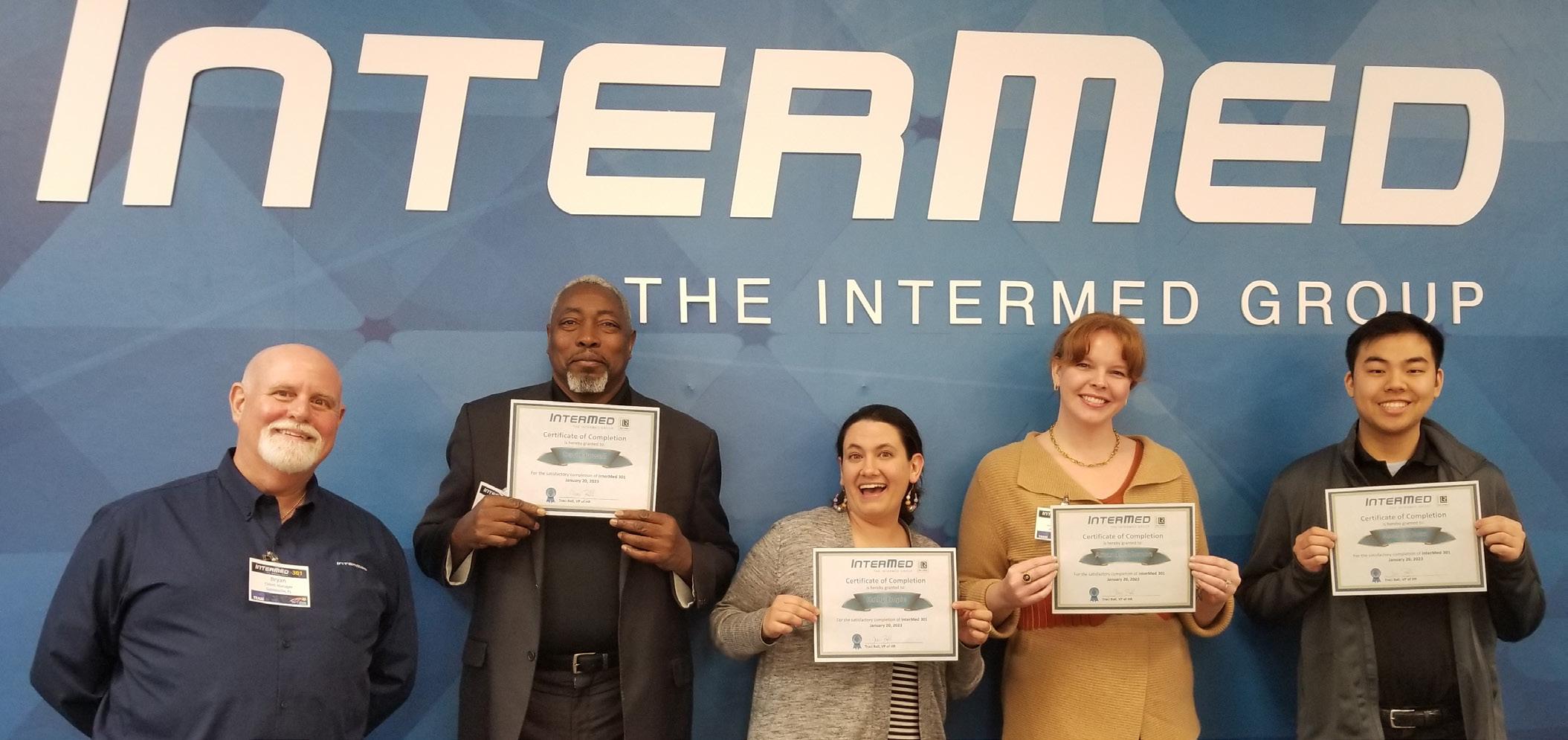
THE INTERMED GROUP
The InterMed Group is a dynamic provider of comprehensive healthcare technology management (HTM) services covering a broad range of client needs. InterMed’s deep-rooted partnership philosophy drives all its offerings, helping to ensure everything it does moves clients closer to achieving their goals.
The most encompassing of their offerings is their HTM services. InterMed acts as the client’s “one-stop shop” for their medical devices – whether that is filling in the gaps for the health care facility’s existing program or implementing a new one. They always bring the best to the client’s devices ranging from the linear accelerators, MRIs and CTs through anesthesia, dialysis, and respiratory therapy, to the patient monitors, infusion pumps and beds and everything in between.
Expanding on this, InterMed also provides field service-based contracts on medical equipment. Roughly 50% of its technical team members are specialized in diagnostic imaging where it provides service contracts for specific devices in hospitals, imaging centers and veterinary hospitals. In service and support of their clients InterMed team members abide by the “Sundown Rule” – they address every challenge or customer service concern by sundown each day, so their clients know their response and when to expect a resolution.
To add value, InterMed is also able to offer new and pre-owned equipment to clients. InterMed provides clients with capital planning reports, through its Tech -
nology Planning Solutions (TPS) offering, along with assessments of existing equipment to identify capital replacements that will best benefit the client. InterMed then also provides the clients with options to consider if they choose to move forward with a replacement.
Many years ago, InterMed established the JumpTeams program as it saw the demand for temporary, highly skilled technical talent. Whether a facility is trying to fill in for vacation time, adding skills for a recall or supplementing staff until a full-time technician is hired, the InterMed JumpTeams can provide partners with qualified staff.

MEDICAL DEVICE SECURITY ENVIRONMENT
There is not a day that goes by without the industry hearing about another health care cyber security attack. The FDA has even issued a warning that health care is being targeted. In addition to their other offerings, InterMed has solutions to create and implement cybersecurity plans for clients, no matter their size or current status in security – a plan that encompasses the life cycle of all medical devices, from procurement onward.
STAFFING CONCERNS AND SOLUTIONS
The industry has been facing a shortage of qualified technical talent for many years, with a large population approaching retirement age and a shortage of new technicians joining the workforce. However, due to the pandemic, those highly experienced technicians of retirement age chose to retire. Now, post-pandemic, InterMed has an increased focus on addressing staffing challenges, and continues to successfully recruit
SPONSORED CONTENT SPOTLIGHT
ADVANCING THE IMAGING PROFESSIONAL 22 ICEMAGAZINE | JULY 2023
and maintain the most qualified individuals as part of the InterMed team. InterMed’s internal philosophy of constant and never-ending improvement is really highlighted by its training, where each employee has a hand in creating their annual training plan, while also educating all employees to be the best at what they do.
THE INTERMED GROUP GROWTH
At The InterMed Group, the team knows the industry will continue to evolve, so InterMed will continue to create solutions for tomorrow’s challenges. That is why their goal is to be the number one independent service organization in the healthcare technology management services industry – bringing the best to as many health care providers as they can, so their partners can focus on what’s important – the patients and their families. •
For more information, visit intermed1.com.
“Every organization has its own mission, and its employees thrive when there is alignment. InterMed is about making health care better and about helping our partners achieve their goals. If you are passionate about health care, it’s a perfect place to grow.”
ICEMAGAZINE 23 WWW.THEICECOMMUNITY.COM
– Larry Hertzler, COO of InterMed


ANNIVERSARY! ahra.org
HAPPY
Premium Quality
Exceptional Value
Impressive Warranty
Replacement X-ray tubes for Radiography, R/F, Mobiles, C-Arms, And Mammography systems


• One-year-old company with 67 years of experience manufacturing and supplying X-ray tubes.
• High-quality components from IAE SpA, Europe’s premium X-ray tube manufacturer for 75 years.
Made in the USA with US and European components.
For a product list and prices contact us at:
Email: info@X-RayAmerica.com
Phone: 1.854.999.6888
Global: IAE.it US Headquarters: X-RayAmerica.com

by

ICEMAGAZINE 25 WWW.THEICECOMMUNITY.COM
Powered
Imaging News
A LOOK AT WHAT’S CHANGING IN THE IMAGING INDUSTRY
SIEMENS HEALTHINEERS, COMMONSPIRIT HEALTH TO ACQUIRE BLOCK IMAGING
Siemens Healthineers and CommonSpirit Health have agreed to acquire Block Imaging. This new acquisition will provide more sustainable options and support increasing demand from U.S. hospitals, health systems and other care sites for multi-vendor imaging parts and services, according to a news release.
“This acquisition, which builds on our existing relationship with CommonSpirit Health, will enable us to offer even more value to our customers and their patients, while promoting efforts to repair and reuse equipment, helping to eliminate waste. Health care providers and industry need to work together to solve our common challenges,” said David Pacitti, president of Siemens Medical Solutions USA Inc. and head of the Americas, Siemens Healthineers.
Under the agreement, Block Imaging will continue supplying parts to providers across the United States to
help create a more sustainable and cost-effective fleet of imaging equipment.
“This acquisition supports our efforts to provide reliable, accessible medical care in the communities we serve, and also aligns with our commitment to sustainable choices for the health care industry,” said Marvin O’Quinn, president and chief operating officer of CommonSpirit Health.
“Through this new agreement, Block Imaging will have the opportunity to expand our reach and accelerate our mission to provide a second chance at life for imaging equipment so that health care providers can provide a second chance at life for patients,” said Josh Block, president of Block Imaging.
The closing of this acquisition is subject to receipt of regulatory approvals and customary closing conditions. •
NEWS
ADVANCING THE IMAGING PROFESSIONAL 26 ICEMAGAZINE | JULY 2023
CARESTREAM LAUNCHES DRXRISE MOBILE X-RAY SYSTEM
Expanding its mobile X-ray portfolio, Carestream Health has introduced the DRX-Rise Mobile X-ray System. This feature-rich, fully integrated digital X-ray unit gives customers an affordable path to digital imaging – or replacement or expansion of their existing DR fleet – without significant capital investment.
“An advanced DR mobile imaging system can dramatically improve patient care, diagnostic confidence and mobile imaging throughput, but the cost of the equipment has been out of reach for some facilities,” said Jordan Berry, global marketing manager, Carestream Health.
“Carestream’s DRX-Rise provides the most important features of a high-end mobile digital radiography system at a price that not only makes sense, but is also justified by noticeable improvements in imaging performance, ease of use and productivity.”
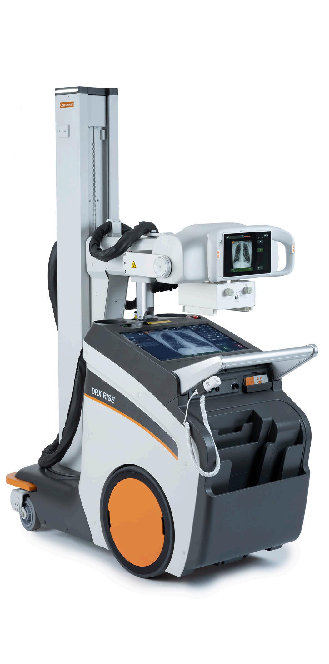
“The system is packed full of efficiency-boosting features. Its state-of-the-art lithium battery and in-bin detector charging allow radiographers to drive farther and complete more examinations on a single charge – saving time and increasing throughput. The system’s two touchscreen displays provide two work zones to accelerate productivity further. At the same time, new drive capabilities allow the radiographer to precisely move the lightweight system into optimal position at the bedside – from the tube head or the wireless remote control,” according to a news release. “The nearly silent DRX-Rise is ideal for use in crowded and sensitive areas such as the ICU and NICU, with the versatility and flexibility to meet the numerous mobile imaging demands throughout a facility.”
The DRX-Rise is equipped with Carestream’s ImageView Software, driven by the Eclipse processing engine with AI.
“The DRX-Rise Mobile X-ray System delivers high-end features that support the most critical needs in medical imaging: exceptional image quality for confident diagnosis; workflow-accelerating attributes for productivity; and a more comfortable imaging experience for patients – all without significantly impacting capital budget,” said Berry. “The new DRX-Rise system places the benefits of an advanced DR mobile system within reach, all from the manufacturer of the industry’s most popular mobile X-ray system, the DRX-Revolution.” •
ICEMAGAZINE 27 WWW.THEICECOMMUNITY.COM
PHILIPS EXTENDS MOBILE C-ARM PORTFOLIO
Royal Philips recently announced the launch of Philips Image Guided Therapy Mobile C-arm System 1000 –Zenition 10, a new addition to the company’s Zenition mobile C-arm series. Based on Philips’ state-of-the-art flat panel detector technology, Zenition 10 helps to expand patient access to routine surgical care and minimally invasive procedures. Now commercially available, the cost-effective, high-utilization, high-image-quality mobile C-arm enables surgeons to treat more patients at a lower cost while helping improve patient outcomes.
“Philips Zenition mobile C-arms have long been recognized for their high-quality imaging and efficiency-enhancing performance. With the introduction of Zenition 10, we are making those benefits available on a C-arm that meets the wide-ranging needs of routine open and minimally invasive surgery, allowing more patients in more parts of the world to receive the high-quality care,” said Mark Stoffels, general manager, image guided therapy systems at Philips.
To help alleviate operating room staff shortages and rising health care costs, Philips Zenition 10 provides a cost-effective imaging solution for routine surgery, delivers the speed and efficiency needed to deal with high patient throughput, while being flexible enough to meet the needs of orthopedics, trauma, and other areas of surgery to maximize utilization. It comes with exceptional C-arm maneuverability, application-specific protocols, and personalized user profiles. At the same time, it delivers the excellent
image quality needed to improve patient outcomes. User-friendly and intuitive to operate, the Zenition 10 supports a fast-learning curve and reduces operating staff training time. Next to this, the new system offers a dedicated low-dose pediatric mode.
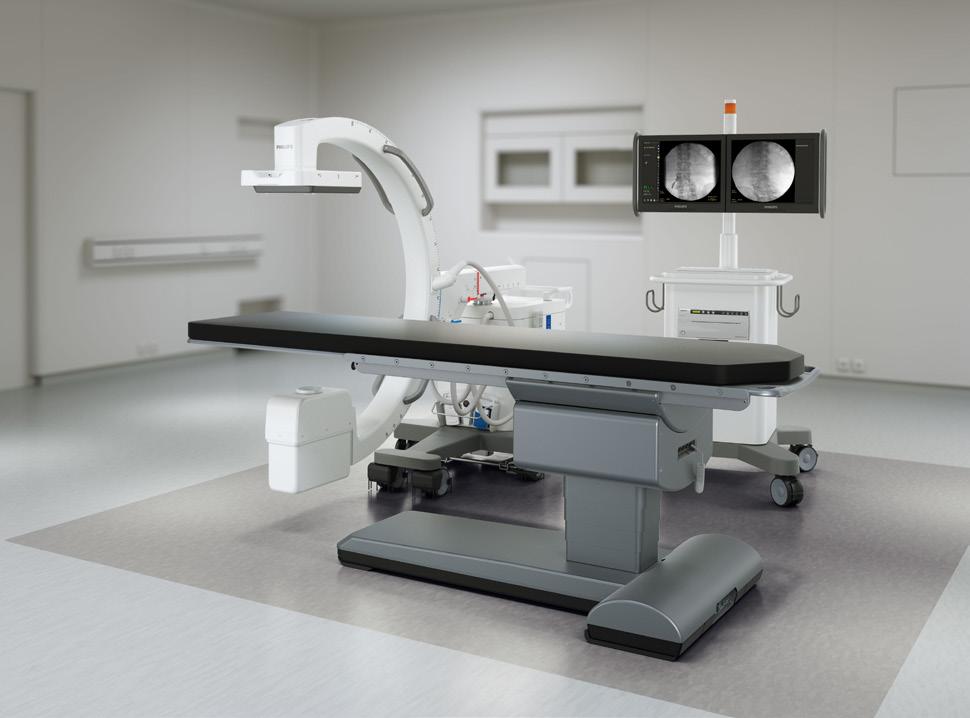
Consistent image quality is facilitated by application-specific protocols, customizable presets, and unique user profiles that automatically adjust Zenition 10 settings to suit an individual user’s preferences whenever they log in. An uninterruptible power supply allows the unit to be moved from one operating room to another without the need to reboot the entire system. Its excellent C-arm geometry provides the maneuverability needed for operating room staff to access target anatomies. Philips BodySmart facilitates dose efficiency by automatically adapting the measuring field to the area of interest.•
NUCLEAR IMAGING COALITION PRAISES FIND ACT
The Medical Imaging & Technology Alliance (MITA), the Council on Radionuclides and Radiopharmaceuticals Inc. (CORAR), and the Society of Nuclear Medicine and Molecular Imaging (SNMMI) have commended Senators Marsha Blackburn and Tammy Baldwin for introducing the bipartisan Facilitating Innovative Nuclear Diagnostics (FIND) Act of 2023 (SB 1544).
Over 20 million Americans benefit from the use of diagnostic and therapeutic radiopharmaceutical drugs, required in nuclear medicine procedures, each year. These drugs are used to diagnose and treat a wide variety of life-threatening conditions, including Alzheimer’s and Parkinson’s disease, breast and prostate cancer, heart disease and neuroendocrine tumors. However, the current Medicare reimbursement methodology applied in the outpatient setting packages diagnostic radiopharmaceutical drugs into nuclear medicine procedure
payments. This reimbursement methodology is flawed and creates a disincentive for hospitals to utilize innovative diagnostic radiopharmaceutical drugs.
S.B. 1544, which reflects companion legislation already introduced in the House (H.R. 1199), offers a bipartisan, legislative fix for this flawed payment methodology which will ensure more patients have access to these innovations.
“The FIND Act would ensure that payment policy for serious conditions – from Alzheimer’s and Parkinson’s disease to prostate cancer and neuroendocrine tumors – keeps up with the progress achieved in medical science,” said Patrick Hope, executive director of MITA. “We applaud Senators Marsha Blackburn and Tammy Baldwin for their leadership on this issue and look forward to working alongside them to expand patient access to innovative therapies.” •
NEWS ADVANCING THE IMAGING PROFESSIONAL 28 ICEMAGAZINE | JULY 2023
RADEQUAL, AAWR SIGN MOU TO ADVANCE OPPORTUNITIES IN RADIOLOGY
Leaders from RadEqual, an organization that fosters networking and mentorship opportunities for leaders in radiology, informatics and IT management, and the American Association for Women in Radiology (AAWR) signed a memorandum of understanding (MOU) at the 2023 American College of Radiology’s annual meeting in Washington, D.C. The two-year MOU will merge both parties into a formal agreement that will support increased collaboration and promote a shared pursuit of creating educational initiatives for the broader radiology community.
“It is part of the AAWR’s mission and purpose to create networking and mentorship opportunities for leaders in the radiology field,” AAWR President Dr. Amy Patel said. “Our new partnership with RadEqual will give us the profound chance to further inspire women within this community, and we’re proud to sponsor future activities that will be the catalyst for amplifying the next generation of leaders.”
In August 2022, RadEqual launched a webinar series sponsored by Intelerad, and now the AAWR, on overcoming the mid-career stall. Webinar attendees have the opportunity to learn from some of the health care industry’s most renowned leaders. The educational series will continue
through July this year.
“Since its founding, RadEqual’s partners and supporters have been at the forefront of creating opportunities for and empowering leaders in the healthcare community,” said Dr. Geraldine McGinty, RadEqual co-founder and professor of clinical radiology and population health sciences, Weill Cornell Medicine. “For over four decades, the AAWR has driven monumental change, and I am proud that today, we can now align our efforts and build a more promising future for leaders in radiology together.”
In November 2016, RadEqual was born when Dr. McGinty, along with other female leaders, noticed that they were often the only women in the room during events. Through networking and open dialogues, women shared similar experiences in the technology sector, where they are often underrepresented in executive leadership roles and corporate board seats. Today, RadEqual’s initiatives are supported by thought leaders in the health care industry, and the global RadEqual community, composed of women, men and nonbinary individuals, has grown to hundreds of individuals who are interested in increasing diversity within the various disciplines related to medical imaging. •
ICEMAGAZINE 29 WWW.THEICECOMMUNITY.COM
TASK FORCE ISSUES DRAFT RECOMMENDATION STATEMENT ON SCREENING FOR BREAST CANCER
The U.S. Preventive Services Task Force (Task Force) has posted a draft recommendation statement on screening for breast cancer. The Task Force now recommends that all women get screened for breast cancer every other year starting at age 40. This is a B grade. More research is needed on whether or not women with dense breasts should have additional screening with breast ultrasound or MRI, and on the benefits and harms of screening in women older than 75.
Breast cancer is the second most common cancer and the second most common cause of cancer death for women in the United States. While the Task Force has consistently recognized the lifesaving value of mammography, it previously recommended that women in their 40s make an individual decision about when to start screening based on their health history and preferences. In this new recommendation, the Task Force recommends that all women get screened starting at age 40. This change could result in 19 percent more lives being saved.
“New and more inclusive science about breast cancer in people younger than 50 has enabled us to expand our prior recommendation and encourage all women to get screened every other year starting at age 40,” says Task Force immediate past chair Carol Mangione, M.D., M.S.P.H. “This new recommendation will help save lives and prevent more women from dying due to breast cancer.”
St Luke’s $30 million investment in 21 GE HealthCare CT systems makes it one of the health system’s largest investments of its kind
Continuous Artificial Intelligence (AI) and software updates will be provided by GE HealthCare’s Smart Subscription, a service to help extend the life of the CT Fleet Chicago, IL – May 25, 2023 – GE HealthCare (Nasdaq: GEHC), a leading medical technology innovator, announced today its largest ever CT deal in the United States – a $30 million order by St. Luke’s University Health Network, a nationally recognized nonprofit healthcare network, to install 21 of GE HealthCare’s innovative CT systems, powered by Artificial Intelligence (AI), across their system. This order builds upon the more than 30year relationship between the two organizations.
The new scanners will include a comprehensive suite of clinical applications, and the latest AI, through GE HealthCare’s Smart Subscription that seamlessly connects and integrates with St. Luke’s existing network. As a result, GE HealthCare will provide St. Luke’s with access to the latest CT technologies and solutions, helping to extend the life of these devices and making it a more consistent experience for patients.
“GE HealthCare is honored to partner with St. Luke’s to provide cutting-edge CT technology across their network coupled with regular software upgrades and updates to keep their fleet of CT systems up to date,” said Catherine Estrampes, President & CEO, US & Canada, GE HealthCare. “These updates will enable greater standardized care for their patients using the latest capabilities available without having to invest in additional new equipment to keep pace with the latest technology.”
St. Luke’s patients scanned on this new CT technology will benefit from faster scans and sharper images, a potential reduction in radiation dose from advancements in technology, the capacity to better detect lesions or tissue abnormalities and to map vascular structures, and the ability to capture fine detail in the head and neck, which is critical in stroke diagnosis. These scanners also are expected to be helpful within St. Luke’s pediatric patient population, trauma cases, and especially in advanced cardiac exams by using GE HealthCare’s SnapShot Freeze technology. That technology, combined with fast rotation speed and wide coverage provided by the GE HealthCare scanners, provides the ability to image the heart with any heart rate in just one beat, which reduces the motion artifacts significantly, thus decreasing the likelihood for additional scanning.
“We can now offer the most advanced CT technology to all of the communities we serve. This provides our patients with access to this technology no matter where they go for their St. Luke’s care,” according to Dr. Hal L. Folander, Senior VP, Chief Medical Strategy Officer, Network Chairman, Department of Radiology at St. Luke’s. “This investment also allows for a faster, more informed and accurate diagnosis, with less inconvenience to patients.” •
NEWS ADVANCING THE IMAGING PROFESSIONAL 30 ICEMAGAZINE | JULY 2023
INTELERAD ANNOUNCES NEW CEO


Intelerad Medical Systems recently announced the retirement of CEO Mike Lipps, who served the company for nearly three years as part of a 20-plus year software industry career, and is handing the organization’s global team and mission over to Jordan Bazinsky as the new chief executive officer.
“We are grateful for Mike’s contributions and leadership that helped shape Intelerad into the medical imaging management leader it is today,” said JB Brian, partner at Hg and Intelerad chairman. “Jordan’s health care experience is coming at a crucial time. His innate ability to efficiently run and significantly scale businesses within this space is invaluable, and we are confident he is the right leader to steer the organization to new heights.”
Bazinsky is recognized for driving breakthrough growth across enterprise health care organizations, and joins Intelerad following over 20 years of rele -
vant experience, most recently serving in executive leadership positions at Cotiviti and Verisk Health. His track record for leveraging technology to improve outcomes for patients and providers brings immediate benefit to Intelerad. Motivated by Intelerad’s mission to improve global human health, Bazinsky’s focus is on the responsibility the organization has to enhance outcomes in health care and bring value to the organization’s global client base.
“I’ve been entrusted with ensuring that Intelerad’s vision aligns with our clients’ most critical needs in supporting their unique health care journeys,” Bazinsky said. “I am intimately tied to this industry, and value the work that has been done before me. Now, we have an opportunity to look ahead, evaluate our position and determine how we can make the biggest impact on the lives of health care providers and the patients they serve.” •


WWW.THEICECOMMUNITY.COM ICEMAGAZINE 31 You’ve
a
revenue source. Let KMG help you secure its future. When you partner with KMG, you’re getting a company that can provide you with full turnkey imaging solutions that can help with patient backlog, continuity of care and ROI. To learn more, visit kingsmedical.com or call 800-854-9061.
built
successful imaging
WHERE KNOWLEDGE, EXPERTISE, AND INTEGRITY MEAN NO WORRIES.


512.477.1500 KEIMEDICALIMAGING.COM
Market Report
of the images that ultimately improves time efficiency and capacity to digitally transfer images.
The global X-ray systems market size was valued at $6.7 billion in 2021 and is anticipated to expand at a compound annual growth rate (CAGR) of 2.3% from 2022 to 2030, according to Grand View Research.
A major factor driving the market is an increase in the demand for early-stage diagnosis of chronic diseases. In addition, continuous technological advancements, an increase in product development, improved fundings, and investments by the government, especially in developing countries, such as India and China, are also expected to contribute to the market growth. For instance, in June 2021 the government of India launched X-Ray Setu, a free Artificial Intelligence (AI) based platform to aid doctors for early COVID interventions.
According to Mordor Intelligence, the digital X-ray devices market is projected to register a CAGR of 8.16% during the forecast period (2022-2027).

The COVID-19 pandemic has turned the spotlight on diagnostic imaging, particularly on digital X-ray devices. Digital imaging plays a key role in the diagnosis of COVID-19 and indicates the affected lung tissue in infected patients. Several key market players had focused on innovations in the production of radiography equipment. For instance, in December 2020, Agfa HealthCare launched its new SmartXR for X-ray Artificial Intelligence (AI) for digital radiography portfolio to aid during the radiology routine, which has proven important during the COVID-19 crisis. Thus, during the pandemic, the digital X-rays devices market is expected to be positively impacted by COVID-19 accurate diagnosis and treatment.
The studied market growth can largely be attributed to factors, such as the increasing occurrence of orthopedic diseases and cancers, the increasing number of serious injuries, the advantages of digital X-ray systems over conventional X-rays, technological advancements, and product development. Digital X-ray devices use digital x-ray sensors instead of films to capture images. This results in an immediate preview
The major advantages of digital imaging are cost-effectiveness and easy accessibility. The hospitals can manage the cost-cutting by lowering the film price, reducing the requirement of storage space, and decreasing the number of people required to run the services and archive sections. The images are also instantly available for distribution to the clinical services without the time and physical effort needed to retrieve film packets and reviewing previous imaging on a patient is much easier. This factor majorly impacts the growth of the digital X-ray devices market. Digital X-rays expose approximately 70-80% less radiation than conventional X-rays. This is hugely beneficial for the long-term health of patients, especially pregnant women or patients who are already suffering from illness, thus ensuring safety. With the help of digital X-rays, dentists can now easily recognize oral issues, which is leading to a declining need for an invasive investigation at the diagnosis stage. Additionally, digital radiography safely stores patient X-rays, resulting in no loss from the holders.
Also, due to the increase in the number of dental disorders, cardiac disorders, cancers especially breast cancer, there is an increased demand for digital X-ray devices globally.
The global digital X-ray market size is expected to reach $22.42 billion in 2030 and register a steady revenue CAGR of 8.1% over the forecast period, according to analysis by Emergen Research. Digital X-ray market revenue growth is primarily driven by factors such as advantages of digital X-ray systems over conventional analog systems. More precise analysis is possible with digital X-ray and it contains quick, accurate and objective automatic slide analysis techniques. Additionally, it provides quick access to earlier related occurrences for scientists, stores data for long-term predictive analytics, and aids in quicker and more accurate detection of serious illnesses such as tumors by doctors. It also offers advantages including mistake reduction, enhanced imaging and increased productivity. Analog X-ray imaging is being rapidly replaced by digital X-ray sensors, which are utilized in place of conventional photographic film. •
STAFF REPORT
PRODUCTS ADVANCING THE IMAGING PROFESSIONAL 34 ICEMAGAZINE | JULY 2023
Focus Product
X-ray
SIEMENS HEALTHINEERS
Multitom Rax
The Multitom Rax Twin Robotic X-ray system from Siemens Healthineers enables a wide range of examinations in multiple clinical areas – from emergency medicine and interventional to pain management and orthopedics, and from conventional radiography and fluoroscopy to 3D bone imaging and full-body slot scanning – all in one room using one X-ray system. Low-dose, full-body slot scanning can be performed in seated, standing or supine positions to benefit orthopedic practices when tracking spinal conditions and surgical planning. The new Real3D bone imaging application for the lumbar spine and extremities enables the acquisition of diagnostically relevant 3D bone imaging, for improved image quality, more stable patient positioning, fewer artifacts and more streamlined exams. This application provides greater physician insights, particularly for weight-bearing examinations. High-Res functionality delivers even higher spatial resolution for images of the hand, wrist and elbow.
*Disclaimer: Products are listed in no particular order.
PRODUCTS
ICEMAGAZINE 35 WWW.THEICECOMMUNITY.COM
1
FUJIFILM
FDR D-EVO III Digital Radiography Detector
FDR D-EVO III is the world’s first glass-free and currently the lightest DR detector based on the standard 14×17 size. It’s engineered to better endure busy imaging departments by eliminating the most fragile component of conventional detectors. FDR D-EVO III incorporates an innovative film-based TFT capture circuitry inside combined with Fujifilm’s patented Irradiated Side Sampling (ISS), which enables exceptional image quality and gentle dose. The detector also includes Fujifilm’s exclusive germ-killing Hydro AG antibacterial coating on its surfaces to aide with ever-important infection controls. The detectors are available in 10x12, 14x17 and 17x17 sizes.
HOLOGIC Fluoroscan InSight Mini C-arm Extremities Imaging System 3
The Fluoroscan InSight FD Mini C-arm imaging system from Hologic is designed to facilitate greater positioning, flexibility and convenient mobility. The system’s flat detector enables imaging of long bones as its rotating detector and collimator enhance surgical positioning. The C-arm is designed with a forward tube source and has a full 120° range of motion, both forward and back, for excellent maneuverability and flexibility.
Hologic’s Fluoroscan InSight FD Mini C-arm imaging system also reduces radiation exposure by offering a low dose mode that is 34% less dose compared to other systems at 7.5 pulses per second. 1 The unit also lowers scatter dose with 50% less scatter at 30 frames per second. 2 The system can improve workflow as its customizable imaging parameters can be set for surgeon-specific preferences.
References
1 Fluoroscan InSight FD system, when using its low dose mode at 15 fps, produces up to 34% less dose compared to OrthoScan FD Pulse’s Low dose mode at 7.5 pps: Dose Study by F.X. Masse Inc., measurements based on an 2014 Orthoscan FD Pulse and a 2018 Hologic InSight FD
2 When scatter radiation measurements are taken at head, waist and knee height, the InSight FD system produces on average 50% less scatter radiation to the operator compared to OrthoScan FD Pulse: Dose Study by F.X. Masse Inc., measurements based on an 2014 Orthoscan FD Pulse and a 2018 Hologic InSight FD.

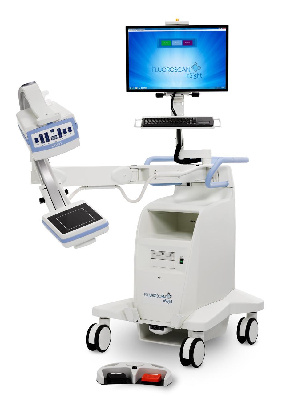
2
PRODUCTS ADVANCING THE IMAGING PROFESSIONAL 36 ICEMAGAZINE | JULY 2023
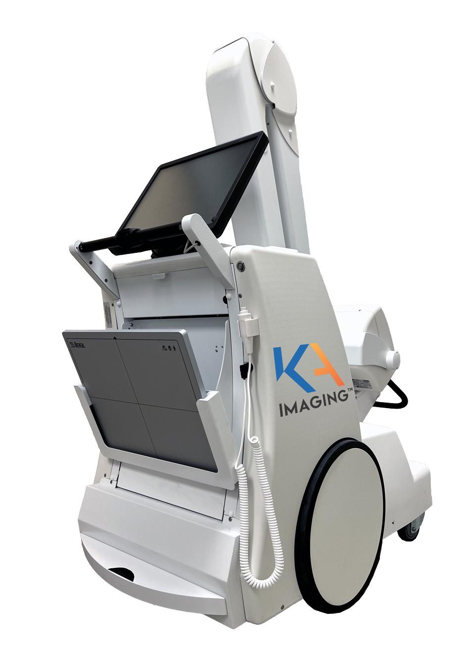
KA IMAGING Reveal Mobi Lite
KA Imaging is growing the Reveal product family with the Reveal Mobi Lite. It’s the company’s first integrated mobile system and is powered by SpectralDR technology. The Reveal Mobi Lite works with the Reveal 35C detector, also sold as a retrofit solution for fixed and mobile systems. KA Imaging’s SpectralDR enables dual-energy subtraction, providing bone and tissue differentiation with a single standard X-ray exposure. The technology uses identical clinical techniques associated with state-of-the-art mobile DR X-ray. The Reveal 35C X-ray detector is FDA cleared and is available for sale in the US. The Reveal Mobi Lite is not available for sale.
CARESTREAM DRX-LC Detector
Carestream’s DRX-LC Detector helps streamline and simplify long-length imaging, making the image capture process more comfortable for pediatric patients and those with limited mobility; and more efficient for radiographers. Its single shot exposure reduces patient hold time and eliminates the need for stitching, leading to improved image quality for increased diagnostic confidence. Plus, its single-shot acquisition reduces dose and cuts down on the need for retakes – both contributing to a more efficient experience. The DRX-LC – which uses ImageView Software, powered by Eclipse – can be wireless or tethered, and is compatible with Carestream’s DR rooms, mobile systems and retrofits.
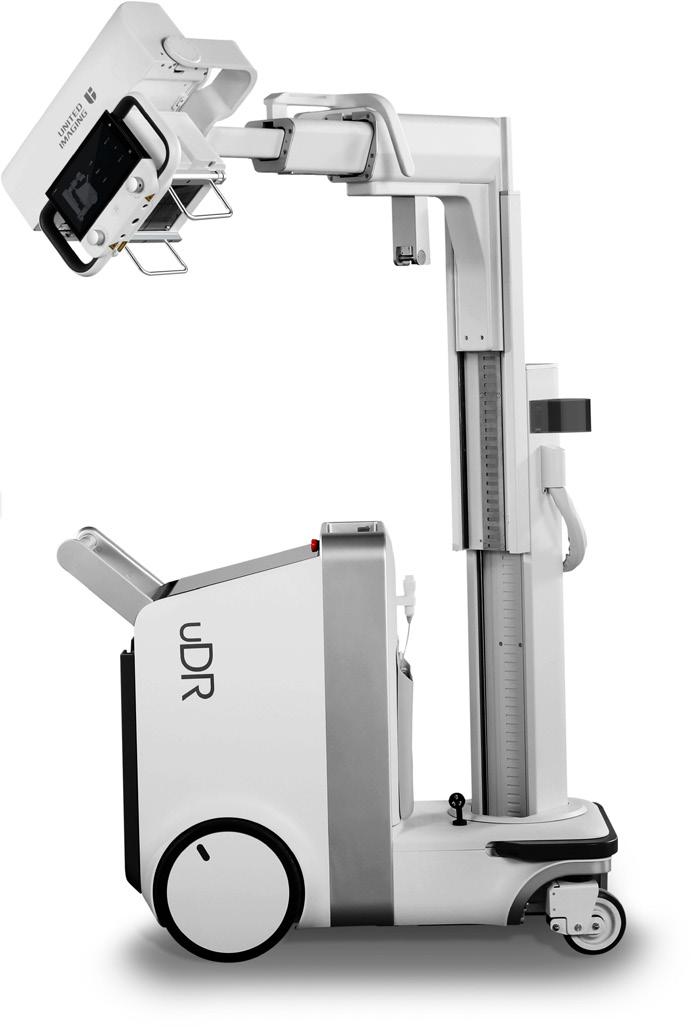
UNITED IMAGING
uDR 380i Pro
This mobile X-ray with beautiful image resolution is a high-performing, agile system that’s ultra-compact and lightweight to fit in tight spaces, is easy to maneuver with motorized power steering, a zero-turn radius and a 13° climbing capability and has a long-lasting battery with up to 800 exposures per charge. With a 50 kW high voltage generator it can image a wide variety of patient types. The uVision Remote Console enables a smart workflow with real-time patient monitoring, voice guidance, remote exposure control and more, redefining the workflow for point of care imaging. Like the rest of our scanners, it comes with All-in Configurations and Software Upgrades for Life.

4
6
5 ICEMAGAZINE 37 WWW.THEICECOMMUNITY.COM

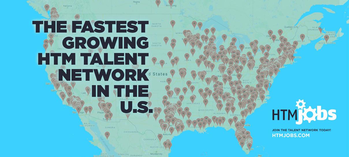

Imaging Jobs NOW AVAILABLE htmjobs.com REGISTER FOR FREE AT HTMJOBS.COM Contact us at htmjobs@mdpublishing.com to learn more about our various posting options! Companies like ours have such a difficult time finding qualified candidates for field service roles that it just made sense to publish our opening with HTMJobs. – K. White, HR/Compliance Manager “ ” LOOKING TO FILL A POSITION? Visit htmjobs.com/start-posting/ to post a job. Companies that post with us: Texas Health Resources, First Call Parts, Associated Imaging Services, InterMed, Renovo Solutions, TRIMEDX, Universal Medical Resources, Medical Equipment Doctor, Agiliti and more!
Our focus at Medical Equipment Doctor is to partner with healthcare administrators to keep their budget and costs in check through providing top-level refurbished equipment sales, service, and rentals, for all their medical equipment needs. This has led our company to be recognized nationally as the affordable solution to purchasing brand new medical equipment.
At RENOVO, we value knowledge, reliability, and integrity in our employees. If you are interested in being a part of a team that is committed to making a difference in healthcare equipment maintenance and healthcare asset and technology management, we invite you to apply for one of the open positions. We are always looking for talented, passionate, hard-working people to join our team.
Imaging Service Engineer II


The InterMed Group is a healthcare technology management company meeting the needs of our customers for over 20 years. InterMed sells and services medical equipment for our customers across the country and is growing quickly. As a result, InterMed is looking for an Imaging Field Service Engineer to perform repairs and preventive maintenance. This individual would be responsible for maintaining, inspecting and repairing imaging equipment for our accounts in the area and must be willing to travel for work.
As one of the largest faith-based, nonprofit health systems in the U.S. we play a huge role in the communities we serve in the greater Dallas Fort Worth area. Our mission is “to improve the health of the people in the communities we serve” and we get it done every day. Imagine the power of 26,000+ team members with a singular focus and determination.
First Call Parts has been providing customers with quality replacement imaging parts since 2009. We pride ourselves in developing a top-notch reputation in the imaging industry as delivering the best in diagnostic imaging replacement parts. We specialize in the sale of refurbished/tested and used, Philips, Siemens, and GE in the Cath/Angio, R/F, and RAD modalities.
Agiliti is a nationwide company of passionate medical equipment management experts who believe every interaction has the power to change a life. Our industry-leading commitment to quality and team of expert technicians helps ensure clinicians have access to patient-ready equipment needed for patient care. Make an impact in healthcare and grow your career with Team Agiliti!
Imaging Engineer I
Universal Medical Resources’ operating principles are based on practicing the core values of Collaboration with the nuclear medicine community; embracing Diversity of ideas, beliefs, and practices; commitment to Excellence in producing the highest quality outcomes; and recognizing our Ethical Community through actions guided by fairness, trust, honesty, and integrity.
Do you want to work in healthcare? Would you like to make a difference in the lives of patients and their families? Do you enjoy a new challenge every day? If you are skilled at servicing medical equipment in a clinical setting, we hope you will join our team!
Associated Imaging Services has been offering nuclear medicine and ultrasound solutions to our customers since 1990. We specialize in the sales and service of new and refurbished nuclear medicine cameras and ultrasound systems throughout Kansas, Oklahoma, Texas, and the surrounding areas.

Ultrasound Probe Repair Technician X-Ray In-House Service Technician II Senior Imaging Engineer Field Service Engineer Imaging Engineer Imaging Field Service Technician III Field Service EngineerNuclear Medicine VIEW FULL DETAILS www.htmjobs.com VIEW FULL DETAILS www.htmjobs.com VIEW FULL DETAILS www.htmjobs.com VIEW FULL DETAILS www.htmjobs.com VIEW FULL DETAILS www.htmjobs.com VIEW FULL DETAILS www.htmjobs.com VIEW FULL DETAILS www.htmjobs.com VIEW FULL DETAILS www.htmjobs.com VIEW FULL DETAILS www.htmjobs.com
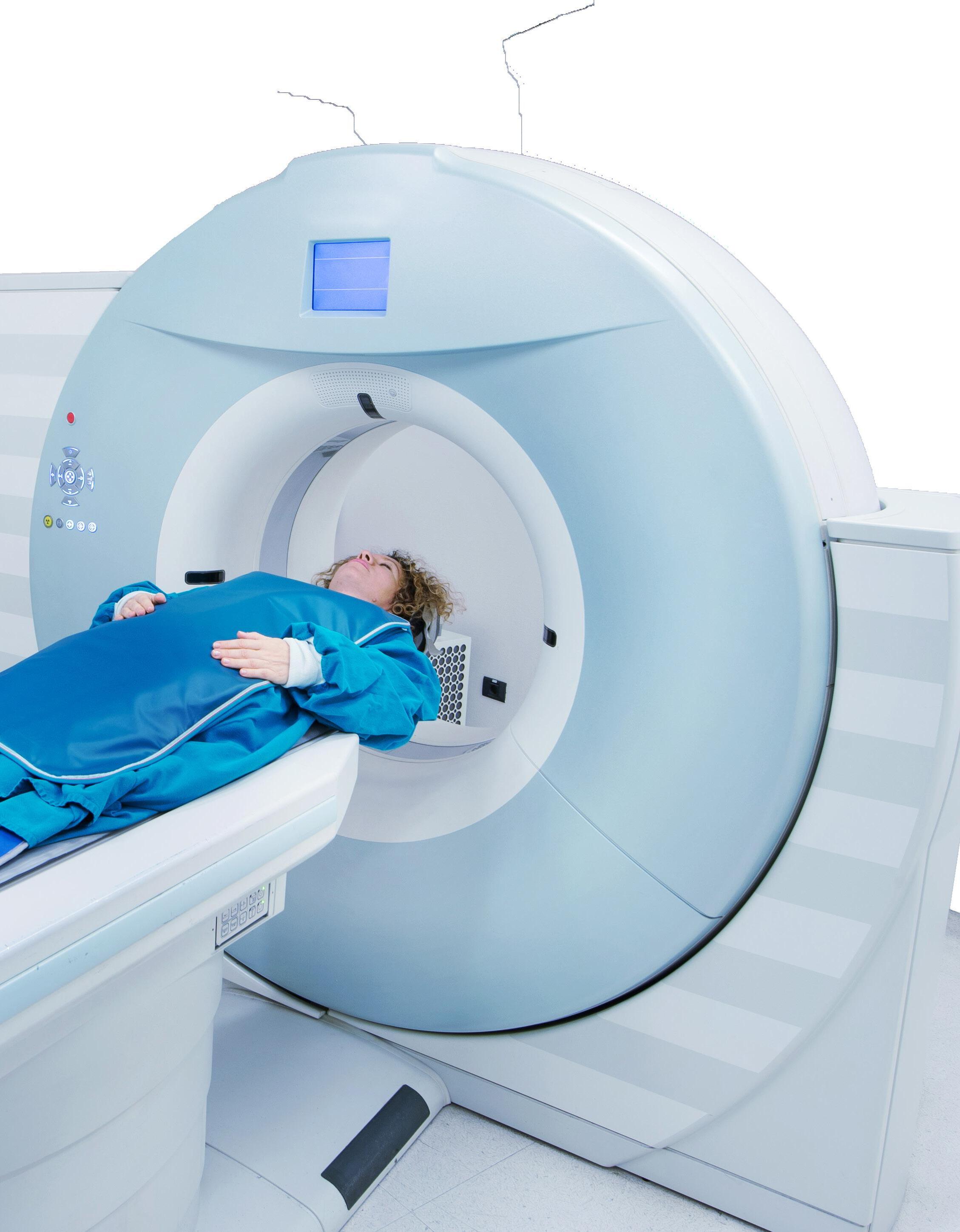
LOWERING SHIELDS THE
 BY MATT SKOUFALOS
COVER STORY ADVANCING THE IMAGING PROFESSIONAL 40 ICEMAGAZINE | JULY 2023
In earlier days of medical imaging studies, patients were routinely given lead aprons to shield the parts of their bodies that weren’t involved in the study from ionizing radiation. At the time, that thinking was governed by concerns about the potentially adverse effects of irradiating sensitive areas of the body to avoid creating hereditary mutations. However, after decades of studies seeking to link radiation and human genetics, research hasn’t shown any evidence for that risk. Now, health care institutions worldwide are dealing with the work of changing their policies around patient shielding.
When a prophylaxis doesn’t offer the protection it once was thought to, how does the rest of the medical field adapt to the new evidence?
Medical Physicist Rebecca Milman, Ph.D., associate professor of radiology-radiological science at the University of Colorado Anschutz Medical Campus, said that, at her campus, the question of patient shielding arose in conversation with some of the school’s radiologic technologist students.

When her colleague’s reply was that the lead shielding “doesn’t really provide any safety for the patients, but it’s the way it’s always been done,” it prompted a conversation about the science behind that thinking. Upon further investigation, Milman said her group determined a number of professional organizations were evaluating the same question in terms of risks and benefits.
One such study, “X-ray shields going by the wayside: What you and your patients need to know,” by Donald Frush, M.D., FACR, FAAP, and Janet Reid, M.D., FRCPC, FAAP, which appeared in the March 2020 issue of AAP News from
the American Academy of Pediatrics, described the institutional reassessment of the benefits of radioprotective shielding.
As recently as 2019, the American Association of Physicists in Medicine (AAPM) issued a position statement recommending that lead shielding of patients’ genitals and pregnancies during X-ray studies need not be a routine practice, as it historically has been, Frush and Reid noted. Technological advancements, better understanding of radiation sensitivity in the gonads – and a lack of uniformity in their location within the body – are among key developments behind the shift in guidance.
From a technological perspective, the practice of shielding imaging patients from ionizing radiation first gained traction in the mid20th century, Milman said. Amid studies of the after-effects of nuclear warfare in World War II, the U.S. Food and Drug Administration began to recommend patient shielding to prevent any adverse hereditary effects of exposing reproductive organs to radiation.
“They were concerned that if you expose the gonads to radiation that it would cause changes in the human gene pool,” she said. “They studied that very closely, and there’s just no evidence that that’s true in humans, even looking at these very large-scale studies in atomic bomb survivors.”
Another major development in the intervening years has been improvements to medical imaging technologies that has decreased the amount of radiation necessary for a high-quality image by more than 90 percent. In systems that use automatic exposure control, image detectors measure how much radiation penetrates the patient’s body to determine the intensity needed
to generate a useful study. Inserting any radiation blocking materials into the field of study – say, a lead apron – could actually have the opposite intended effect and cause the automated process to intensify to attempt to penetrate the shielding.
“There’s this feedback loop that has been demonstrated to optimize how
we use radiation imaging, but also image quality,” Milman said. “If you have a piece of lead in the field of view, it’s trying to look through it, and it actually puts out more radiation. That’s something that technologically didn’t exist in the 1950s, but that’s a concern with current equipment.”
Another concern with lead shielding is that sometimes the lead apron will cover up a part of the body that a technologist is trying to image. Furthermore, attempts to use shielding to cover gonadal areas might not be effective because of the significant natural variation in the body. Among a cross-section of female patients, for example, their ovaries aren’t always located in the
WWW.THEICECOMMUNITY.COM COVER STORY
“A lot of the time, what they really want is some reassurance that someone is paying attention.
When I explain that we’ve got a whole team monitoring the amount of radiation this equipment is using, knowing that someone is paying attention to it, a lot of the time, that is reassuring for patients.”
ICEMAGAZINE 41
- Rebecca Milman
same areas of the body, Milman said.
“You’d have to cover the entire pelvic area,” she said. “If the parts of the body you want to see are covered with lead, then you’d have to either retake the image, which is more radiation, or send the suboptimal image to the physician. If the physician isn’t able to see what they need to see in the image, then there was no point in doing it in the first place.”
closer and closer to that field of view, there’s a greater likelihood that lead ends up in your image,” potentially interfering with the image quality or radiation dose.
But even if lead shielding only has a psychological placebo effect for patients who are concerned about the impact of ionizing radiation, why not just leave things as they are? Milman said conversations in the field about continuing to shield have generally settled into two schools of thought.
“One is that it doesn’t do any good, but it can reassure patients,” she said. “The other side is, if we continue to shield patients when it doesn’t do any good, we’re saying that we’re providing some protection because there’s a risk, although the risk doesn’t actually exist.
next stage of the conversation. In addition to the science behind the refined guidance, Frush and Reid also addressed the other end of the issue; namely, the emotional reaction from patients who have been conditioned for decades to have a healthy respect for the impact of ionizing radiation on soft tissue.
“Exceptions can be made if a parent/caregiver requests a shield, and it is of psychological benefit,” they wrote. “In these situations, the radiology practice should have guidelines and communication strategies to enable the requesting caregiver to understand the benefits and disadvantages of shielding.”
“It’s so deeply ingrained,” Milman said. “It ties into this overall fear of radiation, both in popular culture and our day-to-day lives.”
Additional studies around radiation shielding of pregnant imaging patients led to a similar, potentially counterintuitive conclusion: shielding the fetus from ionizing radiation doesn’t have the intended effect.
“Most of the exposure to the fetus is from scatter radiation generated inside the mother,” Milman said. “Outside that imaging field of view, the amount of radiation falls off quickly. So, in that case, you’re really not able to shield the fetus from the radiation it’s being exposed to. You’re going to get the most benefit if the shield is right up against the imaging field. The problem is, once you get
“This goes back to this overall public perception about radiation, and fear of radiation, and it seems like an academic debate on the surface,” she said, “but the more you talk to radiologists, physicians, medical physicists and technologists, we run into this all the time. The risk from the radiation is much lower than not having an accurate diagnosis.”
“It’s really difficult to watch somebody make a decision based on a fear that we know isn’t a risk, or is such a small theoretical risk that they’re overlooking the bigger picture,” Milman said. “It kills me every time I talk to a patient and they don’t want to have this imaging exam because the risk associated with it is so exaggerated; so deeply ingrained.”
Determining a path forward for patient safety, while both acknowledging the best available scientific evidence, and preserving patient autonomy to make medical decisions based on risks and benefits, is the
“It’s OK to have a gut reaction to something,” she continued. “There are many, many reasons why humans go through that process. Risk perception is a very, very tricky sort of thing, within the health care community especially. It’s about being willing to step back and say, is this still the best thing to do?”
Even throughout the broader medical community, Milman acknowledged “a certain discomfort” with the notion of not shielding patients during X-ray studies, particularly among physicians who refer patients for imaging exams, and who many not have training in radiation safety themselves.
When UCHealth changed its policy regarding patient shielding in 2018, she said that staffers sat down with technologists to script some prepared responses to offer patients who questioned the new procedures. Anecdotally, at least, very few people pressed the issue.
“The technologists said some people asked why we weren’t using shields anymore, and then 98 percent of the time, they’d move on,” Milman

COVER STORY
“It’s really hard to get people to do things that were once deemed very risky, and trying to tell them it’s not. It’s classic human psychology: despite the evidence, it feels like the needle’s barely moving.”
ADVANCING THE IMAGING PROFESSIONAL 42 ICEMAGAZINE | JULY 2023
- Elsa Pearson Sites
said. “There really were very, very few patients who said, ‘I still want to be shielded,’ and in those cases, wherever possible, we’d still shield them.”
“Most of the time, patients wanted to make sure that not using a shield was deliberate,” she said. “A lot of the time, what they really want is some reassurance that someone is paying attention. When I explain that we’ve got a whole team monitoring the amount of radiation this equipment is using, knowing that someone is paying attention to it, a lot of the time, that is reassuring for patients.”
Elsa Pearson Sites, MPH, is policy director for the Partnered Evidence-based Policy Resource Center (PEPReC), a research evaluation center affiliated with the Boston University School of Public Health. Sites described how her facility has worked to translate and disseminate the policies and research around patient radiation shielding, and the efforts that go into driving broader cultural shifts when new evidence changes medical guidance.
“It’s really hard to get people to do things that were once deemed very risky, and trying to tell them it’s not,” she said. “It’s classic human psychology: despite the evidence, it feels like the needle’s barely moving.”
In the past year, Sites pointed to growing acceptance of the shift in thinking around radiation shielding from policy changes at medical organizations like Dartmouth and Yale updating their patient guidance. An interesting outlier persists in the dental community, in which lead aprons are routinely placed on patients during head X-rays. But she believes that if anything’s going to advance the conversation, it’s greater patient education.
“That disconnect between what the medical community is doing and what the dental community is doing is going to be a very challenging public health conundrum,” Sites said. “If you’re getting shielded from head to toe at the dentist, and your doctor is putting you flat on the table, that’s confusing. I’m not sure it’s the medical community’s job to get the dental community onboard, but I think they have to have a seat at the table, and that’s what’s missing: open communication across parties.”

In seeking to unify the variety of perspectives around this issue, or any such issue, Sites said that messaging should emphasize clarity, simplicity, and anticipate questions, concerns, and disagreements with measured respect. She advised a tone that walks the line between addressing concerns without heightening fears.
“Anticipate that people will have questions and concerns and disagree, and respect that, and be ready to stand behind the evidence,” Sites said. “Be able to be clear, concise, simple, and evidence-based, but also respectful of pushback.
“It’s really hard when authority figures in your life change their mind,” she said. “It’s human psychology at work, and it’s just fascinating.” •
COVER STORY ICEMAGAZINE 43 WWW.THEICECOMMUNITY.COM

This will be an iconic meeting filled with education, inspiration, and connections to help you lead through the challenges of today's healthcare environment with confidence. CELEBRATING MEDICAL IMAGING MANAGEMENT'S PAST, PRESENT AND FUTURE. Visit ahra.org/annualmeeting Register by June 15 to reserve AHRA's special conference room rate!
®
OEM-QUALITY CT REPLACEMENT TUBES FOR LESS THE ALTA750 CT TUBE SERIES
Richardson Healthcare engineers form-fit-function CT replacement tubes to enable service providers to improve efficiency and lower the overall cost of healthcare delivery With the launch of the ALTA750G and our legacy product, the ALTA750 , we proudly expand our CT tube replacement solutions for Toshiba/Canon* Aquilion Scanners
LOWER COSTS & MAXIMIZE CT UPTIME - CONTACT US FOR A QUOTE!

Imaging Service Solutions


global provider of OEM quality pre-owned and refurbished medical imaging parts. ISS specializes in supplying parts for Philips and Siemens MRI and CT scanners.


Established in 2009 in Baltimore, MD, we proudly work closely with hospitals, service providers, and manufacturers globally.

SELL US YOUR MRI AND CT SYSTEMS!
We purchase installed systems at fair market value and provide quality replacement parts for your equipment at a great value. We can remove purchased equipment at no cost as well.


•

ICEMAGAZINE 45 WWW.THEICECOMMUNITY.COM Aquilion 8/16* Aquilion 4* Aquilion 16 Large Bore* Aquilion 32/64* ALTA750 compatible on: Aquilion Prime SP* Aquilion Prime S* ALTA750G compatible on: Aquilion RXL* Aquilion CXL* Aquilion Prime Gen 1* Aquilion VeloCT* *All product and company names are trademarks™ or registered® trademarks of their respective holders Use of the trademarks is solely for identification purposes and does not imply any affiliation with or endorsement by the trademark holders ® ® R E L L H E A L T H C A R E . C O M H E A L T H C A R E @ R E L L . C O M 7 0 4 . 7 3 9 . 3 5 9 7
® ®
MRI CT SERVICE,
& PARTS
SALES
(410) 530-5548 | info@imagingss.com | 4001 Washington Blvd | Baltimore, MD 21227 Find us on Dotmed at dotmed.com/webstore/118100 imagingss.com SERVICES
OEM trained service engineers
Repair and maintenance
Professional installation and deinstallation
Rigging
Crating
Worldwide shipping
•
•
•
•
•
•
Trade-in equipment
BY NICOLE DHANRAJ DIRECTOR’S CUT
UNLOCKING THE POTENTIAL OF YOUR TEAM: STRATEGIES FOR EFFECTIVE AND ENGAGING TRAINING
As imaging leaders, we understand the importance of practical training for our staff. But have you ever considered your training methods’ effectiveness and if they resonate with your team? We often let education handle the training, but we must consider whether our department and organizational training methods are effective for our staff because they directly impact our department’s success.
If our staff members don’t feel engaged or motivated during training, they may struggle to retain information and develop the skills needed to perform their jobs effectively. This, in turn, can lead to decreased productivity, lower quality work and potentially negative impacts on our bottom line and becomes a waste of effort with a limited ROI.

So, how can you assess the learning styles of your imaging staff? Here are some tips to help you get started:
OBSERVE THEIR BEHAVIOR
Pay attention to how your staff members interact with information. Do they click through information without paying attention? Are they curious or nonchalant? Do you find them engaging when there are visual aids, like diagrams and charts? Or maybe they ask many questions or prefer
to work independently? Observing their behavior lets you start identifying patterns and preferences and design your training accordingly.
ASK FOR FEEDBACK
Don’t hesitate to ask your team members for feedback on your training methods. This can help you identify areas that need improvement and gain insight into how your staff prefers to learn. This can also work for the material you present in your staff meeting.
USE A LEARNING STYLE ASSESSMENT TOOL
Many assessment tools can help you identify your staff members’ learning styles, such as the VARK questionnaire or the Kolb Learning Style Inventory. These tools can provide valuable insight into the strengths and weaknesses of each individual and help you tailor your training approach accordingly.
ADAPT TRAINING AND PROVIDE EXPOSURE
Once you understand your staff members’ learning styles, it’s crucial to adapt your training approach accordingly. Let’s say you’re a director responsible for training a new manager in your radiology department. Instead of giving them a book or manual or having them wade through a PowerPoint on their own without an opportunity for discussion, assess their learning style and needs. It
INSIGHTS ADVANCING THE IMAGING PROFESSIONAL 46 ICEMAGAZINE | JULY 2023
can be overwhelming for some people to absorb much information through reading or listening alone. The information may not be retained without opportunities to ask questions or apply their knowledge in a practical setting.
You can adapt your training approach to suit their needs when you’ve assessed their learning style and determined that they learn best through interactive discussions and collaboration. You might incorporate more interactive discussions and role-playing scenarios into your training sessions. For example, you might conduct brainstorming sessions with your new manager, asking them to come up with solutions to real-world challenges that they’ll be facing in their role.
You might also pair them up with experienced managers within the department, assigning them to work on joint projects or tasks that require collaboration. This would allow them to learn from experienced managers in a more interactive and hands-on way while also providing opportunities for them to develop their skills and knowledge.
In addition, you might provide them with opportunities to attend relevant conferences, seminars or workshops to expand their knowledge and skills. This would allow them to learn from industry experts, gain exposure to new technologies and network with peers from other radiology departments.

AVOID TIME-CONSUMING ACTIVITIES
You may be thinking this will create a lot of work for you. However, one way to avoid making time-consuming individualistic training is to focus on creating materials that can be easily adapted or customized to meet the needs of different learners. Here are some suggestions:
• Develop training materials that can be combined or rearranged based on the needs of different learners. For example, you could create a series of short training videos or interactive modules that cover various topics and allow learners to choose which topics they want to focus on based on their needs. This is especially great in cross-training individuals.
• Provide opportunities for learners to self-assess their own learning needs and choose the training materials that are most relevant to them. These assessments could be done through a pre-training survey or assessment that asks learners to identify their strengths and weaknesses and then provides recommendations for training modules based on their responses. The suggestions can help you when pairing staff with mentors or onboarding new hires. Provide opportunities for collaborative learning, such as group discussions, role-playing exercises or team-based projects. This collaboration can help learners share their knowledge and skills with others and can also help to build a sense of community and support among learners. This effective team-building exercise can be used primarily on topics such as quality and safety.
Consider using technology to create more efficient and scalable training solutions. For example, you could use virtual reality simulations, online learning platforms or mobile apps to deliver more interactive and engaging training materials. This is an excellent option for new graduates and new hires.
TRY ADAPTING YOUR APPROACH!
By assessing your staff members’ learning styles and adapting your training approach accordingly, you can help ensure that your team members are engaged, motivated and equipped with the skills they need to succeed. So, don’t be afraid to experiment and try new things – your staff will thank you! •
Nicole Dhanraj, Ph.D., SHRM-SCP, PMP, GPHR, CPSS, CRA, R.T(R) (CT)(MR), is an experienced imaging director.
ICEMAGAZINE 47 WWW.THEICECOMMUNITY.COM
BY BRENDAN ELLIXSON
DOSE REGULATION IN A VASCULAR LAB ENVIRONMENT
If you have ever worked in radiology, you have walked past a portable X-ray machine with the exposure switch cable hanging to the floor. The technicians using the portable pull the cable as far as they can when taking X-rays to avoid getting radiated. However, in a vascular lab, the surgeon and nurses are standing tableside performing surgery with the gantry right next to them. What safeties are there to prevent over-radiation of the patient and the technicians, and in what ways can the system help regulate dose? The term ALARA (As Low As Reasonably Achievable) comes into play and will be broken down throughout this article.
COLLIMATION
While performing fluoroscopy and acquisitions (cine), the user can use collimation to reduce scatter radiation. The collimator filters the stream of rays coming from X-ray tube, limiting the beam of X-rays. By “collimating down,” the technician can view just the part of the body being worked on to reduce scatter radiation and radiation in the surrounding environment while increasing contrast of the image for better image quality. When you collimate down, the entrance skin dose rises because the image is darker, and the DAP can be reduced greatly. However, with collimation, less of the patient is exposed to radiation, reducing overall patient dose. The
blades are made of lead, which completely blocks radiation, narrowing the field of view to the anatomy needed. This focuses the radiation and increases the clarity and depth, so the image does not have “fog” or white space in the image.
USE OF FLUOROSCOPY VS. CINE
The best way to regulate dose in a vascular lab is to use best practices with using fluoroscopy versus cine. Regardless of which is being used, the patient should be closer to the detector and not beyond the isocenter. By moving the detector close to the patient, you reduce patient skin dose and increase radiation received by the detector, resulting in better image quality. Cine should be used sparingly, as it can produce up to 15 times more radiation than fluoroscopy. With the invention of “Last Image Hold” option it makes using fluoroscopy a much safer alternative. It allows the user to review the last flouroscopy run as long as necessary to perform the next steps in surgery. There is also an option for pulsed fluoroscopy which uses small millisecond pulses and can be varied by frame rate.
DOSE MONITORING

Monitoring dose during surgery for the patient and staff is important. For the patient, this is done electronically. DAP (Dose Area Product) comes from an ionization chamber mounted onto the collimator inside of the tube cover. This is an important number to ensure there are no stochastic effects. The
INSIGHTS
SPONSORED CONTENT ADVANCING THE IMAGING PROFESSIONAL 48 ICEMAGAZINE | JULY 2023
other concern to patient safety is PSD (Peak Skin Dose), which is controlled by the operator keeping an eye on air kerma levels. The number considers angulation of the gantry angles and table position. Air kerma acts as a real-time safety indicator for the operator and staff. There is a dose structured report that can be used to help improve procedures. It should be noted that measuring Air Kerma and DAP is not perfect and doesn’t always take into effect the physicians use of the system, table and positioning angles when displaying, among other factors. The trick is pre-planning and using ALARA.


During procedures, staff wear dosimeters on the outside of the lead aprons, as well as under the lead apron. The dosimeter on the outside of lead apron helps estimate the dose that goes into exposed skin. The dosimeter worn under the apron is used to estimate the operator’s effective dose. There is a calculation made to determine the radiation absorbed, which is monitored throughout the year to calculate operator risk.
LEAD BLOCKERS


Lead blockers are used to reduce radiation from staff and the patient. There are a few different pieces that are mainly used to help regulate the dose exposure. The staff can put a lead blocker over the patient areas where surgery is not being performed. Generally, the lead blocker will go underneath the patient to block X-ray coming from the tube that is usually below the patient table. There are also a few options

for lead shields, some hang from the ceiling and others roll on a stand, blocking all radiation for the user behind it. The tableside also can have a swinging lead shield to block the user’s legs from radiation. There are also lead shields that staff wear. Lead aprons can be worn to protect all vital organs. A thyroid shield is also worn to protect the neck area, while lead shield glasses are worn to protect the eyes. There are also lead shields that can be worn on the head and hands. The use of lead blockers combined with dosimeter badges worn by everyone working in the surgery room can help protect everyone from too much radiation exposure over time, as well as improving radiation reduction practices.
CONCLUSION


Dose regulation is an incredibly important factor for anyone who works in radiology spaces or being treated in one. Knowing some steps to reduce dose will prolong your life and others, as well as reduce chances for side effects. By remaining dedicated to adhering to best practices, Avante strives to surpass our customers’ expectations, enabling them to deliver the utmost level of care to their patients. As we continue to evolve and innovate, we will remain steadfast in our commitment to quality and safety, and we are proud to have the best industry experts supporting our mission. •


X-R AY T UBES & P ARTS FOR M EDICAL I MAGING E QUIPMENT Email: sales@w7global.com Toll Free: 855-W7GLOBA (974-5622) Online: www.w7global.com X-Ray Tubes for CT, Cath/Angio, and X-Ray • Tubes for All Major Equipment Brands • New, Used, and Refurbished • OEM Replacement Parts FOR 2022 Parts Vendors Certified ICEMAGAZINE 49 WWW.THEICECOMMUNITY.COM
Brendan Ellixson is a Field Support Specialist serving Georgia with 13 years of electronics experience.
BY VERLON E. SALLEY DIVERSITY
MY 4-STEP PLAN
Every other month I have written an article about diversity and/or health equity. After this article, I plan to take a timeout from writing to focus more my role as vice president of health equity at University of Alabama Birmingham (UAB) Health System. This work is so important currently that I want to dedicate even more time to it. I want to improve health equity, especially here in Alabama. How can I help to improve health equity in Alabama? First by recognizing my organization’s influence. UAB Health System is ranked the number one health system in the state of Alabama. We are also the number one employer in the state. What we do has ripple effects across the state. Therefore, I am going to ask all of you to keep me honest. In this article, I am going to divulge my personal four step plan of attack (see below).
1. Insight
2. Discovery
3. Action
4. Outcomes
INSIGHT
I will study the social determinants of health in my target area (Jefferson County, Alabama). We have created our own social determinant
INSIGHTS ADVANCING THE IMAGING PROFESSIONAL 50 ICEMAGAZINE | JULY 2023
• 7 Domain index
• 4 regions within Jefferson County

• 164 Census tracks were ranked
• Zip codes chosen are housed within census tracks
• Target Population: Un & Underinsured
Vulnerable Zip Code/ Community Index
index where we identified and ranked the communities in the county using a Likert scale within seven domains. The seven domains are defined:
• Income – percent of population in a census tract that is below the poverty line.
• Unemployment Rate – percent of population in a census tract that is unemployed.
• Education – percent of population in a census tract that does not have a high school diploma.
• Health Insurance – percent of population in a census tract that does not have health insurance.
• Life Expectancy – is the average number of years an individual is expected to live.
• Food Insecurity – a community with at least 500 people and/or 33% of the census tract’s population residing more than one mile from a supermarket or large grocery store in urban areas.
• Affordable Housing – percent of monthly housing cost as a percent of household expenses for household incomes at or between $35K-$50K.
Because of this process, we have focused on the top 10 vulnerable/marginalized ZIP codes in the county. In all initiatives we create, we will attempt to apply them to one or all of these top 10 areas.
DISCOVERY
In our discovery phase, we will focus on our internal clinical data. We will analyze our utilization, valued-based care metrics, community health needs assessments and other health surveys. We will filter these data sets by race, payor and locations. The goal is to find differences and disparities in preventive care, access and clinical outcomes.
ACTION
Once the prior two phases are complete, we will partner with community-based organizations and payors to create initiatives that will have a positive and equitable impact on the communities we want to target. Currently, our mission as a health system is to provide quality and effective care to the community we serve. Moreover, we are not equipped to solve social determinants of health. Instead, we can partner with organizations that can provide transportation for care. We can partner with organizations that have a voice and trust with our targeted marginalized communities to deliver preventative care (i.e. – vaccines, health education and awareness).
OUTCOMES
The final phase is to create a dashboard to measure the success of the initiatives we create through our own means and partnerships. As a manager of an outpatient imaging center I once said, “If you don’t measure it, you can’t manage it.” This mantra of mine still applies to my new responsibilities. We will create a dashboard to ensure we are moving in a positive direction to achieve health equity in our target area. Overall, I am happy in my role and responsibilities. I cannot wait to resume writing in this publication to share successful endeavors. Stay tuned. •
Verlon Salley is the vice president of community health equity at UAB Health System.
Income Life expectancy Unemploy ment Rate Food Insecurity Education
Affordable Housing Health Insurance
Data used is from the Jefferson County Health Department and CDC
ICEMAGAZINE 51 WWW.THEICECOMMUNITY.COM
BY MARK WATTS PACS/IT
THE MENTAL STRESS FOR RADIOLOGISTS AND MEDICAL IMAGING AI
Irecently watched the award-winning documentary movie “AlphaGo.” It is on YouTube and easy to access.
I was struck by the complexity of the game GO and the pride of the highest-level players (9 dot, world champions). This movie made me focus on a point of view I had missed. What is it like as a human to watch a machine do a task better than yourself? Specifically, what is the human toll for radiologist psychology.
Artificial intelligence (AI) has been rapidly advancing in various domains, revolutionizing the way tasks are performed. In the realm of games, AlphaGo, developed by DeepMind, stunned the world when it defeated legendary Go player Lee Sedol in 2016. This event highlighted the mental stress experienced by Sedol, as he faced an AI opponent that seemed to possess an unparalleled level of strategic prowess. In a similar vein, radiologists are encountering their own set of challenges as AI algorithms increasingly assist them in interpreting medical imaging. In this article, we delve into the mental stress experienced by both Lee Sedol and radiologists, exploring whether the presence of AI fosters a greater sense of humanity for the latter.
LEE SEDOL: BATTLING WITH AI IN THE GAME OF GO
When Lee Sedol faced off against AlphaGo, he encountered an opponent that forced him to confront the limits of human capability in the intricate game of Go. Sedol’s mental stress stemmed from the realization that AlphaGo’s algorithmic calculations were surpassing his own strategic intuition and experience. The pressure to maintain the pride and honor associated with being a world-class player created immense stress, as Sedol struggled to adapt his game to counter AlphaGo’s unconventional moves. Ultimately, Sadol’s defeat highlighted the emotional turmoil experienced when humans face the rise of AI in domains traditionally dominated by human expertise.
RADIOLOGISTS: COLLABORATING WITH MEDICAL IMAGING AI
Medical imaging AI algorithms have shown promise in aiding radiologists by automating certain aspects of image analysis, such as highlighting potential abnormalities or suggesting potential diagnoses. However, this integration has introduced its own unique set of chal -
lenges and mental stressors for radiologists.
Rather than replacing radiologists, AI has the potential to enhance their capabilities and efficiency. However, the need to collaborate with AI algorithms has placed additional demands on radiologists. They must learn to navigate and trust the outputs of AI systems while continuing to exercise their clinical judgment. This creates a complex interplay between human expertise and machine assistance, adding an extra layer of responsibility and stress. Radiologists must balance the reliance on AI with their own interpretation skills, ensuring that they provide accurate and reliable diagnoses.
While both Lee Sedol and radiologists experience mental stress in the presence of AI, the implications for their sense of humanity differ. For Sedol, the encounter with AlphaGo highlighted the uniqueness of human intuition and creativity in the game of Go. Sedol’s struggle against AI demonstrated the emotional depth and the importance of human touch in activities that were once considered exclusively human domains.
In the case of radiologists, the integration of medical imaging AI may not necessarily enhance their sense of humanity. Rather, it challenges them to adapt their skills and mindset to work collaboratively with AI systems. The stress faced by radiologists stems from the need to balance human expertise with machine-generated insights while upholding their responsibility towards patient care. Although AI can improve efficiency and accuracy, radiologists must continually strive to maintain the human connection, empathy, and holistic approach that AI may struggle to replicate.
The mental stress experienced by both Lee Sedol in playing AlphaGo and radiologists working with medical imaging AI highlights the evolving relationship between humans and technology. While Sedol’s encounter underscored the value of human intuition and creativity in a game steeped in tradition, radiologists face the challenge of navigating the collaborative landscape of AI in medical imaging. The integration of AI does not diminish the humanity of radiologists but rather places greater demands on theirs. This is the true test of success of medical imaging AI and the radiologist. •
INSIGHTS
ADVANCING THE IMAGING PROFESSIONAL 52 ICEMAGAZINE | JULY 2023
Mark Watts is an experienced imaging professional who founded an AI company called Zenlike.ai.
Cap your imaging parts expenses.
WHAT IS A CAPITATED PARTS COVERAGE (CPC)?
• Unlimited coverage of all parts and X-ray tubes
• Certified Pre-Owned Glassware
• Includes Technical Support and System Training
• Forward Stocking
WHAT ARE THE BENEFITS TO OUR CUSTOMERS?
• Cap risk on parts expenses
• Improved access to parts & tubes
• Faster access to critical parts with our forward stocking locations
Under our unique Capitated Parts Coverage (CPC), you will have unlimited parts and glass coverage for your imaging equipment. You will also receive premium access to our technical support team and technical training classes.
Call us today to learn more.
Don’t risk it. Go with the parts experts.
855.401.4888 | triimaging.com Located in Nashville, TN • ISO 13485:2016 certified
SOLUTIONS
EMOTIONAL INTELLIGENCE
BY DANIEL BOBINSKI
RESOLVE TO RESOLVE CONFLICT
When disputes arise in healthy relationships, the issues in question are put on the table and discussed with objective language. Each party is empowered to state his or her position with confidence that the other party is genuinely listening and wanting to understand. Possible solutions are explored with open minds, and ripple effects are considered and weighed for each issue discussed.
It’s an easy process to understand, but as most of us know, it can be incredibly difficult to do. After all, people want what they want, they value what they value and they believe what they believe. Each person has a mental picture of how things ought to be or how a problem is best solved.

With that, it can be difficult to imagine how the pictures of our solutions could be different than how we imagine them. It’s especially difficult to let go of an idea if we’re emotionally attached to it.
For many, the stretch of trying something different is uncomfortable, so rather than negotiating through to a solution that doesn’t match our ideal, some people develop different strategies for dealing with conflict. Some won’t even bring up problems and the solutions they think will work, instead just coping as they go and doing whatever someone else suggests. This behavior could be called passive.
PASSIVE BEHAVIORS INCLUDE:
• Nodding in agreement instead of expressing disagreement
• Finding something else to do or changing the topic instead of discussing an issue
• Getting upset after a decision is made despite not offering any input into the discussion
Some people become aggressive and try to intimidate
or bully others. Aggressive behaviors include:
• Using positional power to squelch discussion of an issue
• Raising one’s voice or increasing vocal intensity so as to intimidate by creating an atmosphere of power and control
• Blaming others for a problem’s existence and insisting that if others would change, the problem would go away Even worse, some people become passive-aggressive, doing or saying things that get in the way of forward progress but doing so with plausible deniability. Several examples of passive-aggressive behavior include:
• Deliberately working inefficiently and/or avoiding responsibility
• Refusing to discuss problems while claiming that everything is fine
• Verbally sniping at someone but claiming it is a compliment Unfortunately, many people develop passive-aggressive mechanisms as survival or coping strategies and do not even realize they are doing them. Whether it’s at work or at home, passive-aggressive behavior almost always damages relationships by perpetuating and exacerbating unresolved conflict.
The healthiest mindset to have when purposing to resolve conflict is one of assertiveness. People commonly conflate assertiveness with aggressiveness, but specific differences exist. Think of an aggressive person as someone with a win-lose mindset. Such a person seeks to win at all costs, and you and your ideas will not. Someone can also be aggressive by holding a “win” mindset. This is when the aggressive person doesn’t care if you get what you want or not, but that person will definitely get what he or she wants.
The mark of assertiveness is thinking win-win. Such a person wants a result where everyone is good with a solution and no one – not even the passive person – has given anything up.
INSIGHTS ADVANCING THE IMAGING PROFESSIONAL 54 ICEMAGAZINE | JULY 2023
CREATING THE RIGHT ATMOSPHERE
Since human beings are complex creatures, each with untold experiences and emotional imprints that unconsciously impact our thinking, it helps to build a culture that rewards actions and attitudes that are commonly known to foster teamwork. Regardless of whether you are the team’s leader or the lowest-ranking person, you can help construct a framework for healthy conflict resolution by doing a few simple things.
Build trust and respect by being trusting and respectful. The best way to build trust is to first be trustworthy. This includes being truthful and honest, following through on what you say you will do and refusing to participate in gossip or negative talk. Being respectful includes listening to others actively and attentively, expressing appreciation for others’ efforts, and leaving room to show some compassion when others are expressing frustrations or disappointments.
1. Being respectful also includes using polite and courteous language such as “please” and “thank you.” One time I had a valuable employee who was quite efficient, but I drew a line when she asked that we dispense with phrases like “please” and “thank you” in the office. She stated she thought the team could be more efficient if we didn’t waste time using such phrases. Although I agreed that urgent times do exist when such words can slow down productivity, the truth is that such words create a healthy and appreciative work environment that enhance feelings of respect and cooperation.

2. Foster a culture of accountability. In its simplest form, having a culture of accountability means
not letting things slip through the cracks. But how do we create a culture like that without people feeling pressured or bullied? One practice is to be in regular conversation with people about the deliverables that are expected. Rather than assuming everyone knows all the details, ask questions to clarify the who, what, where, when and how. Find out who will be doing which tasks. Ascertain where the resources will be, or who has them and, if necessary, how to get them. Be sure everyone is on the same page for expected timelines, methods and definitions of success.
These conversations shouldn’t be interrogations, but rather curious inquiries propelled by a desire for integral teamwork. Having the resolve to resolve conflict requires having determination and commitment to reach agreeable solutions so everyone on the team can stay engaged and productive. It requires an understanding of human nature, but also an understanding of the conditions that foster forward-thinking teamwork. Do you have the desire to be part of a team that investigates and analyzes options so problems and disagreements can be resolved to everyone’s satisfaction? It’s not just a leader’s responsibility. Everyone plays a part. •
Daniel Bobinski, who has a doctorate in theology, is a best-selling author and a popular speaker at conferences and retreats. For more than 30 years he’s been working with teams and individuals (1:1 coaching) to help them achieve excellence. He was also teaching Emotional Intelligence since before it was a thing. Reach him by email at DanielBobinski@ protonmail.com or 208-375-7606.
ICEMAGAZINE 55 WWW.THEICECOMMUNITY.COM
ROMAN REVIEW

GIVE THEIR TIME BACK
Ijust found the TED.com talk by Jim VandeHei, cofounder of media companies Axis and Politico. The talk is titled “How to write less but say more.” VandeHei explains that, with the advent of modern communications such as cellphones and the Internet, people just do not pay attention to reading the entire message. This is even though they might share it with others.
He then explains that we are mostly showing off when we write. We write about what we care about and for as long as we want to write about it. We don’t think about the purpose and what the audience actually cares about and wants to know. We are selfish with our writing. We need to reverse the way we think about communicating. He proposes five steps to take.
• Stop Being Selfish, Audience First. What do they actually need to know? What do they actually care about?
• Grab Me. No matter what you’re writing what is the most important item, the reason for your writing? What would you say if you only had 26 seconds to say it so they remember it?
• Keep It Simple. One sentence is better than two, one paragraph is better than two, etc. Use simple strong
words in a simple sentence structure.
• Be Human. Don’t show off in the writing. If you were talking in a bar, your words and descriptions would be simpler. (At least until the alcohol kicks in at which time you elevate to incoherent – My words, not his)
• Just Stop. The greatest gift both for you and the audience is to give the time back. Use few words to clearly provide the message and stop.
OK, so now my take on the above. This is excellent advice and I will make honest attempts to implement this procedure. I love the idea of the Just Stop and giving back time to me and you.
Here’s the however: I am given a page to fill when I write this stuff. I have to embellish, not to show off, well maybe a little, but to fill the requite space. You might have noticed that I was actually done at the Just Stop and that I am now searching to fill space since I have a great deal of space to go.
To heck with it. Let them place an informative ad in the emptiness. I will Just Stop.
Wait, I have more filler. Ruth has always told be to “Just Stop.” I now wonder if she is asking for her time back. •
Manny Roman, CRES, is association business operations manager at Association of Medical Service Providers.
MANNY ROMAN
INSIGHTS ADVANCING THE IMAGING PROFESSIONAL 56 ICEMAGAZINE | JULY 2023
When you need a partner who can confidently handle every aspect of your contrast injector program, look no further... Althea-US has you covered.
Largest aftermarket parts inventory.
Largest fleet of loaner injectors.
Onsite PM support.
Tech teams ready to travel to you. Onsite, hands-on training.
BANNER IMAGING IS HIRING


Come experience everything Arizona and Colorado have to offer. Life doesn’t get much better than 300 days of sunshine, hiking, biking, lakes, rivers, and so much more. We invite you to our out patient imaging centers where your feedback and life work balance are valued.


Contact us at (888)652-5974 or us.sales@althea-group.com
FIND OUT HOW WE CAN SUPPORT YOU & YOUR INJECTOR PROGRAM. CALL 888.652.5974 OR EMAIL US.SALES@ALTHEA-GROUP.COM
THE INDUSTRY’S MOST COMPREHENSIVE INJECTOR PROGRAM.
ICEMAGAZINE 57 WWW.THEICECOMMUNITY.COM
apply today at bannerhealth.com/careers
FEBRUARY 18-20, 2024

IRVINE, CALIFORNIA SAVE THE DATE
“I think it has been very well put together and very informative. I highly recommend it to anyone in the imaging field.”
– B. Wilson, TRIMEDX
CALL FOR PRESENTERS

You are invited to submit a proposal to share your experience and expertise with colleagues at ICE 2024 in Irvine, CA.
To submit a proposal, visit attendice.com/call-for-presenters.
LEADERSHIP
To apply, visit attendice.com/ice-18.
FOR MORE INFORMATION, VISIT ATTENDICE.COM.
SUMMIT APPLICATIONS OPEN!
BOOTH RESERVATIONS OPENING SOON!


JOIN AT THEICECOMMUNITY.COM/SUBSCRIBE Subscribe to ICE Magazine and stay up to date on industry trends, news, webinars and conferences! FREE SUBSCRIPTION LOVE FREEBIES? HOW ABOUT A Use the QR code to sign up today!


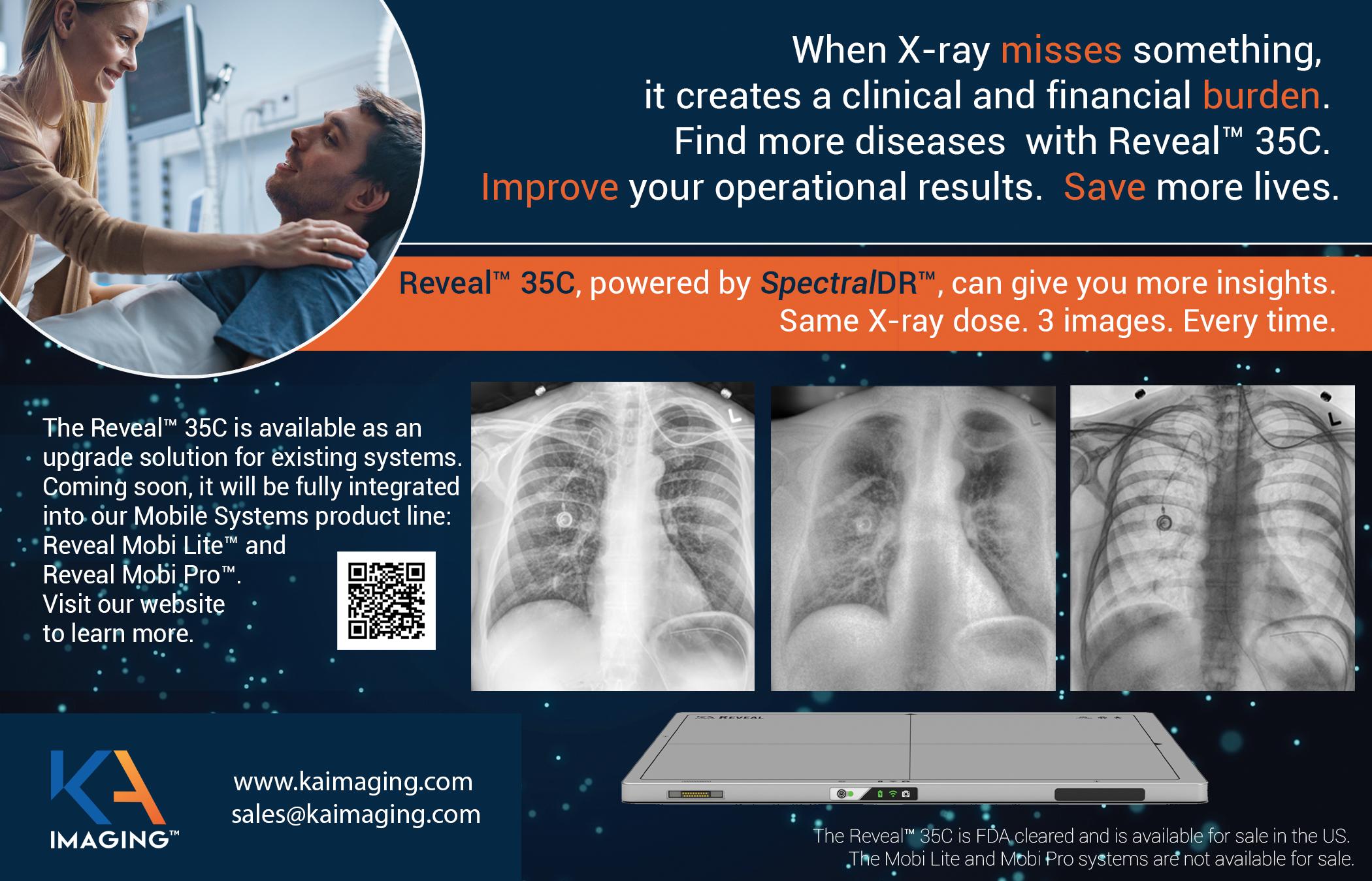
FOLLOW US ON SOCIAL MEDIA! COME VISIT US AT AHRA BOOTH 329 800-541-0632 brandywineimaging@comcast.net Offering the best reliability and support in the medical industry, just for you. Digital Imaging, X-Ray Sales and Service Company ICEMAGAZINE 61 WWW.THEICECOMMUNITY.COM

The Biergarten at Rathskeller Restaurant | 401 E. Michigan St. Indianapolis, IN 46204 Scan the QR code to RSVP for Drinks on ICE, sponsored by KMG. Then stop by AHRA booth #1228 or #1133 to pick up your item for entrance into the Drinks on ICE event. SPONSORED BY: MONDAY, JULY 10 7:30PM, DURING AHRA YOU ARE INVITED TO DRINKS ON ICE


ICEMAGAZINE 63 WWW.THEICECOMMUNITY.COM Contrast Injector Training • 100s of Error Codes not found in OEM Lit • Online and Onsite Training Available • Training BMETs since 2008 • Up to $5,500 in FREE Parts, PM Kits and Service • Massive Troubleshooting Library WWW.MAULLBIOMEDICAL.COM | 440-724-7511 | STEVE@MAULLBIOMEDICAL.COM ON-SITE AVAILABLE EXCLUSIVE CONTENT, INCLUDING: • BREAKING NEWS • DIGITAL ISSUES • WEBINARS • PRODUCT SPOTLIGHTS MAGAZINE FOLLOW US ON SOCIAL MEDIA!


ADVANCING THE IMAGING PROFESSIONAL 64 ICEMAGAZINE | JULY 2023 INDEPENDENT X-RA Y QUALITY ASSURANC E years of X-ray safety & quality Forty years of innovation and research to be the world-leading manufacturer of X-ray testing and QA solutions we are today. View our portfolio of multimodality solutions at www.rtigroup.com! Contact us for Trade In Promotion! CT ● Dental ● Rad/Fluoro ● Mammo Contact us RTI Group North America sales.us@rtigroup.com Phone: +1 800-222-7537 www.rtigroup.com

ICE Break Visit theicecommunity.com for the solution. IMAGING CROSSWORD LEADERSHIP SUMMIT APPLICATIONS OPEN! To apply, visit attendice.com/ice-18. FEBRUARY 18-20, 2024 • IRVINE, CA IMAGING CONFERENCE & EXPO
Across 1 One of the problems for medical imaging employers 8 Method of angiography, abbr. 9 ____ field gradient 11 Post-E.R. area 12 Viewpoints 17 Observe 18 Patient’s name and date of birth, e.g. 19 Concerned with identifying the cause of an illness 23 Are situated 24 Vein related 26 Type of scan 28 Increase 29 Plug completely or partially blocking a blood vessel 31 Org that develops and publishes international standards 32 It lies between the caecum and rectum 33 Positively charged particle: fundamental component of all atoms Down 1 Unit of equivalent dose 2 Small quantities 3 Big Apple, for short 4 Publicist’s concern 5 __ negotiable 6 Radiology output 7 Camera setting 10 Community connecting radiology professionals 13 ____ flag: indicator of danger or trouble ahead 14 Sudden uncontrollable wave of electrical activity on the brain, causing involuntary movement or loss of consciousness 15 X-ray, MRI, CT, for example 16 The equivalent of 100 rem 20 Disease also known as hyperthyroidism 21 Meet as a board 22 Abnormal sacs 25 _____meter: device which measures the volume of air movng in and out of the lungs 26 Arterial blockage 27 Gold symbol 30 Floor cleaner 31 Popular ICEMAGAZINE 65 WWW.THEICECOMMUNITY.COM
ICE2024
626 Holdings

9
Advanced Health Education Center
25 AHRA
44
ADVERTISER INDEX
ICE Webinars p. 18


AllParts Medical


Imaging Academy p. 2 Imaging Service Solutions p. 45 Intermed p. 29 International X-Ray p. 19


PM Imaging Management p. 19 Radon p. 19 Ray-Pac® Ray-Pac p. 68


57
Banner Imaging

57
Brandywine Imaging
61 CM Parts Plus
47 Engineering Services p. 3 HTMJobs.com
38
KA Imaging p. 61

KEI Medical Imaging p. 32 KMG p. 31


Maull Biomedical p.63 Metropolis International p. 64

MW Imaging Corp. p. 15
RIchardson Healthcare p. 45 RSTI p. 67

RTI Group North America p. 64
Technical Prospects p. 4
SOLUTIONS TriImaging Solutions p. 53 W7 Global, LLC. p. 49 X-Ray America p. 25
p.
p.
p.
p. 5 Althea p.
p.
p.
p.
p.
INDEX ADVANCING THE IMAGING PROFESSIONAL 66 ICEMAGAZINE | JULY 2023
RSTI is committed to improving the quality of diagnostic imaging service and helping students advance their careers through knowledge, education and hands-on, technical learning.
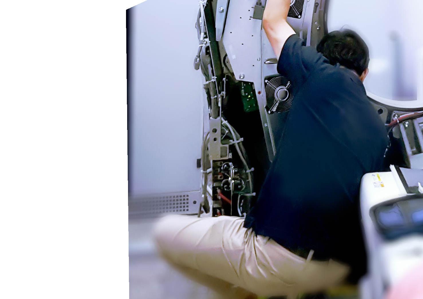
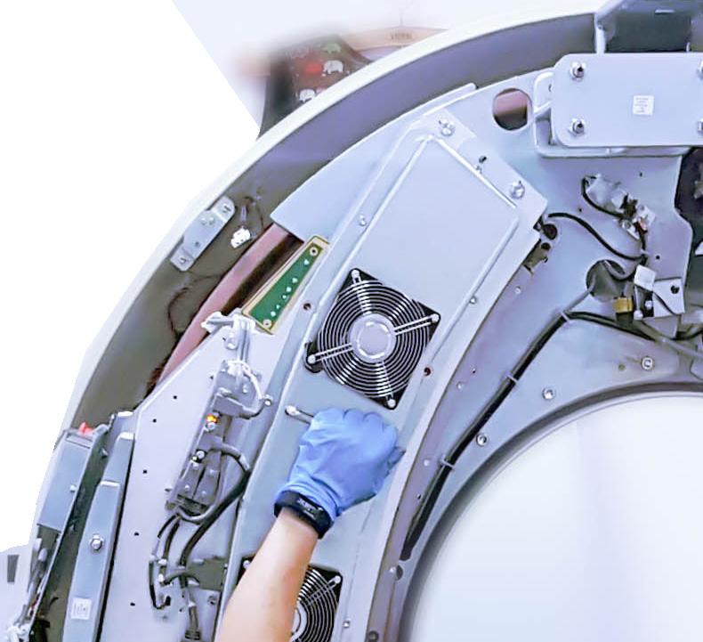
“Having completed Phase I: Principles to Servicing Diagnostic X-Ray Systems at RSTI was a large factor in getting selected for my current position with Mayo Clinic in the Medical Imaging Department.”
– Tim Z., RSTI Alumnus
“RSTI teaches you how to work on a modality, not single machines…learning the theory then the process of how the machine works and how to service it gives you a good foundation and makes you ready for any model.”
 – Alex P., RSTI Alumnus

ISO 9001:2015 CERTIFIED (IQC CERTIFICATE NO. Q-1158) STATE OF OHIO REG. NO. 93-09-1377T
COMPLETE SUPPORT PARTNERFOR OUR SERVICE CUSTOMER: • Training • Imaging Parts
Service Support
Technical Support • Equipment Options COMMITMENT TO OUR STUDENTS (440) 349-4700 parts@rsti-exchange.com registration@rsti-training.com CONTACT US:
TRAINING THE INDUSTRY SINCE 1985 – ALMOST 40 YEARS! Visit us at these shows! July: CFBIS (Central Florida) quarterly meeting August: CABMET (Colorado) symposium HEARTLAND (Nebraska) symposium NCBA (North Carolina) symposium Complete list of classes that we offer can be found at rsti-training.com
•
•
ENGINEERED FOR LEARNING ™
www.Ray-Pac.com 843-767-8090 Check Out Our NEW Website
