Volume 1, Issue 1
2010 Annual Symposium Lights, Camera,
SciENcE!
BuildiNg BoNE
from Stem Cells
Engineered Nanomaterials
nbt
Welcome As directors of Johns Hopkins Institute for NanoBioTechnology (INBT), we welcome you to our fourth annual symposium: Environmental and Health Impacts of Engineered Nanomaterials. The symposium is chaired by Jonathan Links, member of INBT’s executive committee and a professor in the Bloomberg School of Public Health, our co-host.
Peter Searson Director, INBT Joseph R. & Lynn C. Reynolds Professor, Materials Science and Engineering
Denis Wirtz Associate Director, INBT Director, Engineering in Oncology Center Theophilus H. Smoot Professor, Chemical and Biomolecular Engineering
For this morning’s talks in Sheldon Hall, Dr. Links has invited a diverse group of speakers including faculty from across Johns Hopkins who are currently investigating the various ways engineered nanomaterials may affect our health or that of our environment. Speakers will discuss topics such as the chemical characterization and environmental transport and fate of nanomaterials. They will address the ways that humans become exposed to nanomaterials and introduce us to state-of-the-art methods to assess exposure. Finally, we will consider the implications that consequences of exposure may have on the development of policy for science and industry. During the afternoon, please join us for a poster session in Feinstone Hall. Here, you will have an opportunity to browse a display of 100 posters, representing the multidisciplinary work in nanoscience from many of our more than 200 INBT affiliated faculty laboratories. The graduate and undergraduate students and postdoctoral fellows associated with this research will be on hand to discuss their work, and you will no doubt discover something fascinating in the disciplines of engineering, medicine, public health and the basic sciences. We also would welcome you to the inaugural edition of our new NanoBio Magazine. On pages 4 through 8, the magazine serves as a program for today’s events. In the pages that follow, NanoBio Magazine provides a broad overview of some of the newest innovations and discoveries coming out of INBT laboratories. We have designed a magazine that we hope will educate and inspire you with engaging stories and stunning photography highlighting our most recent achievements and expanding programs. As a companion to our quarterly newsletter NanoBio News, we hope NanoBio Magazine will become a publication to which you will refer again and again until we see you at the annual symposium in the spring of 2011. We would like to thank our corporate partners for their ongoing support of our endeavors both today and throughout the year. We also gratefully acknowledge our media sponsors for making this event and publication possible. Again, welcome and thank you for joining INBT for today’s symposium. If there is anything that we can do to make your experience with us here today more pleasant, please alert one of us, or an INBT staff member or student volunteer.
Visit us online: inbt.jhu.edu
Editor-in-Chief Mary Spiro mspiro@jhu.edu
Photography Will Kirk Mike Keung
Director of Corp. Partnerships Tom Fekete
Administrative Manager Susannah Porterfield
Art Director Yvonnia Martin yvonnia@ylmediallc.com
Contributing Writers Ashanti Edwards Tom Fekete Jacob Koskimaki Adam Shelley
Web Director Martin Rietveld
Senior Education Prog. Coordinator Ashanti Edwards
Photography Vasudev Bailey Rich Folkers
Research Service Analyst Christie Johnson
NanoBio Magazine is published annually and produced by
nbt NEB, 1st Floor 3400 North Charles Street Baltimore, MD 21218
All contents © copyright 2010 Johns Hopkins Institute for NanoBioTechnology. All rights reserved. Any use of the contents of this publication without express written permission of the publisher is strictly prohibited. NanoBio Magazine is an independent journal published annually by the Johns Hopkins Institute for NanoBioTechnology and is affiliated with Johns Hopkins University.
2 | Johns Hopkins NanoBio Magazine
Contents Volume 1, Issue 1
Symposium
Research (cont’d)
4 Symposium Agenda
20 Engineering in Oncology Center
5 Speaker Bio Highlights 6 Symposium Feature: Risk/Benefit
21 Mitochondria Mysteries
In Every Issue
2 Welcome Letter 32 Coming Attractions
ON THE COVER:
Corporate Partnerships
Education
10 Tool Repository
22 Science in the Summertime
12 Corporate Partner Program
24 REU
13 Profile: ARmark
25 Fall Retreat
Research
Outreach
14 Artificial Cilia
25
20
13
16 Bone from Stem Cells
26 Lights, Camera, Science!
18 Let’s Fold Small
27 Animation Studio
Cadmium Selenide quantum dots like these will fluoresce in a range of colors depending on their size. But the small size of nanoparticles also could cause them to be a potential hazard to health and the environment. Photo: Rich Folkers/NCI
6
2010 Environmental and Health Impacts of Engineered Nanomaterials | 3
Environmental and Health Impacts of Engineered Nanomaterials Thursday, April 29, 2010 Johns Hopkins Bloomberg School of Public Health 615 N. Wolfe Street
Greetings from the Symposium Chair Thank you for participating in the annual symposium of Johns Hopkins Institute for NanoBioTechnology! As one of the co-founders of INBT, I have always been excited to be part of an endeavor that, in my opinion, optimally approaches the research and development of the novel technology that nano represents. From day one, this approach has explicitly included consideration of its health and environment effects. Past symposia have focused primarily on potential benefits of nanobiotechnology … and there are many! This year, we focus on challenges that nano may pose to health and the environment. Past symposia have included these issues, but this is the first to focus exclusively on this important topic. Why? Because only now has the field matured to the point where meaningful conversation can take place. I am particularly pleased at how many distinguished Hopkins faculty are qualified, engaged, and willing to speak about the range of challenges nanotechnology presents. Today’s speakers and topics cover the “waterfront” of issues, testimony to the critical mass of efforts occurring at Hopkins. I hope you enjoy the day! Jonathan Links Professor and Deputy Chair, Johns Hopkins School of Public Health Department of Environmental Health Sciences; Director, Center for Public Health Preparedness; Deputy Director, Office of Critical Event Preparedness and Response
4 | Johns Hopkins NanoBio Magazine
8:00 a.m.
Registration, Monument Street Entrance
8:30
Welcome and Introductions, Sheldon Hall W1214 Jonathan M. Links, Professor, Environmental Health Sciences Bloomberg School of Public Health
8:45
Potential Impacts of Engineered Nanomaterials: What we know; what we need to know Ellen Silbergeld, Professor, Environmental Health Sciences, Bloomberg School of Public Health
9:30
Modification and Characterization of Engineered Nanomaterials D. Howard Fairbrother, Professor, Chemistry, Krieger School of Arts and Sciences
9:50
Environmental Transport, Transformation, and Fate William P. Ball, Professor, Geography and Environmental Engineering Whiting School of Engineering
10:10
Break, Courtyard
10:30
Exposure Assessment of Nanomaterials Patrick Breysse, Professor, Environmental Health Sciences Bloomberg School of Public Health
10:50
Transport of Nanomaterials Across Mucus Membranes Justin Hanes, Professor, Ophthalmology, School of Medicine
11:10
Example Toxicity Assessment: Neurotoxicity Tomás Guilarte, Chair, Dept. of Environmental Health Sciences Columbia University Mailman School of Public Health
11:30
Policy Implications of Nanomaterial Risk Assessment Ronald White, Associate Scientist, Health Policy and Management Bloomberg School of Public Health
12:00
Break
1:30
Poster Session/Poster Judging, Feinstone Hall E2030
3:00
Award Ceremony and Adjourn
Speaker Bio Highlights Jonathan M. Links is a medical physicist, professor and deputy chair of the Department of Environmental Health Sciences in Bloomberg School of Public Health with joint appointments in Radiology and Emergency Medicine at the Johns Hopkins School of Medicine. He directs the CDCfunded Johns Hopkins Center for Public Health Preparedness, a training center for frontline public health workers, and is principal investigator of the CDC-funded Johns Hopkins Preparedness and Emergency Response Research Center.
Professor Patrick Breysse is director of the Division of Environmental Health Engineering and director of the Occupational and Environmental Hygiene Academic Program at the Bloomberg School of Public Health. He also directs the Center for Childhood Asthma in the Urban Environment. His work focuses on evaluation and control of chemical, biological, and physical factors that can impact health or well being and includes research on risk/ exposure assessment.
Ellen Silbergeld, professor of Environmental Health Sciences at the Bloomberg School of Public Health, has served as scientific advisor to federal agencies including CDC, EPA, DOE, and OSHA, Maryland and New York, World Bank, ILO, UNEP, and PAHO. Her research bridges science and public policy, focusing on incorporation of mechanistic toxicology into environmental and occupational health policy, including those related to the cardiovascular risks of arsenic, lead, and cadmium; immunotoxicity of mercury compounds; and health and environmental impacts of industrial food animal production.
Justin Hanes is a Professor of Ophthalmology in the Johns Hopkins School of Medicine. He also has appointments in Biomedical Engineering, Chemical and Biomolecular Engineering, Environmental Health Science, Neurosurgery and Oncology. His research interests are at the intersection of nanotechnology with medicine and public health.
Howard Fairbrother is a professor of chemistry in the Krieger School of Arts and Sciences. Fairbrother’s research focuses on surface chemistry with an emphasis on the role of interfacial phenomena in the deposition of nanostructured materials and the environmental fate and impact of carbon based nanomaterials in aquatic environments. One aspects of his research aims to develop an artificial plasma environment to study the microscopic surface processes that are collectively responsible for the deposition and structural characteristics of nano-scale carbonaceous thin films.
Tomás R. Guilarte, formerly of the Bloomberg School, was recently appointed chair of the Department of Environmental Health Sciences at Columbia University’s Mailman School of Public Health. His research focuses on mechanism-based neuroscience and neurotoxicology using behavioral, cellular, and molecular approaches, ranging from studies using primary culture of neural cells to the application of brain imaging technologies.
Environmental engineer William P. Ball is a professor of Geography and Environmental Engineering at the Whiting School of Engineering with a joint appointment in Civil Engineering. Among his research interests, Ball seeks a better understanding of the potential environmental impact and fate of carbonbased nanomaterials in aquatic systems, with focus on multi-walled carbon nanotubes. Ball is an adjunct research professor for the Chesapeake Research Consortium.
Ronald White is an associate scientist in the Bloomberg School’s Department of Health Policy and Management with joint appointments in Environmental Health Sciences and Epidemiology. White is deputy director of the Risk Sciences and Public Policy Institute where his research seeks to improve the science base for risk assessment, cultivate better risk assessment methods, and enhance the risk management process. His research areas include health effects of air pollution; environmental health policy and risk assessment.
2010 Environmental and Health Engineered Nanomaterials |5 2010 Environmental andImpacts Health of Impacts of Engineered Nanomaterials |5
Researchers Seek to Assess All Aspects of Nanotech Use
By Mary SpIro
S
cientists and engineers have referred to nanotechnology—science at the scale of just a few atoms—as an “enabling technology” with the ability to improve applications in industry, medicine and basic scientific research. Engineered nanomaterials, such as carbon nanotubes (CNTs) and nano-silver, are revolutionizing many commercial technologies and are found in more than 800 products including cosmetics, cell phone batteries, and sporting equipment, as reported by the Project on Emerging Nanotechnologies at the Woodrow Wilson International Center for Scholars. According to Nanoparticle News Review, the market for engineered nanomaterials could exceed $27 billion over the next five years. However, the small scale of these materials coupled with their unique physicochemical properties may cause some engineered nanomaterials to pose potential risks to human health and the environment. Nanomaterials exist at a sub-cellular scale, on the order of one-billionth of a meter, allowing some of them to slip through the blood-brain barrier. Nanoparticles also have been detected in rivers and streams and in the tissues of aquatic species. Consequently, intense scientific research is now
6 | Johns Hopkins NanoBio Magazine
being directed at understanding the possible environmental, health and safety risks posed by engineered nanomaterials. The fourth annual symposium for Johns Hopkins Institute for NanoBioTechnology (INBT) explores nanotechnology in terms of human and environmental risk assessment, management and policy. “Environmental and Health Impacts of Engineered Nanomaterials,” held April 29 in collaboration with the Johns Hopkins Bloomberg School of Public Health (SPH), brings together faculty experts engaged in various aspects of risk assessment and management research. Jonathan Links, professor in the department of Environmental Health Sciences at the School of Public Health assembled the slate of speakers from across four divisions of the university. This diversity reflects the multidisciplinary approach needed to effectively address questions about how nanomaterials move through and interact with the environment and how they may impact biological organisms, including humans. Links said, despite some concerted efforts to assess risk, many questions remain unanswered about how engineered nanomaterials and nanoparticles impact
human health and the environment. “Without these data, we are flying blind. But when risk assessment is performed in tandem with research into beneficial applications, it helps researchers make better decisions about how nanotechnology is used in the future,” Links said. Links points to historical cases where research and development failed to recognize risks to health and the environment until after a beneficial advancement was already in widespread use. For example, the fibrous substance asbestos made construction materials flame retardant but is now linked to lung diseases. Chlorofluorocarbons made effective air conditioning coolants but are now associated with the depletion of the ozone. “By studying potential risks to human health and the environment handin-hand with benefit-driven research and development, it gives us the best chance to reduce risk proactively, while maintaining the benefits” Links said. INBT has supported research into the potential health and environmental impacts of nanomaterials from its inception. Institutional research in this realm aims to answer four primary questions:
How do nanomaterials enter and move through the environment? How are humans exposed to the materials? How do these nanomaterials affect humans? Who is most at risk? Symposium presenter William P. Ball investigates the environmental transport, transformation and fate of engineered nanoparticles, specifically multi-walled carbon nanotubes, one of the most commonly used engineered nanoparticles. “Industry values carbon nanotubes because they are exceptionally strong and possess unusual chemical, optical and electrical properties,” said Ball, a professor in the department of Geography and Environmental Engineering at the Johns Hopkins Whiting School of Engineering. “But CNTs also are capable of binding a great deal of toxic heavy metal on their surfaces because they have an extremely high surface area to mass ratio,” said Ball. “Even small amounts of CNTs have the potential to transport relatively large amounts of adsorbed toxins.” This property, Ball said, “turns carbon nanotubes into ‘Trojan horses,’ bringing toxic contaminants to locations that they may not otherwise reach. If the nanotubes wind up in the food chain, they could deposit toxic metals in the stomachs of animals or humans,” Ball added. Howard Fairbrother, professor of chemistry in the Krieger School of Arts and Sciences, has collaborated with Ball Next page
Quantum dots, like these made of Cadmium Selenide could be used to detect cancer. But scientists and engineers want to know about their potential risks as well. Photo: Rich Folkers/NCI
While researchers ought to devote plenty of resources toward developing beneficial medical and biological applications of nanotechnology, they must also realize that this is only part of the story...”
2010 Environmental and Health of Engineered Nanomaterials |7 2010 Environmental andImpacts Health Impacts of Engineered Nanomaterials |7
Risk/Benefit, Continued from page 7 on carbon nanotube research, particularly with regard to ways the material’s surface chemistry affects its ability to adsorb these toxins and modify its colloidal stability. Manufacturing processes and functionalization strategies often result in the carbon nanotubes (CNTs) being “decorated” with different types of chemical functional groups, Fairbrother said. Among the most important surface modifications is the introduction of an oxygen-containing functional group. “When oxygen-containing functional groups are added to the surfaces of the carbon nanotubes, their stability in water greatly increases, which influences their fate and transport in aquatic environments. Our studies have shown that even comparatively small changes in surface chemistry can exert significant
Hanes has been working on getting drug-loaded nanoparticles through the mucus barrier to treat disease. Recently, his group devised a biodegradable nanoparticle that successfully slips through natural human mucosal secretions, such as those found in the lungs or female reproductive tract. This technology could be used as a drug delivery system to treat cystic fibrosis or small cell lung cancer, for example. However, Hanes also realizes that, even as effective as mucus is at keeping unwanted things out, the mucus mesh is not a perfect barrier. “Its netting contains many small holes, just big enough to let the smallest particles pass through,” he said. But by chemically manipulating the mucus mesh, Hanes devised a method to block the passage of some harmful parti-
When we know what the potential problems are at an early stage, we can adjust our research and development pathways to mitigate risk and maximize benefit.” changes in both the colloidal stability and sorption properties of CNTs,” Fairbrother explained. Fairbrother discusses these issues in his symposium talk on the modification and characterization of engineered nanomaterials. The findings of studies like those of Ball and Fairbrother will no doubt impact the future decisions of policy makers and regulatory agencies about the use of nanoparticles in products that come into contact with water and of scientists and engineers who are trying to develop “safe by design” nanomaterials. But nanoparticles can enter and move through the environment in other ways as well. For example, nanoparticles may become airborne as a result of manufacturing and pollution. Justin Hanes, a professor in the Department of Ophthalmology at the School of Medicine, investigates nanoparticle transport through human mucus for both beneficial purposes and as a measure of potential risk. Hanes, who also has an appointment in the Whiting School, discusses this topic at the symposium. 8 | Johns Hopkins NanoBio Magazine
cles. A technology like that could reduce worker exposure to nanoparticles in a manufacturing setting or prevent harmful viruses from transmitting disease. “We used a detergent common to many personal care products to shrink the holes in the mucus mesh so that nanoparticle movement slowed dramatically, especially in the 200- 500 nanometer range,” Hanes said. The effect may be sufficient to provide protection from exposures to nanoscale materials—from viruses expelled in a cough or sneeze to particles aloft in exhaust fumes. The theme of this year’s symposium, Links said, demonstrates that “while researchers ought to devote plenty of resources toward developing beneficial medical and biological applications of nanotechnology, they must also realize that this is only part of the story. When we know what the potential problems are at an early stage, we can adjust our research and development pathways to mitigate risk and maximize benefit.” ®
Thank you INBT would like to thank our Corporate Partners for supporting us today and throughout the year:
BIg RESEaRCH
Tool ToolRepository RepositoryOffers OffersSmall SmallSolutions Solutionstoto
QuESTIONS By Mary SpIro
T
o be useful, a tool should be where you can find it--like neatly organized in a tool box. Nanotools are no different, and many useful ones with applications for science, engineering and medicine are made right here at Johns Hopkins University. So that other researchers at Hopkins, and elsewhere, can easily find these tools, the Johns Hopkins Institute for NanoBioTechnology (INBT) recently launched a nanobiotechnology “tool” repository. This online site provides an overview of nanobiotechnology related devices, materials and methods available from researchers at various campus divisions. The repository aims to be a one-stop-shop for researchers looking for small scale solutions to use in their research projects. “For applications such as diagnostics, targeted drug delivery, and the study of cellular interactions at the molecular level, a nanoscale tool can provide important new data,” says INBT director Peter Searson, the Reynolds professor of materials science and engineering in the Whiting School of Engineering. The idea for the repository grew out of comments made by Chi V. Dang, vice dean for research at the School of Medicine and professor of cell biology, oncology and pathology. “A site like the tool repository is one step toward increasing and improving cross-disciplinary research efforts at Johns Hopkins,” says Dang. Each entry in this virtual nano-tool box includes an image, a short description, suggested applications, relevant publications, and contact information for the tool developer. The tool entries are linked to the personal profiles of the
10 | Johns Hopkins NanoBio Magazine
INBT affiliated faculty members who may post items in the repository. That way, visitors to the tool repository can find out what other types of research the developer is involved or interested in. Although only INBT affiliated faculty may add tools to the site, anyone can browse the entries in the tool repository through the institute’s website. “Even investigators who had not previously considered using nanobiotechnology may find just what they are
Cadmium Selenide quantum dots of different sizes fluoresce in a rainbow of colors. Photo: Searson Group
looking for in the NanoBio Tool Repository,” Searson adds. Researchers also can use the repository to identify potential collaborators within research groups synthesizing novel materials or developing methods for nanobiotechnology research. In addition, the repository serves as a way to facilitate industry collaboration with university researchers. “These tools will increase the university’s potential for collaboration with industry,” says Denis Wirtz, associate director of INBT and Smoot professor of chemical and biomolecular engineering. “I expect that Hopkins investigators, as
well as researchers from industry, will search the tool repository to find nanomaterials as well as potential faculty collaborators for a variety of projects. Such interaction with commercial enterprises provides avenues for future investigation, for commercialization of Hopkins-made technologies, and establishes networks that will help graduates of Johns Hopkins find jobs.” Currently about a dozen tools are listed in the tool repository, but this number is expected to increase rapidly once faculty become aware of it, Wirtz adds. Entries include items such as quantum dots made of cadmium selenide and cadmium telluride from the Searson Lab that can be used to image live cells. The Searson lab also has posted a method for creating an artificial double layered membrane that behaves much like a membrane in a living cell. David Gracias, associate professor of chemical and biomolecular engineering, has developed “tetherless” micro-grippers that can be controlled biochemically that could have potential surgical applications. David Goldberg, associate professor of chemistry in the Krieger School of Arts and Sciences, has synthesized nanoparticles that mimic the shape and chemical properties of the certain organic compounds, such as porphyrin. INBT’s leaders hope that as more people become aware of the tool repository, more faculty members will add their own nanobio tools to its catalog. As the listings grow, they anticipate the interest and demand for the repository’s tools also will increase. ®
inbt.jhu.edu | 11
INBT’s I Corporate Partnership Program paves pathway between business and bench By ToM FEkETE
Doctorial student Janice Lin uses Langmuir-Blodgett deposition. Photo: Rich Folkers/NCI
In my role as the Director of Corporate Partnerships for Johns Hopkins Institute for NanoBioTechnology (INBT), I meet regularly with business and industry leaders. Scientists and engineers from industry are eager to learn about the Institute, but why would a corporation consider partnering with INBT? INBT possesses a unique multidisciplinary research structure and educational philosophy. It is focused on creating new knowledge at the interface of nanoscience and medicine. Over 200 researchers from across Hopkins, representing the School of Medicine, Whiting School of Engineering, Krieger School of Arts and Sciences, Bloomberg School of Public Health and Applied Physics Laboratory are engaged in dozens of programs linking basic engineering and science with cellular and molecular dynamics to find new approaches to therapeutics and diagnostics for a wide variety of diseases. Elements of nanotechnology, novel tools and techniques, new approaches to surface and molecular engineering form shared approaches to solve complex problems. Along with faculty researchers, INBT also supports more than 30 graduate fellows and postdoctoral fellows. These young people, with their creativity, energy and drive, rev the engines of innovation at INBT and bring fresh perspectives from diverse disciplines to collaborative corporate partnerships. They also form the next generation of scientists, physicians and engineers who will be at the forefront of developing new technologies whether in academics or industrial laboratories. From the very beginning, INBT’s leaders recognized that successful translation of basic research into viable tools, techniques, and devices requires a further broadening of our multidisciplinary approach and a robust outreach and collaborative partnership with industry and government. At the same time, corporations in market sectors such as pharmaceuticals, medical devices, medical instrumentation, sensors, microelectronics, advanced materials, and chemical products see the potential in nanobiotechnology and nanomedicine. Technology invented in INBT laboratories offers dramatic opportunities for new products and treatments to benefit society. Enhanced external focus and collaborative industrial partnerships
12 | Johns Hopkins 12 | 2010 JohnsNanoBio Hopkins Magazine INBT annual Symposium
Justin Galloway, doctoral student, synthesizing CdSe quantum dots. Photo: Rich Folkers/NCI
are vital ingredients required to successfully transition technologies created at the university to the marketplace. Our Corporate Partners play a crucial role in helping INBT research teams define objectives and identify barriers to commercialization. They also work with academic researchers to identify products for end-users and are involved in the development of techniques for large-scale manufacturing processes. The Corporate Partnership Program establishes a framework to facilitate interactions with companies interested in a mutually beneficial relationship with INBT. We recognizes that our partners have diverse interests and objectives. Every partnership agreement is tailored to the needs of both the academic and the corporate researchers involved. Partnerships can focus on collaborative research projects including joint grants, sponsored research, accelerated access to new knowledge as well as establishing better personal ties and bridges with undergraduate and graduate students, researchers and faculty through internships, sabbaticals, information exchanges and technology transfer. The road from bench to bedside is treacherous. It is just as important to breakdown internal barriers to collaboration between disciplines as it is to collaborate with the commercial sector to traverse the difficult translational pathway to technology use and to do good basic science. INBT’s Corporate Partnership Program acknowledges and embraces this challenge. For answers to questions about INBT’s Corporate Partnership Program, call Tom Fekete at 410-516-8891 or contact him via email at tfekete1@jhu.edu.®
ARmark manufactures these 100% solid extruded microspheres using High Definition Micro Extrusion (HDME). Photo: ARmark.
Corporate Partner Profile:
aRmark Indisputable By Mary SpIro
F
ingerprints are one way to prove that a person is who he says he is. But how does one authenticate an electronic device, a document, or pill without interfering with its function or purpose? That’s the job of Pennsylvania-based company ARmark Authentication Technologies, LLC , a subsidiary of Adhesives Research and corporate partner of Johns Hopkins Institute for NanoBioTechnology (INBT) since 2009. ARmark develops made-to-order authentication systems that help organizations protect their brands and prevent counterfeiting. Through a proprietary technology, ARmark developed markers called “information centric microtags” that have become valuable to the pharmaceutical, medical device, luxury goods, secure documents, foods and clothing industries. The microtags can display any type of information required for authentication, such as logos, batch codes, or pharmaceutical formulas. The company claims the microtags work so well, their results are “indisputable,” and they use the word as part of their trademark. Although the microtags are small, that’s not the reason ARmark has an interest in working in nanobiotechnology. The company’s latest endeavor involves a uniquely devised Microvector™ Drug Delivery Technology. “More than 75 percent of our corporate research focuses on health care,” says Peter Gabriele, a technical director at ARmark and co-inventor of the microvector technology. “We are one of three divisions of Adhesives Research. Adhesives Research has been involved in the development of other drug delivery systems such as transdermal patches and microfluidic devices. Our ARx division developed the dissolvable drug delivery technology for Theraflu strip.” By marrying the technologies used in the microtag to their
knowledge of drug delivery, Gabriele says, the next logical step was to create something like the microtag with a therapeutic cargo. “If we could load the microtag up with data, why not a drug?” he says. Under Gabriele’s direction, the company’s ARmicron Life Science section fabricated the Microvector™ via a process called high definition micro-extrusion (HDME). Gabriele describes this process as something like a “vertical dot-matrix printer.” The Microvector™ material builds up into a long strand that can be sliced into shorter sections. When the sections contact liquid, they ball up into uniformly shaped spheres that encapsulate their pharmaceutical payload. “It’s a very stable microparticle that holds nanoparticles like passengers on a ferry,” Gabriele says. The HDME method can create Microvector™ of different shapes and with different properties. Just by changing the materials used, Microvector™ that heat up or shine when imaged can be quickly made, Gabriele says. ARMark sees numerous opportunities in their affiliation with INBT, and he says the corporate partnership program is a perfect union of forward-thinking and pragmatism. “All of a sudden, it makes the dream concept a little more realistic because the INBT researchers recognize what is merely theoretical and what has practical potential,” Gabriele says. “The idea of having a commercial entity working with an academic institution generates the type of conversation that gets you to your endpoint faster.” To read more about ARMark’s Microvector™ Drug Delivery Technology, go to http://www.rmark.org/login.html®
2010 Environmental and Health Engineered Nanomaterials | 13 2010 Environmental andImpacts Health of Impacts of Engineered Nanomaterials | 13
Naturally inspired:
Nanoparticles Self-assemble Into artificial Cilia By Mary SpIro
B
chanical energy into electrical energy.” In a fluid filled environment and supported by a silicon wafer, the filaments are built up, nanosphere-bynanosphere, much like adding beads to a string. However, no string holds the 25 nanometer diameter beads together. Instead, they order, self assemble and cling together via magnetic attraction. Even when the magnetic force fields are removed, the artificial cilia remain intact. Because of this construction, the fabricated cilia are thin, long and flexible. “A single filament can be as many as 2000 particles in length but just one particle in diameter,” Benkoski said. Benkoski said the need for such a material developed from the fact that “even though microchip and microsensor technologies were advancing rapidly, the technology for mechanical actuators associated with them lagged far behind. It limited what you could do.” Working with Jeffrey Pyun and colleagues at the University of Arizona, the APL group used a cobalt particle with thin polymer coating to construct the cilia. They next applied a magnetic field to the solution of particles, which helped draw and align the particles into continuous linear structures. “But even before we applied the magnetic field we noticed that the particles were starting to form chains,” he said. Benkoski also said the manner in which the Mixtures of 250 nm magnetite (red) and 25 nm cobalt (grey) nanoparticles artificial cilia self assembled organize into flagella-like assemblies. The “head” plus “tail” structures flick back mimicked the way that actin and forth under the influence of an alternating magnetic field to swim much like spermatozoa. Images: JHUAPL fibers grow in cells.
acteria flick long tails, called flagella, to get around. Cells that line your lungs, stomach and that help you hear wave their cilia to push fluids and molecules across their surfaces. Now, scientists at the Johns Hopkins Applied Physics Laboratory (APL) and the University of Arizona want to mimic these naturally occurring structures and motions to create a synthetic version, built from just a few nanoparticles. Researchers hope these biomimetic cantilevers and swimmers will be used as microsensors in devices that detect biological agents, identify voices and even swim through the body to deliver drugs. Jason Benkoski, a senior scientist at APL and an affiliated faculty member of Johns Hopkins Institute for NanoBioTechnology, has been taking nanospheres of cobalt and magnetite, applying magnetic forces and creating self-assembling artificial cilia and flagella that undulate and wave much like the real thing. “It’s the simplest kind of actuator,” Benkoski said. “We can envision this being used in fluid transport, locomotion, acoustic detection, and transferring me-
14 | Johns Hopkins NanoBio Magazine
The fibers wave and swipe in response to applied magnetic fields. Benkoski said technology like this could be developed into nanoscale “windshield wipers” for self-cleaning surfaces or micropumps to produce flow in microfluidic devices. Beyond the construction of artificial cilia, Benkoski’s group also has fabricated a structure resembling a bacterium with a flagellum—or tail. These “artificial swimmers” are made of a larger 250 nm diameter head made of superparamagnetic magnetite with an attached 25 nanometer diameter ferromagnetic cobalt tail several micrometers in length. Like the cilia, the flagellum of this fabricated bacteria bend and flip much like the real thing, albeit in response to a magnetic pulse. Movies of the devices show how easily they propel themselves through fluid. “Like E. coli, they don’t use much energy to get around,” Benkoski said. Although nanotools like the artificial cilia and swimmers are likely decades away from being used in living organisms, applications for devices like sensors probably are not that far off. Benkoski said. “You could make them out of any material as long as it was magnetic and biologically compatible.” ®
For further reading: Dipolar assembly of ferromagnetic nanoparticles into magnetically driven artificial cilia. Jason J. Benkoski, Ryan M. Deacon, H. Bruce Land, Lance M. Baird, Jennifer L. Breidenich, Rengaswamy Srinivasan, Guy V. Clatterbaugh, Pei Yuin Keng and Jeffrey Pyun. Soft Matter, 2010, 6, 602-609
Be remarkable.
And maximize your unique perspective alongside today’s most brilliant minds. Your individuality makes you a valuable team member here. Because shaping some of the world’s most critical technologies is a highly collaborative endeavor. The Advanced Technologies, Advanced Materials Group at BAE Systems is focused on developing technology solutions in chemical and biological sensing and detection, alternative power and energy, and imaging. Located at the Steiff Silver building near the Homewood campus of JHU and in Columbia, MD, the group consists of senior scientists from venerable institutions like MIT, Harvard, NIH, NIST amongst others and offer a gold mine of learning opportunities. Our scientists come from a variety of disciplines; chemistry, biology, materials science, physics and electrical engineering and have a range of expertise from nanomaterials to laser systems. At BAE Systems, we are looking for outstanding research engineers and scientists across a broad range of disciplines, especially those who have been cross trained in institutes like the INBT and can work in multi-disciplinary areas involving chemistry, physics, biology, and other areas of science. Common qualities required in each discipline are a complete and thorough understanding of the field, the ability to confidently address a broad range of research questions, the ability to work effectively in small interdisciplinary teams, and excellent interpersonal and communications skills. For senior positions, a track record of success in research and development, and experience working with customers to define and execute research programs are also required. We are always interested in increasing the depth and breadth of our group! With more than 100 new inventions every year from a global workforce of 100,000+ highly skilled people and 2008 sales of $34.4 billion, BAE Systems is the 3rd largest global defense company to offer unprecedented opportunities worldwide. Applicants selected may be subject to a security investigation and must meet eligibility requirements for access to classified information. U.S. citizenship is required. BAE Systems offers excellent benefits. For detailed information and for a full listing of all opportunities, please visit our Web site at www.baesystems.jobs EOE, M/F/D/V.
Building Bone from Stem Cells Bioreactor Could Propel Regenerative Medicine to the Next Level
By JaCoB koSkIMakI
R
Custom-designed perfusion bioreactor for cultivation of anatomically-shaped human temporomandibular joint (TMJ) bone grafts. Photo: Keith Yeager
16 | Johns Hopkins NanoBio Magazine
egenerative medicine may offer hope for those afflicted with injury, birth defects or disease by having damaged tissues and organs replaced by those grown in cell culture. Yet, the complex biological and engineering challenges required for making a functional organ in a laboratory have limited advancements in the field. Using a specially designed bioreactor and stem cells harvested from a patient’s own body, Warren Grayson, an assistant professor of Biomedical Engineering at Johns Hopkins University School of Medicine, may have cleared a hurdle on some of those limits to grow one of the most complex joints in the human body. Grayson, an affiliated faculty member of Johns Hopkins Institute for NanoBioTechnology, and his colleague Gordana VunjakNovakovic from the Columbia University Laboratory for Stem Cells and Tissue Engineering, designed a method for using stem cells to grow a biologically viable temporomandibular joint (TMJ) The TMJ is the joint linking the jaw to the base of the skull. The method, developed prior to Grayson joining the Johns Hopkins faculty, uses a bioreactor design that can be tailor-made to the anatomy of individual patients.
At the world’s first TMJ conference in 2006, it was reported that TMJ disorders affect nearly 25 percent of the population, yet current methods to correct defects are limited to autologous tissue grafting, where bone is transplanted from one part of the body to another. This method requires harvesting tissue elsewhere in the body and is painful and invasive for patients. According to Grayson, “other approaches, such as injecting bone marrow or stem cells directly into damaged regions, are severely limited in their ability to regenerate large bone defects.” Synthetic materials have shown promise, yet remain difficult to integrate with host tissue. Engineering scaffolds that allow for host cells to regrow may be the best way to integrate engineering design with the body’s own cells and ability to heal. Grayson’s approach uses patientderived adult stem cells, and a scaffoldbioreactor system. The bioreactor is essentially a mini-biological ‘womb’ mimicking the physiological environment required for development allowing cells to develop into viable structures. “Bioreactors train cells to form functional constructs through the application of tissue-specific biophysical and biological cues,” he explains. By carefully providing
TMJ encircled. Image: Grayson Lab
growth factors, mechanical forces, and temperature/pH control, the cells can grow into a new physiologically-viable TMJ bone graft, which Grayson hopes will one day be used for implantation in patients. Published in the Proceedings of the
Warren Grayson, Assistant professor, biomedical engineering.
Bioreactors train cells to form functional constructs through the application of tissue-specific biophysical and biological cues...”
National Academy of Sciences, his early studies have shown healthy bone cells and mineral deposition, indicating they are “fully osteogenic;” that is, expressing the proteins and genes sufficient for implantation. The thing that sets Grayson’s bioreactor apart is that the grafts cultivated can be geometrically accurate and patientspecific. Using high-resolution data from computerized tomography to define exact dimensions and shape of the joint and cells harvested directly from patients, this method can be used to tailor bone grafts to each patient’s unique anatomy. “Geometry is a big part of their function. This affects how they articulate with neighboring bones and is a significant component affecting how the joint actually performs,” Grayson says. Aside from patient-to-patient variation, the TMJ has a complex shape, and reconstructing it serves as a proof of principle that this technique could be employed to develop other bone constructs, each of unique anatomy and size. This technique may be especially advantageous for patients with facial trauma, where appearance is a primary concern. Being able to provide anatomicallyshaped grafts would be a considerable advantage that would change how these patients are currently treated, Grayson explains. Grayson’s lab also researches basic stem cell biology, trying to understand exactly how populations of stem cells develop into multi-cellular tissues by creating novel methods to alter the signals the cells see at different points in the bioreactors and over the cultivation period. Varying such conditions can impact how different cells within the
constructs organize themselves into hierarchical structures similar to healthy bone tissue. Understanding stem cell constructs for bone will allow expansion to other tissues such as cartilage, which also can be grown in bioreactors in tandem with bone and which could potentially lead to the growth of entire biological joints, Grayson suggests.
geometry is a big part of their function. This affects how they articulate with neighboring bones and is a significant component affecting how the joint actually performs.” Grayson hopes his constructs will bridge the gap between basic biology studies of stem cell properties and translational clinical research. He also has started using mathematical models to better understand and more finely tune the complex developmental process in making functional bone tissue from stem cells. The next step, he says, is to test the joints in animals to determine host integration with tissues before moving to clinical testing. ® Jacob Koskimaki is a NanoBio IGERT doctoral fellow in biomedical engineering working in the Popel Lab.
2010 Environmental and Health Impacts of Engineered Nanomaterials | 17
SMaLL
Pushing the limits of nanofabrication into the third dimension
By Mary SpIro
W
Tetherless thermobiochemically actuated microgrippers Gracias Lab/JHU
18 | Johns Hopkins NanoBio Magazine
hen Johns Hopkins University engineer David Gracias started building self-assembling structures that were millimeters to tens of microns in size, he knew he’d developed something with staggering potential. Self-loading micro-containers developed by members of his lab might one day carry life-saving drugs or protect transplanted cells from the immune response. Gracias, an associate professor of chemical and biomolecular engineering and a member of the Johns Hopkins Institute for NanoBioTechnology (INBT), wondered how small he could make these objects. How far could one push the limits of nanofabrication, especially into the third dimension? It turns out that the answer to that question is pretty darn small. Gracias and his colleagues recently constructed a 100 nanometer cubic container with precisely patterned surfaces. One nanometer is one-billionth of a meter in length or about 1/100,000th the width of a human hair. “These cubes are about 15 times smaller than anything we’ve constructed. They are ten times smaller than a bacterium and are the typical size of viruses or small organelles within eukaryotic cells. Previously, no one has been able to sculpt a 3D nanoparticle with precisely patterned surfaces, so this is a big leap in human engineering,” Gracias said.
Research A report on this achievement was published in Nano Letters in 2009 and describes the work of chemical and biomolecular engineering postdoctoral student Jeong-Hyun Cho. Constructed in two-dimensions upon a silicon wafer support, Cho’s nanoboxes are made using a three step process that enables the creation of miniaturized versions of pat-
Self-folding structures (a) Nanoscale boxes with precisely patterned surfaces (b) Self-folding metamaterials with large numbers of hinges (c) A self-folding patterned tissue scaffold that spontaneously rolls up into a cylinder. Photos: Gracias Lab
terned macro objects found in our daily lives. “Everything has multilayer patterns on it; look around you,” Gracias said. “A closet may be thought of as a box made of ‘A,’ with hinges made of ‘B,’ and a handle made of ‘C’.” For Cho’s nanoboxes, electron beam lithography laid down a coating of nickel (or alumina) in precise spots to create the box panel faces in two dimensions. Then, a layer of tin was added to pattern the hinges. Finally, the faces of the panels were chemically etched in gold—in this case with the letters J-H-U—using line widths as small as 15 nm. The etching process caused the box panels to rotate slightly, lift from their silicon support, and fold on their own. Making such precise 3D patterned objects at this scale is technically challenging. One of the hardest parts was getting the panels to fold at a 90 degree angle. “No one had shown even a single folded panel at a right angle on this size scale, so this was completely uncharted territory,” he said. “Present day nanoparticles have very limited patterns; for example nanowires can have ring patterns, but it is hard to imagine constructing too many things, especially in 3D, with only these building blocks,” Gracias continued. Multilayer patterned 3D polyhedra increase the possibilities.” Recently, the Gracias laboratory also has been successful in creating curved nanoparticles such as patterned nanotubes. This work was published in Advanced Materials.
The idea that you can make such intricate structures out of a simple, flat, piece of paper drove us to see if we could create intricately structured materials at the micro-scale...” But why is patterning important? “Circuits have patterns, so do cell membranes and viral surfaces. Patterns can code information for biological recognition or templating. Patterns can also interact with electromagnetic fields and enable heterogeneous integration of electronic devices,” Gracias explained. Dielectric polyhedra with metal patterns are expected to interact with light in novel ways, Gracias said, so he also envisions applications in optics. “We are also exploring the creation of circuits on the surfaces of these polyhedra to enable ‘smart’ nanoparticles. Also, the containers have the volume of a typical virus, so we could put small molecules, proteins, or DNA inside.” Surface patterns functionalized with molecular receptors can then code for precise docking or recognition.
Nanoscale Boxes
Members of the Gracias team wanted to see what else they could fold at the nanoscale. Inspired by the Japanese art forms of origami (paper-folding) and kirigami (paper-cutting) and even children’s pop-up books, doctoral students Noy Bassik and George Stern fabricated “metamaterials” able to self-fold at 90 degree angles in two directions. “The idea that you can make such intricate structures out of a simple, flat, piece of paper drove us to see if we could create intricately structured materials at
the micro-scale,” Gracias said. “In Japan, pop-up books are not just for kids. Some of them are quite complicated; people have replicated cathedrals, landscapes and faces. We initially started with a simple pop-up structure modeled after ones in books to see if we could achieve folding in both directions at the same time.” Similar to Cho’s nanoboxes, the faces of these folded structures can be modified, in this case with square or triangular pores. However, unlike Cho’s structures, Bassik and Stern’s metamaterial have thousands of hinges. “Yet remarkably, they fold up precisely on their own,” Gracias said. Bassik and Stern refer to this method as “hands-free” origami and the article “Microassembly based on hands-free origami with bidirectional curvature” appeared in Applied Physics Letters in late 2009. It took some trial and error, but after tinkering around with various combinations of materials with differing thicknesses, the team developed a pattern of copper and chromium layers that would create the self-assembling 3D cubic structures. “Even sophisticated tools are not small enough to fold up these structures at small size scales, so we are pretty much forced to engineer self-folding structures at these size scales,” Gracias said. Like the nanoboxes and curved nanostructures, the self-folding metamaterials possesses could enable novel optical and structural properties. “The goal for optics is to precisely guide the light waves where you want them to go. This enables optical functionality useful for sensing, communication and defense needs. The concept of using periodocity metamaterials to shape optical properties is not new, however, our approach enables the creation of 3D and isotropic metamaterials, which have previously been challenging to fabricate” Gracias said. Another possible application for self-folding metamaterial is tissue culture, Continued on page 28
2010 Environmental and Health Impacts of Engineered Nanomaterials | 19
Engineering in Oncology Center Physical scientists partner with physicians to tackle cancer By Mary SpIro
EOC hosts fellowships, professorships Johns Hopkins’ Engineering in Oncology Center is recruiting recent MDs and PhDs to its Physics of Cancer postdoctoral fellowship training program. Fellows train across disciplines including engineering, physics, and cancer biology to develop new models for cancer research and technologies to enable an inside-view of cancer cell functions. EOC also hosts a short-term Distinguished Visiting Professorship in Physics of Cancer. Participants in this program conduct interdisciplinary and collaborative research at the interface of physics, engineering, and cancer biology. Faculty with expertise in engineering, physics, biophysics, cancer biology, and developmental biology are encouraged to apply.
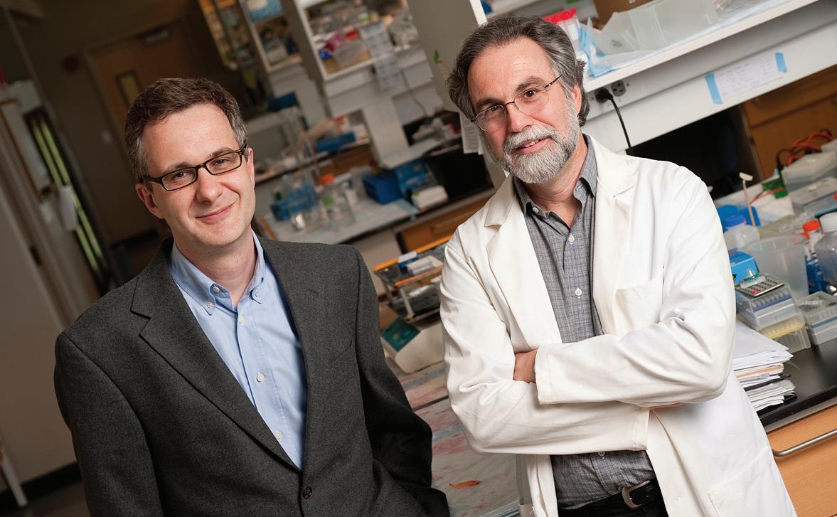
Denis Wirtz (left) and Gregg Semenza (right) direct the new Johns Hopkins Engineering in Oncology Center. Photo: Will Kirk/JHU
J
ohns Hopkins new Engineering in Oncology Center (EOC) brings together physical scientists and clinicians to unravel the physical underpinnings of cancer spread—metastasis. Funded with $14.8 million from the National Cancer Institute’s (NCI) Physical Sciences-Oncology Centers (PS-OC) program, EOC researchers hope to discover never-before-considered approaches to cancer therapy and diagnostics. EOC is one of 12 NCI-funded PS-OCs across the nation designed to bring a new cadre of theoretical physicists, mathematicians, chemists and engineers to the study of cancer. “Cancer cells break free from primary tumor, penetrate into the bloodstream, evade host defenses, become stuck to the interior walls of blood vessels and travel to other organs, where they set up new cancer cell colonies. Tumor cells push on and are pushed by mechanical forces within their microenvironment. If we can better understand this process, we may find better ways to treat cancer,” said center director Denis Wirtz, Theophilus H. Smoot professor of chemical and biomolecular engineering in the Whiting School of Engineering.
20 | Johns Hopkins NanoBio Magazine
Gregg L. Semenza, C. Michael Armstrong Professor in Medicine and founding director of the School of Medicine’s Institute for Cell Engineering vascular program, serves as EOC’s associate director. Semenza and Sharon Gerecht, assistant professor of chemical and biomolecular engineering, lead a research project analyzing the makeup and physical properties of extracellular matrix, the 3D scaffold in which cells live. Wirtz and Greg D. Longmore, a cancer cell biologist at Washington University in St. Louis lead a project to study the physical basis for cancer cell adhesion and de-adhesion and how it increases the chance that cancer cells will break free, move into the bloodstream and migrate to other tissues. Konstantinos Konstantopoulos, professor and chair of the Whiting School’s Department of Chemical and Biomolecular Engineering, and Martin L. Pomper, who holds appointments in the School of Medicine’s Department of Radiology and the Kimmel Cancer Center lead the center’s third project team to investigate fluid mechanical forces at different oxygen tension microenvironments on tumor cell signaling, adhesion and migration.
Network Investigators Meeting During NCI’s First Annual Physi-
Bryan Smith (Stanford) and Christopher Hale (JHU) shared in PSOC’s Young Investigators Trans-Network Award. Photo: Mary Spiro
cal Sciences-Oncology Centers Network Investigator’s Meeting, April 5-7, in Washington, D.C., EOC director Denis Wirtz presented a tutorial on particle tracking, presented a talk on mechanobiology, and chaired a panel discussion on cancer cell mechanics. In addition, Wirtz’s doctoral student Christopher Hale and his postdoctoral fellow Daniele Gilkes both received Young Investigators TransNetwork Project Awards for their poster presentations. More than 22 Johns Hopkins PS-OC people (faculty, postdocs, and students) attended. ®
To find new therapeutic targets in cancer cells, the Hill Lab studies how proteins, such as fission 1 (red), control shape and distribution of mitochondria (green). DNA shown in blue. Images: Cara Marie Manlandro
Toward Solving the
Mysteries of the Mitochondria By adaM SHEllEy
O
ne of the grandest works of the modern age is our understanding of the cell. Medicine, biology, and much of human health and wellness rests on the details of that smallest of machines. But how well do we understand what powers those cells? And what happens when that power source bites the bullet? Blake Hill, associate professor of biology in the Krieger School of Arts and Sciences at Johns Hopkins University and affiliated faculty member of Johns Hopkins Institute for NanoBioTechnology, came to this question from an unusual angle. Originally a student of biochemistry, he worked under William DeGrado at the University of Pennsylvania on a far different problem: the nature of proteins inside the cell, and if it was possible to make artificial proteins. “DeGrado’s research focuses on building proteins from scratch,” Hill explained. “We should be able to design functioning, native proteins.” These attempts at protein design led Hill to work heavily in understanding why a protein folds the way it does and the difficulties one encounters: in particular, a sometimes overlooked problem where a single protein can be folded in multiple ways, giving it different properties. When designing proteins these multiple folding arrangements are a great difficulty that must be overcome as it prevents easy prediction of the final structure. At other times though, when studying the cell’s own proteins, these transformations from one arrangement to another are key to understanding what’s
going on. One such time when proteins rearrange themselves is in the mitochondria, the power plant of the cell, when the cell is dying. “That’s what led us to the fundamental biological question: how mitochondria maintain their size and shape in living and dying cells,” Hill said. The mitochondria are a part of the cell that has always been an oddball in their behavior. While they are critical for keeping us alive, transforming sugar and raw materials into energy, they are a world apart from the rest of the cell. With their own separate genetic code, hundreds of unique proteins and an unusual process of joining together and splitting apart from the rest of the cell’s cycle, the mitochondria behave like few other things. In addition to their oddities in life, their mechanisms for dying are equally unique. In a process currently under study by many, including Hill, some proteins on the mitochondria’s surface are “multi-state proteins’ which can re-fold into a new shape and be absorbed by the mitochondria. In the case of one protein, Bcl-xL in particular, BAX, we know that in the end this creates large changes in the mitochondria as a whole and leads to the mitochondria’s death. However, the ‘how’ and ‘why’ of this protein’s behavior is still in many ways a mystery. As Hill admitted, “We have no idea what they’re doing at the mitochondrial membrane. It’s one of the most studied proteins in the last ten years, and we still don’t know.” Here, the question of how and why it folds the ways it does could yet bring great insight into the
field, and this question is a deadly serious one. With a firm understanding of how cells die, and how they are told to die, new horizons of treatment options for a variety of diseases may open up. “Cells have an innate suicide mechanism.” Hill explains. “The body needs to maintain tight control of this process.” Too many cells being told to die can kill off the wrong cells and lead to diseases like Alzheimer’s. If there are too few cells dying, there are many other problems. Cancer, almost by definition, is what occurs when cells do not respond to signals to die. Also virtually every viral defense the body has relies on instructing the right infected cells to die at the right time, while the virus tries to stop this process. If these key mitochondrial proteins are part of how a cell is told to die, then by activating them or suppressing them at the right times we may yet be able to tell the body how to correct a situation once it’s gone wrong. Currently one of the proteins under study in the Hill Laboratory, Bcl-xL, is a target for drug discovery with clinical trials under way. Other aspects of mitochondrial behavior during cell death are further off from final medical applications, however, the potential is astounding and so work continues. It’s quite likely that it will only be a matter of time and of investigation, until these questions can be put to rest. ® Adam Shelley is a NanoBio IGERT doctoral fellow in physics working in the Edidin Lab.
2010 Environmental and Health Impacts of Engineered Nanomaterials | 21
A Plaza in Leuven, Belgium.
Visiting Cologne, Germany.
Science in the SuMMERTIME INBT/IMEC program sends students to Belgium for research internships
By Mary SpIro pHoToS By MIkE kEUNG
A
select group of students from Johns Hopkins University will spend 10 weeks during the summer of 2010 in Leuven, Belgium conducting research at The Inter-University MicroElectronics Centre (IMEC). Faculty affiliated with Johns Hopkins Institute for NanoBioTechnology (INBT) worked cooperatively with researchers at IMEC to choose students to participate in projects of mutual interest to both institutions. INBT will fund four students this summer through an IRES (International Research Experience for Students) grant received from the National Science
22 | Johns Hopkins NanoBio Magazine
Foundation. Students work at IMEC’s world-class microfabrication facility and learn to design, fabricate and test a wide range of biomedical devices. This is the second year that INBT has offered the IRES program at IMEC. During the summer of 2009, INBT sent Mike Keung to work at IMEC. Keung recently completed a bachelor’s degree in chemical and biomolecular engineering with a minor in entrepreneurship and management and is now working toward a master’s degree. While an undergraduate student, Keung assisted in the laboratory of associate professor David Gracias in the
department of chemical and biomolecular engineering and accumulated nearly two years of experience working in micro- and nanofabrication. He also had participated in previous summer corporate internships, most notably at Exxon-Mobil. When Keung left for Belgium in May 2009, he had fairly narrow expectations for what he wanted to achieve. “I hoped to learn new fabrication techniques and ways of perfecting the current ones I was already familiar with. By bringing these back to the Gracias lab, I hoped to improve our own fabrication methods as well as learn more about
Microfabrication
. EC. Photo: IMEC
laboratory at IM
The Eiffe
l Tower,
The
EC e to IM
of cours
e!
nc entra
The people at IMEC are very dedicated to their research. It was nice being able to spend my entire day working on one project. also, I had time to be exposed to the research and ideas of those around me...” the biological side of nanotechnology,” Keung said. But upon his return, Keung said the internship experience was far richer than he had imagined. “The environment at IMEC is similar to an academic environment,” Keung said. “There are many young people of all ethnicities, and most of them speak English. The dress code is casual. There are many people working on postdocs or PhDs.” But, since IMEC is a corporate research and development organization, there also were some major differences. For example, the facilities at IMEC were state-of-the-art with devices he had not used before, such as a scanning electron microscope. But when Keung wanted to use a specific piece of equipment for an experiment, he had to schedule the experiment with the technician in charge of operating that machine. “At a university, you can run your experiments pretty much when you want to,” Keung said. “But at IMEC the work day was the typical 9 to 5, so it could take a while before the technician could get to your sample.” Although he was not able to operate the equipment himself, he was happy to have the results from highly trained technicians who had had years of training and experience using the advanced tools. Keung also took advantage of his time in Europe. On
weekends, Keung took short trips and was able to tour major cities such as Paris, London, Amsterdam, Cologne, Antwerp, Brussels, Ghent and Bruges. He took many photographs, most of which can be seen at the blog he kept of his work and travels. Read his blog at http://keungatimec.blogspot.com/. Keung said his time at IMEC was an amazing research experience. “The people at IMEC are very dedicated to their research. It was nice being able to spend my entire day working on one project. Also, I had time to be exposed to the research and ideas of those around me, just by walking around and chatting with my coworkers. It helped me become more independent and definitely gave me a taste of what working in a corporate environment would be like,” Keung said. “I also learned that the food markets close early and that a bicycle is absolutely necessary to get around. Nearly everyone bikes in Belgium,” Keung added. Both undergraduate and graduate positions are available for the IRES program at IMEC. Students from underrepresented racial/minority groups and women in science and engineering are encouraged to apply. The application process for Summer at IMEC 2011 will open in December 2010. ®
Environmental and Health Impacts of Engineered Nanomaterials | 23
Summer Program Draws
Diverse, Talented Future Researchers
By aSHaNTI EdwardS
T
he National Science Foundation (NSF) reported in November 2009 that over the last decade about 10 times more white students earned doctoral degrees in science and engineering disciplines than did minority students. Acknowledging this fact, yet resolving not to accept it as status quo, Johns Hopkins Institute for NanoBioTechnology (INBT) employs aggressive measures to increase the number of individuals from under-represented groups who apply to its educational programs. INBT embraces the challenges of recruiting minorities. As INBT’s Senior Educational Program Coordinator, every year I go out to several conferences across the country to recruit the best diverse students to INBT’s many programs. These meetings include the Historically Black Colleges and Universities Undergraduate Program National Research Conference, the annual meeting of the Society for Advancement of Chicanos and Native Americans in Science, among many others. INBT has so many unique educa-
tional programs that we are able to pull students in through diverse pathways. We have graduate and postdoctoral programs and an international research experience program that is open to both graduates and undergraduates. One of the most successful and popular programs for INBT has been its NSF funded summer internship program—the NanoBio Research Experience for Undergraduates (REU). Now beginning its third year, INBT’s REU has consistently attracted the best and the brightest students interested in research from top universities across the nation. The REU program was launched as a conduit to attract highly talented and motivated students to pursue academic careers in research, particularly women and minority scholars. INBT’s REU provides housing and a stipend for students so that they can come to Johns Hopkins for 10 weeks in the summer to conduct research under the guidance of faculty and graduate student mentors. The program is highly competitive. For summer 2010, the number of applicants for the 10 slots
Students spend 10 weeks conducting research in INBT faculty labs. Photo: Vasudev Bailey
in the program rose to more than 400, almost twice what it had been the year before. We have been told that this is a high number of applicants. Why has the REU program at INBT been so successful? Many college recruiters, program administrators, admissions directors, and faculty find it a challenge to recruit a diverse selection of qualified students, but we have found at INBT that our summer REU has been a great tool for recruiting. We are building a pipeline, starting with our summer program. We have found that the minorities and female students who enroll in our summer program, not only participate in national and scientific conferences after their summer with us, but most go on to apply to several graduate programs and even to Johns Hopkins University. We receive a lot of applicants to our program, but there is a certain type of student we are after. To gauge the ideal REU candidate, you have to understand the goal of our REU and that is to give the students a graduate experience in interdisciplinary research in hopes that they in return to Hopkins to pursue graduate studies after their undergraduate degree. We are looking for wellContinued on page 29
24 | Johns Hopkins NanoBio Magazine
), Terrence Binder (NTCM eakers. s (IGERT), Zev us at between sp Ke ch t T) at ER M t, (IG au at From lef Kh m ya HMI) and Sh Dobrowsky (H
INBT Doctoral and Postdoctoral Fellows
‘Retreat’ from the Lab By Mary SpIro
G
raduate students and postdoctoral fellows affiliated with Johns Hopkins Institute for NanoBioTechnology (INBT) clock many hours in the laboratory. INBT gives them a little break at the annual Fall Retreat. The most recent gathering, held November 21, 2009 at the Mount Washington Conference Center, focused on the theme “Career Pathways for Scientists and Engineers: What happens after the PhD?” Participants come from INBT’s various graduate and postdoctoral programs, including the National Science
Postdoc Daniele Gilkes (center) listens to MedImmune’s Cindy Oliver during discussion session.
From left, Jesse Placone, Andrew Wong, Charli Dvoracek and Meghan Vellotti.
Foundation funded Integrative Graduate Education and Research Traineeship (IGERT), the National Institutes of Health T-32 Nanotechnology for Cancer Medicine (NTCM) postdoctoral training program, and the Howard Hughes Medical Institute (HHMI) NanoBioMed graduate fellowship. INBT currently hosts more than 30 students through these programs. Participants heard Courtney Stelling, a senior scientist at Becton Dickinson Diagnostics of Sparks, Maryland and recent PhD graduate, explain the career development pathway provided to “newbies” at her company. Cindy Oliver, vice president for development at MedImmune in Gaithersburg, added her perspective as a woman scientist with many years of experience at all levels of the corpo-
rate world. The faculty presentation came from Assistant Professor Warren L. Grayson from the Department of Biomedical Engineering at the School of Medicine. You can read about Grayson’s bone tissue bioreactor on pages 16 and 17 of NanoBio Magazine. Lively discussions followed the presentations and students broke out into groups: one set of those nearing graduation who met with Stelling and Oliver and another set of students just starting out in the programs who met with INBT director Peter Searson and associate director
Laura Ensign (HHMI) has a comment. Photos: Mary Spiro
Denis Wirtz. The new students learned about graduation requirements and discussed developing courses in entrepreneurship and teaching. Participants in the group with postdocs or soon-to-be PhDs discussed the current job market and opportunities in academia and industry. The value for professional development that resulted from this informal and conversational approach at the retreat was best summed up in a comment from IGERT fellow Julia Swavola, doctoral student in chemical and biomolecular engineering working with the Bevan Group. “I knew that becoming affiliated with INBT would be different, but I had no idea that it would help me figure out my future. This was really great!” Swavola said. ®
2010 Environmental and Health Impacts of Engineered Nanomaterials | 25
Lights Camera
SCIENCE! By Mary SpIro
E
verything about movie making seems so glamorous. From beautiful stars to special effects, making films might appear magical. But actually, when you break it down, shooting a film is not unlike performing experiments in a lab. And, just as reading the script would be far less entertaining as seeing a film, reading a protocol might be confusing until the steps were performed in real life. That’s the philosophy behind a new effort at Johns Hopkins Institute for NanoBioTechnology: produce short films describing recently published research and the protocols that go with them. The movies are produced collaboratively with INBT’s science writer Mary Spiro, INBT’s Animation Studio director Martin Rietveld, and the scientists and engineers involved. The INBT Animation Studio already has several research-oriented films to its credit. The animation skills of Rietveld and his student crew have taken us inside a lipid bilayer and carried us along a fiber of collagen. INBT also has produced several video news releases using the talent of students in the annual science communication course. Recently, however, INBT produced its first film describing a protocol from Nature Methods. Investigators Bridget Wildt, a PhD in materials science and engineering, Peter Searson, Reynolds Professor of Materials Science and Engineering, and Denis Wirtz, Smoot Professor of Chemical and Biomolecular Engineering, served as technical consultants for the production. Materials science and engineering PhD candidate Yu-Ja Huang performs each step in assembling the Hopkins team’s device and demonstrates how to conduct programmed cell detachment experiments. “Studying cell detachment at the subcellular level is critical to understanding the way cancer
Photos: Mary Spiro
26 | Johns Hopkins NanoBio Magazine
Photos: Mary Spiro
INBT web director Martin Rietveld worked with predoctoral student Yu-Ja Huang, Materials Science and Engineering, to develop the shots for each step in the experimental protocol film, “Programmed subcellular release for studying the dynamics of cell detachment.”
cells metastasize,” Searson said. “Development of scientific methods to study cell detachment may guide us to prevent, limit or slow down the deadly spreading of cancer cells.” Using a draft script developed by Wildt and Searson, Spiro simplified the text further for narrator, materials science and engineering PhD candidate Andrew Wong. Rietveld recorded Huang as he performed the protocol and refined the script further during filming. Viewing the final cut, Wong was able to read the script in a conversational and friendly tone. You can watch the version of this new protocol video on INBT’s YouTube channel. The film may never earn an Academy Award, but we hope it will help specialists, and even the general public, to understand this unusual and complex procedure. http://www.youtube.com/user/JHUINBT
Eye-catching animations that Help
Visualize the Nanoworld By Mary SpIro
T
he Animation Studio at Johns Hopkins Institute for NanoBioTechnology (INBT) continues to expand and produce more eye-popping animations to help describe science too small to see. Animation studio director Martin Rietveld currently supervises the work of two students formally enrolled in the independent study course, Animation in Nanotechnology and Medicine. Two other previously enrolled students have continued on in the studio as assistants. “Exciting visuals that show exciting science will keep being useful for INBT in its education as well as its outreach efforts,” Rietveld said. “This also applies to the newly launched Engineering in Oncology Center. The ability to communicate abstract concepts in mere seconds is strongly present in animation. It is therefore an excellent means for telling the story behind any type of science but in particular the kind that takes place on the nanoscale.” Still images from recently completed projects can be seen here. Upcoming projects include an illustration of the process of cancer metastasis based on the work of Denis Wirtz, director of the Engineering in Oncology Center and Smoot Professor of Chemical and Biomolecular Engineering. Another movie will show the differentiation of stem cells into blood vessels, based on the research of Sharon Gerecht, assistant professor, also in Chemical and Biomolecular Engineering. Students who have enrolled in
the Animation in Nanotechnology and Medicine course have come from diverse departments at Johns Hopkins University, as well as from the Maryland Institute College of Art. Rietveld expects participants to be self-starters and self-directed learners, using online tutorials to get up to speed on the various software programs needed to make the animations. Prior to the development of each animation, students learn about the research through interviews with the faculty members and graduate students involved in the work and by reading the current literature on the subject. The course follows the basic animation pipeline from concept to post production. Students at all levels and from all disciplines with an interest in art and animation are invited to enroll in the course. For more information, contact Martin Rietveld at Rietveld@jhu.edu.®
From the film “Self-folding cubes” by David Filipiak and Martin Rietveld and based on the research of David Gracias, Chemical and Biomolecular Engineering, Whiting School of Engineering. Cross-sectional illustration of collagen fibers from the film “Collagen Mimetic Peptides” by Ella McCrea (MICA), Nathan Weiss, Martin Rietveld, Jay Corey and Mary Spiro and based on the research of Michael Yu, Materials Science and Engineering, Whiting School of Engineering.
Based on the work of Blake Hill, Biology, Krieger School of Arts and Sciences, Ammon Posey and Martin Rietveld created this animated film describing Bcl-xL, a transmembrane molecule of the mitochondria.
2010 inbt.jhu.edu 2010 Environmental Environmental and and Health Health Impacts Impacts of of Engineered Engineered Nanomaterials Nanomaterials inbt.jhu.edu || 27 27
Let’s Fold Small, Continued from page 19 especially as the 3D geometries designed become more complex. A recent paper in the journal Biomaterials by Mustapha Jamal, an INBT IGERT fellow in chemical and biomolecular engineering, explores the concept of a “selfassembling” scaffold. “Hybrid structures of cells and supports can form complex, yet anatomically relevant, geometries, such as cylinders, spirals and bidirectionally folded sheets. These geometries are found in the vasculature, the cochlea of the ear, and the intestines, respectively. Mustapha’s work represents a first step towards the construction of self-assembling tissues,” said Gracias. These are just a few of the self-assembly technologies coming out of the Gracias Lab. Many more can be found on the Gracias Lab web page and several have been written about previously on the INBT website and in the popular science press. To find out more, visit the Gracias Lab page at http:// www.jhu.edu/chembe/gracias/® References: Curving nanostructures using extrinsic stress, J.-H. Cho, T. James and D. H. Gracias Advanced Materials (2010) DOI: 10.1002/adma.200904410. Self-assembly of lithographically patterned nanoparticles J. H. Cho and D. H. Gracias, Nanoletters 9, 12, 4049–4052 (2009). Directed Growth of Fibroblasts into Three Dimensional Micropatterned Geometries via Self-Assembling Scaffolds, M. Jamal, N. Bassik, J.-H. Cho, C. L. Randall and D. H. Gracias Biomaterials (2010) DOI:10.1016/j.biomaterials.2009.11.056. Reversible actuation of microstructures by surface chemical modification of thin film bilayers J. S. Randhawa, M. D. Keung, P.Tyagi and D. H. Gracias, Advanced Materials (2010) DOI:10.1002/adma.200902337. Tetherless thermobiochemically actuated microgrippers, T. G. Leong, C. L. Randall, B. R. Benson, N. Bassik, G. M. Stern and D. H. Gracias, Proceedings of the National Academy of Sciences (PNAS) 106, 703-708 (2009). Self-assembled three dimensional radio frequency (RF) shielded containers for cell encapsulation B. Gimi, T. Leong, Z. Gu, M.Yang, D. Artemov, Z. M. Bhujwalla and D. H. Gracias, Biomedical Microdevices 7 (4) 341-345 (2005).
28 | Johns Hopkins NanoBio Magazine
Undergraduate, Continued from page 24 rounded students, who are versed in their discipline, either through current research or through advanced studies, but who are not afraid to cross the boundaries of disciplines. Ideally, we pair our REU students with a lab that has one of our INBT graduate training grant or postdoctoral fellows. These more advanced scholars serve as mentors. This is a perfect union because the student or postdoctoral fellow is able to truly interact with the summer student and show them interdisciplinary research and the graduate experience, as well as the INBT environment. Through several organized activities, our REU participants also have a chance to meet students in other summer research internship programs at Hopkins. At the end of the 10 weeks, all the students from the summer research programs from across campus come together to present the results of their work at a collaborative poster session held at the School of Medicine. A few have even made arrangements to work a few more weeks in their faculty labs before the fall semester starts. The REU application process is 100 percent electronic. Applicants must have at least a 3.0 grade point average and be pursuing an engineering or science degree. The applicant simply has to answer two questions, send in an unofficial transcript and then have two recommenders send in a form with a letter of recommendation. INBT’s 2010 REU students will arrive on campus to begin their research on June 1. Applications for the summer 2011 INBT REU will be available online after December 1, 2010 with a deadline in mid February 2011. ® For more information about the INBT REU program or any of our educational programs go to: http://inbt.jhu.edu/education
T H E N AT I O N A L A C A D E M I E S P R E S S
EXPLORE NaNOtEchNOLOgy REsOuRcEs from THE NATIONAL ACADEMIES | Advisors to the Nation on Science
A Matter of Size: Triennial Review of the National Nanotechnology Initiative
Implications of Nanotechnology for Environmental Health Research
search 11752
search 11248
Review of Federal Strategy for NanotechnologyRelated Environmental, Health, and Safety Research
NAKFI Designing Nanostructures at the Interface between Biomedical and Physical Systems
search 12559
search 11317
download these and more for free at www.nap.edu
2010 Environmental and Health Impacts Engineered Nanomaterials | 29 2010 Environmental and Healthof Impacts of Engineered Nanomaterials | 29
Outreach
Over Over its 110-year its 110-year history, history, BD BD hashas led led thethe medical medical technology technology industry industry by partnering by partnering with with leading leading organizations organizations to address to address global global health health issues issues thatthat areare devastating devastating to human to human life.life.
Partnering Partneringfor forhealth health Helping Helping to provide to provide relief relief in the in the wake wake of natural of natural andand man-made man-made disasters. disasters. Helping Helping to eliminate to eliminate deadly deadly diseases diseases likelike measles, measles, maternal maternal andand neonatal neonatal tetanus tetanus andand TB. TB. Fighting Fighting thethe spread spread of diabetes. of diabetes. BD BD serves serves healthcare healthcare institutions, institutions, lifelife science science researchers, researchers, clinical clinical laboratories, laboratories, industry industry andand thethe general general public public every every day.day. Around Around thethe globe globe BD BD manufactures manufactures andand sellssells a broad a broad range range of medical of medical supplies, supplies, devices, devices, laboratory laboratory equipment equipment andand diagnostic diagnostic products. products.
1
1 2 2 ™ ™ FORTUNE, FORTUNE, March March 2010,2010, Ethisphere Ethisphere Magazine, Magazine, AprilApril 20102010 Please Please visit visit www.bd.com www.bd.comBD and BD and BD Logo BD Logo are trademarks are trademarks of Becton, of Becton, Dickinson Dickinson and and Company. Company. ©2010 ©2010 BD BD
Named Named oneone of the of the World’s World’s Most Most Admired Admired ®1 Companies Companies as®1 well as well as one as one of the of the World’s World’s Most Most 2 Ethical Ethical Companies, Companies, BD2 BD is privileged is privileged to work to work with with governments, governments, non-profit non-profit groups groups andand other other organizations organizations to address to address broad broad health health issues, issues, develop develop an array an array of responses of responses andand pioneer pioneer new new technologies. technologies. BD BD – Helping – Helping all people all people livelive healthy healthy lives. lives.
INBT Coming Attractions Professional Development Seminars
Visit INBT at USA Science & Engineering Festival
Johns Hopkins Institute for NanoBioTechnology (INBT) will host four professional development seminars for scientists and engineers this summer. These seminars aim to expand student’s knowledge of issues and ideas relevant to, but outside of, the laboratory and classroom experience. Topics include intellectual property, commercialization and entrepreneurship, science journalism and publishing, ‘life after college,” and much more. Talks will be held at 11 a.m., on June 16, June 30, July 14 and July 28 at a location to be determined. All JHU/JHMI and APL faculty, staff and students are invited to attend these free seminars designed to promote discussion and interaction with scientific and engineering professionals. Please visit http://inbt.jhu.edu for more details on speakers and locations closer to the scheduled dates.
Two weeks of free events in the Washington, D.C. area lead up to a two-day Science Expo on the National Mall, October 23-24, 2010. Johns Hopkins Institute for NanoBioTechnology will join more than 350 of the nation’s leading scientific organizations for this first-of-its-kind science and engineering extravaganza. INBT will present an exhibit on nanobiotechnology and self assembly. More than 750 exhibits spanning aerospace, green energy, medicine, biotechnology, climatology, robotics, nanotechnology, botany, neuroscience, genetics, and many other scientific disciplines will be free to all. The festival is hosted by Lockheed Martin. For a complete list of sponsors, partners and exhibitors go to www.usasciencefestival.org.
Institute for NanoBioTechnology NEB, 1st Floor 3400 North Charles Street Baltimore, MD 21218
nbt